Almost every person has moles, or skin changes. Usually we treat them calmly, but even the smallest formations can become dangerous if they degenerate into malignant tumors. Histological analysis will help determine the degree of danger of a particular mole. Let's figure out why they send it for histology, whether any mole needs to be sent for analysis, and how the degree of danger is determined in each case.
The essence of the procedure
The histology of a nevus consists of examining a small part of it. The examination will reveal the presence or absence of cancer cells in the mole.
Histology is especially necessary if there is a border nevus. It belongs to a more dangerous formation.
To undergo the test, a referral from an oncologist or the person’s own desire is required. A biopsy is taken using the following methods:
- A piece of biomaterial intended for research is taken during surgery. The excised material is transported to the laboratory in a special container.
- Using the needle method. An area of nevus tissue is excised using a dermopanch. Before the procedure begins, the person is given an anesthetic.
- The excisional method is performed during surgery. The entire body of the mole is removed. The intradermal nevus is sent for histology along with the healthy cells that were located near it.
After collecting the material or completely removing the mole, it is immersed in a container with formaldehyde, where it can be stored for up to 25 days.
The duration of storage does not affect the quality characteristics of the material. The condition for this is the amount of formalin in the container: it should be several tens of times greater than the volume of the nevus.
What to do after analysis
If the analysis does not reveal any malignant cells in the nevus, you can sleep peacefully. But if melanoma cells were found in a mole, the patient needs to contact an oncology clinic to determine further treatment. It must be remembered that in the early stages, melanoma and other malignant skin processes are well treated. It is important to start treatment on time!
We recommend that you familiarize yourself with the video material we have prepared on the topic of diagnosing nevi, performing tests and histology:
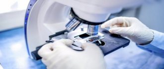
Stages of histological examination
There are several stages of histological examination. I won’t go into detail about their details, I’ll just list them and briefly explain the essence:
- Biopsy, i.e. removal of a mole. The main thing I want to note here is that there is never “too little” material for research. They often write to me: “The doctor said that the formation was too small, there would be nothing to send for examination, so I did not have a histology.” In my practice, there have been cases when a normal conclusion was received for a mole up to 1 mm in size.
- Fixing the material. To prevent the mole from decomposing while it is being transported to the laboratory, it is necessary to destroy microorganisms and deactivate the enzymes of the mole cells. This is done using special solutions. Immediately after removal, the mole is placed in a formaldehyde solution. You can replace it with alcohol, but this option is less common.
- Washing the material. To remove traces of the fixing liquid, the material is washed, as a rule, for several hours in running water.
- Dehydration . Preliminary preparation for paraffin filling.
- Compaction (filling). The washed and dehydrated mole is filled with liquid paraffin. This is necessary so that the thinnest sections can be made, suitable for examination under a microscope. The result is paraffin blocks like these.
- Preparation of sections. Using a special device - a microtome - sections are made with a thickness of about 0.05 mm.
- Coloring the material. This is necessary to distinguish different cells and tissues under a microscope. In an unpainted material, all structures refract light equally and nothing can be seen.
- Conclusion of sections. This is the name for the arrangement of colored sections between two glasses.
All stages of histological examination take no more than 10 days. The fastest stage, in my opinion, is the examination of the preparations by a pathologist. In some advanced laboratories, due to the high automation of all of the above processes, histological examination is completed within 48 hours.
And sometimes I am surprised by patients who tell me that the histological examination took two months or more. If this happens to you, you should call the laboratory and find out what the difficulties are.
Scraping and puncture of a mole - what's the difference?
I write these two words side by side because... both scraping and puncture are just two different ways of obtaining cells
moles for research.
Scraping
used in rare cases when there is weeping or ulceration on the mole. As a rule, using a spatula (or scalpel), individual mole cells are simply “scraped off” from the weeping or ulcerated surface, applied to glass and sent to the laboratory.
There, the material is stained and examined by a cytologist under a microscope, after which the doctor gives an opinion about the mole - whether it is benign or malignant.
Puncture
suspicious mole is carried out if there are no wounds on the mole, i.e. its surface is not ulcerated. The needle of a simple syringe is inserted into the mole, the cells are “sucked” into the syringe as the piston is pulled back.
Then the cells are “sprayed” onto the glass and, as in the first case, go to the laboratory.
One mole – one container with full name
If histological examination of the mole reveals melanoma, the removal site should be re-excised more widely. The capture of healthy tissue is determined depending on the Breslow thickness.
I would like to draw your attention to the fact that if all removed moles are placed in one container, they will be able to undergo histological examination. But in this situation it will no longer be possible to establish where exactly the melanoma was and it will be necessary to do a wide excision at the sites of all removed moles.
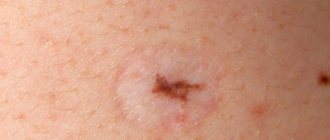
To have a correct idea of the volume of tissue removed, you can look at the photo and see what an average scar looks like.
If you have several moles removed, when sending them for histology, the location of each nevus must be clearly indicated.
But it’s not worth removing so many moles at once, as in the photo. Why? I'll tell you in the following publications.
What does poor histology of moles mean?
If glomus angioma is not confirmed, but cancer is detected, we are talking about melanoma. When it occurs, doctors recommend surgical treatment. Conservative tactics are not used. The removal procedure is performed in a clinical setting using the following strategies:
- laser excision,
- electrocoagulation,
- radio wave method,
- using an electric knife.
If surgery is not performed in a timely manner, there is a risk that infected cellular elements can form metastases to other organs.
It is important to know! Some time after surgery, you need to see a doctor so that he can eliminate the possibility of relapse.
“Can I not take away the answer? Will they call me if something is wrong?”
As far as I know, there is no law according to which the morphology laboratory must notify the patient that cancer has been detected.
At the end of the conclusion after the examination, moles may be written: “Re-examination with the results of histological examination.” But no one reads these conclusions.
And then everything happens according to the classic scenario, even if in an institution calls of this kind are in the order of things, the human factor comes into play. For example, the person responsible for this quit, or someone replaced him, or he got sick, didn’t make an appointment, it was a birthday, etc. - in the end, no one called, and the patient didn’t even know that they could have done this. Remember that your health is yours and no one owes you anything.
Please always take the result of the histological examination and always show it to the oncologist who performed the removal.
Histological answer, what to do with it?
No. If you received the result of the study in paper or electronic form, you must show it to the oncologist who performed the removal. Let me give you an example.
Here are two diagnoses:
- Dysplastic nevus with severe dysplasia. There are tumor cells at the resection margin.
- Squamous cell papilloma with inflammatory component. Removed within unaffected tissues.
In the first case, repeated excision of the mole removal site is required, but in the second - not. Therefore, do not be lazy, show the conclusion to the doctor.
Histology results
As a result of histological examination, the oncodermatologist will determine whether the mole is malignant or benign. In addition, based on the histological report, the doctor is able to determine what type of birthmark the birthmark belongs to, the severity of the disease and structural indicators. You will not be able to decrypt it yourself. If a mole has been removed and the histology is poor, it is necessary to undergo surgery to remove the nevus as soon as possible.
The analysis can reveal the degree of cancer, which determines what treatment will be used. In the first stage, the formation is removed within the unaffected tissue. The second stage is based on the removal of the growth and lymph nodes that are located near it. The third stage involves excision of all affected nevi and lymph nodes that are adjacent to them, followed by chemotherapy. If the fourth stage of cancer is detected, the patient has a minimal chance of a positive outcome.
If there is no danger, it is recommended to excise the stain. This will help avoid pathological processes in education. Modern removal methods are painless and safe. If surgical intervention to remove a malignant growth is not carried out in a timely manner, the risk of developing and spreading metastases increases. They are able to penetrate vital organs, disrupting their functioning and destroying their structure. In medical practice, there are cases when, after removal, a mole appears again. This serves as an alarming signal that requires an immediate visit to a dermatologist or oncologist.
Are there errors in the histological examination of nevi?
Yes, unfortunately, they are possible. I wrote about this. Histological examination is carried out by people, and they can make mistakes. If you have the slightest doubt about the results of the conclusion, take your histological preparations and send them for review to another laboratory. The preparations include both paraffin blocks with mole tissue and glasses with sections.
You ask: “How can I go and pick something up? They’ll send me…” If they don’t give you histological preparations, then you need to go to the head doctor and demand it. In my experience, after that everyone gives away.
Indications
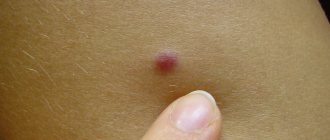
If you want to remove a skin pigment spot just because it does not look aesthetically pleasing, then histology of the mole will most likely not be necessary. Nevi often need to be checked if they hurt, itch, or cause physical discomfort. For example, a so-called “hanging” mole requires analysis much more often than ordinary pigment spots. But in addition to the general sensations and appearance of the mole, there are other signs of degeneration of the mole, in the presence of which it is necessary to carry out tests:
Nevi should be checked if they hurt, itch, or cause physical discomfort.
- mole injury;
- rapid growth of nevus;
- the appearance of plaque-like spots around the nevus;
- pain or itching;
- peeling or change in the appearance of the nevus;
- bleeding from a mole;
- change in the color or structure of a mole.
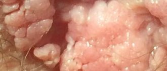
Why are papillomas not sent for histology?
The main property of a malignant tumor is uncontrolled cell division. Papilloma is a small (1–3 mm) formation on a thin stalk of soft consistency.
If the papilloma cells were malignant, they would grow a “leg”, and the papilloma would lose it. In addition, the consistency is softer than the surrounding skin, in my opinion, always indicates good quality.
This case is the only one when, in my opinion, the material need not be sent for histology.
Indications for histology of a mole
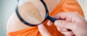
During the visual examination, the doctor has the right to refer the patient for additional tests.
These include:
- study at the molecular level,
- skin examination,
- radioisotope type scanning,
- CT,
- histology.
It is the latter option that is considered the most acceptable and preferable. This is due to the fact that the technique helps to identify malignant cells and prescribe timely treatment measures.
Here are the indications, when they occur, the doctor will refer you to a histological procedure:
- intensive growth of the tumor,
- changes in structure
- the appearance of tubercles,
- the appearance of a shiny crust on the mole,
- excessive bleeding
- dark plaque or darkening of a mole,
- spreading of the stain over the surrounding skin areas,
- the occurrence of pain due to mechanical impact.
If glomus angioma is suspected, tactics are rarely performed; it is appropriate only if a cancerous tumor is suspected.
Price, location and duration of analysis
Many people worry that a mole examination plus the surgery itself will be much more expensive than laser mole removal. Moreover, the pain from the surgical method can be many times greater: the operation itself, even under local anesthesia, cannot be called pleasant, and rehabilitation after removal of a nevus is longer and more difficult than after laser removal. The cost of surgically removing a large “hanging” mole is 2000-3000 rubles. This is approximately two times cheaper than laser removal. The price of the surgical operation also includes a histological examination. So in terms of price, surgical removal is more profitable.
The price of the surgical operation also includes histological examination.
The answer to the question of where to check moles is even simpler. You don’t need to worry about this and run around the city with a jar of materials: a surgeon in any clinic will independently send the mole for analysis, and then inform you about the results. So, first of all, you need to look for a highly qualified surgeon who will perform the operation at the proper level, will not leave scars in place of the mole and will support you during the rehabilitation period.
The analysis takes a short amount of time. Typically, a week is allotted for its completion, but in reality, histological analysis can be carried out much faster: it all depends on the number of patients in a particular laboratory. For example, in a large private clinic you can get results within two to three days.
Why and how it is carried out
Biopsy is a minimally invasive technique for research and treatment of various diseases. Allows you to establish the correct diagnosis. The concept of “biopsy” is not identical to the diagnosis of “cancer”. The invention of this method has made it possible to reduce the frequency of extensive surgical interventions and the use of aggressive medications.
No specialized preparation is required for skin examination. It is advisable to stop taking any medications, especially blood thinners, 1 week before the procedure. On the day of sample collection, stop smoking and drinking alcohol. If you have a history of a chronic disease, you should inform your doctor.
The procedure is performed under local anesthesia. The patient does not feel pain.
The tissue is excised, the bleeding is stopped with the help of hemostatic drugs. If the size of the wound surface is significant, then suturing the edges of the wound or applying a skin implant is indicated.
Why should the removal of skin tumors be performed by an oncologist?
Moles, nevi, warts, papillomas and other neoplasms often become an unpleasant cosmetic defect. Removing undesirable effects on the skin can solve the problem not only from an aesthetic point of view, but also prevent a malignant disease. However, in order for the operation to proceed without consequences, it is necessary to contact an oncologist.
Moles and other neoplasms can pose a potential health hazard from an oncological point of view. Therefore, they cannot be removed in the office of a regular surgeon, dermatologist or cosmetologist. Only related specialists—oncologist surgeon and dermatologist-oncologist—can accurately distinguish “good” from “evil.” At the same time, excision of tumors is carried out according to certain rules and strict indications.
Patients often come to us with benign formations - viral warts, raised moles, nevi, pedunculated papillomas. Primary consultation is provided by oncologists of two directions - a surgeon or a dermatologist. They examine not only the “problem” area on the skin, but also the patient completely. By the way, this is also an important aspect - an ordinary surgeon, cosmetologist or dermatologist may not pay close attention to all the patient’s skin tumors. Recently a woman came to me with a traumatized nevus, and upon examination she discovered melanoma on her eye.
After the examination, the doctor may prescribe cytology - a test for the presence of cells with signs of malignant degeneration. If there are no malignant cells, then removal can be done using a laser or radio wave device. If the result shows “evil,” then the oncologist surgeon excises the formation within healthy tissue and sends the material for histological examination to his own laboratory of the National Medical Research Center of Oncology named after. N. N. Petrova.
An ordinary cosmetologist does not conduct a cytological examination, which can lead to dire consequences. Sometimes patients come to us who have had a tumor excised incorrectly or incompletely and have experienced a relapse. And this is an additional risk of cancer.
There is no need to be afraid of the oncologist's office - now specialists can help the patient in one-day surgery, easily and painlessly fix the problem and provide qualified assistance.