Common benign tumors include lipomas, fibromas, angiolipomas, benign fibrous histiocytomas, neurofibromas, schwannomas, hemangiomas, tendon cell tumors, and myxomas. Benign soft tissue lesions rarely metastasize but are often large and deep. However, some formations behave very aggressively. Diagnosed invasion of nearby tissue increases the chance of incomplete removal and the possibility that the tumor will return. In adults, the most common benign soft tissue tumor is lipoma. In children - Baker's cyst. Most often, lipoma and hemangioma are observed in both adults and children.
Danger of illness
Fibroids on the inner thigh are dangerous if the size of the tumor increases rapidly. This indicates the absence of a capsule that inhibits tumor growth. If not removed in time, the neoplasm grows into the surrounding tissue and reaches a volume of several cubic centimeters.
Fibroids located in the groin or folds of skin are subject to friction, become damaged and may bleed, causing pain.
The main problem with having fibroids in people with hormonal disorders is the tendency to grow and increase in size.
Classification of neoplasms
Fibromas on the legs differ in their shape, growth rate, localization and specificity of the cells that make up the tumor. The doctor must not only correctly identify the pathology, but also correctly qualify it; it is on this that in the future the methods of treating the tumor and the possibility of removing them will completely depend.
Based on the rate of formation, it is customary to distinguish two categories of fibroids:
- Diffuse ones are not limited in growth, are localized in the muscles and legs, and as they develop, they can involve nearby glandular and vascular tissues in pathological processes.
- Limited hip fibromas are characterized by a slow growth rate, their size does not exceed 10 centimeters, which is due to the presence of a restraining capsule.
The growth rate and structure of cells can be determined by conducting an appropriate examination, including computed tomography, which makes it possible to determine the presence or absence of pathological processes in healthy tissues located near the tumors.
Depending on their location, fibroids on the legs can be divided into two types:
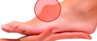
- Dermafibromas can be located on the legs, located on the inner surface of the thighs and lower legs.
- Plantar pain is located on the arches of the feet and is characterized by pain when pressing and walking.
Neoplasms are often observed in women during menopause and after pregnancy, which is explained by hormonal imbalances.
Therefore, it is imperative to undergo general diagnostics of the body, making it possible to prevent various dangerous diseases.
Causes of soft tissue fibroids
Scientists have identified several reasons for the development of fibroma of the soft tissues of the thigh. The main version of the formation of fibrous growths is hormonal imbalance. According to doctors, the risk group includes:
- pregnant women.
- patients with diabetes mellitus;
- people with thyroid and adrenal problems.
In second place in terms of prevalence is traumatization of skin areas - friction, bruises, squeezing. In people who are overweight, a fibromatous nodule may appear in folds of skin that do not breathe well.
In third place is heredity. The presence of fibroids in one of the immediate family makes it possible for growths to form in the next generation.
As you age, your risk of developing fibroids under the skin on your thigh increases.
Lipoma
The tumor can occur in the soft tissues of the thigh. A formation called lipoma is common. The onset of the disease is manifested by the formation of a small tumor, which gradually grows. The formation has a soft consistency and can easily move when pressed. Lipoma sometimes reaches 10 cm and affects nearby areas. The location is in the muscles or directly under the soft tissues. Formed due to enlarged fat cells.
The disease can be recognized by the presence of dull painful sensations when moving. There remains a danger of tumor transformation from benign to malignant. To avoid this, it is recommended to consult a doctor in a timely manner and take the necessary countermeasures. You must not damage the lipoma, otherwise it will increase in size, making the process of getting rid of it more difficult.
For correct therapy it is necessary to undergo diagnostics. As a rule, the operation becomes effective. Wen can be removed using a laser.
Types of tumor
Fibroids are distinguished by consistency:
- soft;
- hard.
According to the tendency to grow:
- diffuse;
- limited.
The soft type of fibroma consists of fatty inclusions. It is more typical for women who have a thicker subcutaneous fat layer. The soft fibroma is wrinkled and can be easily moved when palpated. Soft neoplasms sometimes have a stalk, repeating the shape of a mushroom. They most often form in the armpits, groin, under the breasts - these are the places that are least ventilated and are subject to increased sweating.
Hard growths appear in places that do not have a deep subcutaneous layer of fat - the outer surface of the thigh, face, neck. They have a round shape and do not move well during palpation.
Leading clinics in Israel
Assuta
Israel, Tel Aviv
Ikhilov
Israel, Tel Aviv
Hadassah
Israel, Jerusalem
Solid fibroma, or dermatofibroma, is the most common form. Most often it forms on the face, back, limbs (on the arm in the area of the shoulder and forearm, on the leg under the skin in the area of the foot, lower leg), and also affects the mucous membranes (fibrohemangioma). It has a dense structure and smooth surface.
Clinical picture of the disease
Fibroids are painless, except when they are injured by awkward movements. The color of the tumor can vary from flesh-colored to dark blue. It depends on how blood circulation occurs inside the tumor. Sometimes, when the fibroid stalk is twisted, the nutrition of the tumor is disrupted and it dies on its own.
The surface of the growth is uneven. There are additional formations and dents. Hard varieties have clear edges, soft ones are looser and shapeless.
Fibroma in children
Fibroids can appear on the skin of the extremities of children starting in infancy. The reason is heredity and metabolic disorders. Some types appear more often in male infants, while others appear with equal frequency in both sexes.
There are:
- Congenital forms that appear before the age of one year. They are located on the skin singly or in multiple groups - nodes.
- Tumors that appear after five years of a child’s life.
The most dangerous phenomenon of fibromatosis is malignancy - malignant degeneration of tissue, therefore neoplasms are removed using gentle methods so that no trace remains on the child’s body.
Most types of single benign tumors do not recur. After removal, the tissue is sent for histological examination to exclude the presence of atypical cells.
Types of childhood fibroma:
- Hamartoma - affects boys more often; relapses are rare, sometimes occurring up to a year. The most common age is 2 – 4 years.
- Gingival fibromatosis is thickening of the gums due to tissue overgrowth. It does not go away on its own, sometimes it recurs.
- Digital infantile fibromatosis - manifests itself as growths on the lower extremities in infants and older children.
Until the age of 10, children have tumors removed under general anesthesia, although operations with a laser or radio knife are practically painless.
Possible complications
Soft tissue fibroma is not a life-threatening or health-threatening disease. Most often it is perceived as a cosmetic defect, which in certain cases worsens the quality of life.
Complications can occur in cases where the fibroid is injured. Constant razor damage and friction from clothing. This causes pain, sensitivity and bleeding.
Damaged fibroids can also become infected. In certain cases, tissue necrosis is observed.
On this topic
Formations formed on the surface of the skin and mucous membrane, in rare cases, degenerate into cancer. This process is provoked by the influence of a number of negative factors. Fibroma malignancy occurs with rapidly growing formations. They can reach significant sizes.
Drop-shaped neoplasms may also appear on the body. They are dangerous because they spread throughout the body. But they occur in exceptional cases.
Diagnostics
Diagnosis of fibroma is carried out for the sake of the patient’s peace of mind and in order to clarify the origin of the benign tumor. To do this, the following procedures are carried out:
- Biopsy is the removal of a piece of tissue for examination under a microscope. It can be performed under local anesthesia or without anesthesia - at the request of the patient. The doctor gives a referral for a biopsy if the tumor grows rapidly or the person experiences pain. Discoloration and redness of the skin around the fibroid is also an indication for a biopsy.
- Subcutaneous tumors are examined using an ultrasound machine.
- If the presence of multiple nodes is suspected, examination of internal organs using ultrasound is recommended. For women, a mandatory examination by a mammologist and gynecologist is required.
When diagnosing, it is important to distinguish fibroids from other skin diseases.
Osteomyelitis
A tumor in the hip joint is expressed in acute and chronic osteomyelitis. Caused by the bacteria staphylococcus, streptococcus and salmonella, it affects bone tissue with the formation of inflammatory processes. The tumor can affect external and internal tissue due to a leg injury or a viral disease.
Symptoms appear within 4 days. The patient feels a lack of strength in the legs, pain in the thigh and aching in the hip joint. The temperature rises. A complication of the disease is possible, which is expressed by the appearance of purulent formations and sepsis. There is swelling of the thigh.
You should consult a traumatologist. The doctor has the right to conduct an examination using palpation and visually assess the degree of neglect of the situation. Then the doctor sends the patient for general blood and urine tests. The level of leukocytes, indicating the onset of inflammation, is taken into account. X-rays are used for diagnosis. The examination will show how much bones and joints are affected. Tomography and radioisotope scanning are widely used to make a diagnosis.
Treatment occurs with individually selected antibiotics. Therapy lasts 5 weeks, accompanied by injections locally into the muscle tissue. In advanced cases, surgical intervention is performed.
Treatment methods and prevention
Treatment of fibromatous neoplasms begins with normalization of nutrition and drinking regimen. When using medications, you can completely get rid of tumors located in a visible place, but the growths can recur. One of the best is considered the drug Diprospan, which stimulates the reverse development of fibroids, but has a large number of contraindications and side effects. In addition, Diprospan cannot be used during pregnancy due to high toxicity.
The safest and fastest is surgical removal of the tumor. Several methods are used:
- laser, which does not require anesthesia;
- radio wave method, which does not leave marks on the skin;
- conservative - with a scalpel, in which the wound takes longer to heal, but the effect is positive.
The first two methods are more expensive, since they require the presence of modern expensive equipment and a trained specialist. The second is cheaper or free if carried out in a public clinic.
Traditional methods involve treating fibroids with various homemade ointments and taking tinctures orally to normalize general well-being and hormonal levels, and reduce intoxication.