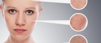
Removing a mole is a serious procedure, after which the area of skin requires special care. Usually the wound heals quickly, but if after removal of the mole a tubercle or other neoplasm appears, it is recommended to immediately contact a specialist in order to examine it and exclude or prevent melanoma.
The recovery period after mole removal may be accompanied by changes in the areas.
Healing
Any method of removing a nevus leaves a mark on the skin - a wound, the diameter of which depends on the original size of the mole. It becomes covered with a crust that forms a small tubercle and performs a protective function (protects the vulnerable area from infection). For this reason, it is forbidden to remove the crust from the wound; you must wait until it falls off on its own: the process takes about 2 weeks.
The young skin formed at the site of nevus removal (it is colored pinkish) should be protected from the harmful effects of ultraviolet rays using a protective cream with an SPF factor of 60 or more.
Otherwise, there is a possibility that the area will turn into a pigmented spot. The color change from pink to normal occurs within one to two months. However, if a slight discomfort remains, do not panic. Complete healing sometimes takes about 6 months, during which the area where the nevus was located is palpated and causes discomfort.
An almost imperceptible scar may remain at the site of the removed mole if the excision was carried out using liquid nitrogen, electric current, the radio wave method, sometimes a laser leads to this result. Scarring is a natural process, this is how the body reacts to a violation of the integrity of the integumentary tissue. Since some people have a higher ability to regenerate, removal can take place without leaving a trace.
Scars that look like small tubercles may remain when a nevus is excised surgically, since the operation involves the application of cosmetic sutures: damaged tissue is cut off with a scalpel, healthy tissue is stitched. It is almost impossible to avoid noticeable marks in such a situation.
How to get rid of hardening
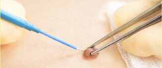
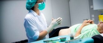
If a mole is hard and itchy, doctors advise removing it. Operations are performed in a hospital. Surgeons use the following methods:
- Surgical. The doctor uses a scalpel to excise the protrusion on the dermis and send the tissue for histological examination. The operation is used to remove large tumors with a deep root system and is performed under anesthesia.
- Laser. The doctor acts on the thickening with a laser. The method is painless, healing occurs in a short time.
- Cryodestruction. Liquid nitrogen is applied to the neoplasm. It becomes covered with a crust, which gradually disappears. The disadvantage of the procedure is manifested in possible damage to healthy areas of the skin.
- Electrocoagulation. A high-frequency current is applied to the mole and lump; the procedure is performed with local anesthesia. The tumor is sent for histology.
- Radio waves. The tubercle is removed using a radio knife.
If there is a lump under a mole and it hurts, you cannot try to eliminate it yourself using traditional medicine.
Thickening of the dermis under the birthmark can also be observed in a child, in which case the formation should be seen by a doctor immediately.
Consequences
Sometimes removing moles leads to unexpected unpleasant consequences that require taking certain measures. May cause concern:
- A malignant tumor that occurs in the area where the removed nevus is located. Detection of melanoma requires surgery, which removes not only the tumor itself, but also healthy tissue (a small area) around and under it. Afterwards, the excised edges are sutured and the cut material is examined.
- The formation of a scar (keloid scar), which is a red bump that rises above healthy tissue. There are 2 cases in which such a mark may appear on the skin: if the operation was performed by an inexperienced doctor; when the mole being removed has a large diameter.
- The appearance of edema. The reason is the involvement of the lymphatic vessel during the excision of a mole. When lymphatic collaterals (bypasses) form, swelling usually disappears.
- Compaction detection. If enough time has passed after the operation to remove a mole for the excision area to heal, but instead a lump has formed, it is necessary to visit an oncologist to rule out malignant degeneration of the cells.
- Re-formation of nevus. Relapse is the most common result of mole removal, in which it can reach a larger size than it was before the operation.
Why do they appear?
At birth, a child often does not have a single mole on his body, with the exception of birthmarks caused by heredity.
- All information on the site is for informational purposes only and is NOT a guide to action!
- Only a DOCTOR can give you an ACCURATE DIAGNOSIS!
- We kindly ask you NOT to self-medicate, but to make an appointment with a specialist!
- Health to you and your loved ones!
- Every month there are more moles, this may be due to the baby’s exposure to the sun, as well as the development of hormonal levels.
- By the end of puberty (puberty), most of the hereditary and natural nevi are already on the human body.
The appearance of new birthmarks after puberty and their growth in size can be observed:
- first of all, on open areas of the body when exposed to the sun;
- as well as hormonal imbalances and taking certain drugs that affect the production of melanin by melanocytes.
The nature of the tubercles formed
The tubercles that may appear at the site of excision of nevi, in most cases, represent an infiltrate that develops with severe trauma to tissue structures, poor hemostasis (blood clotting), or infection. They look like cellular structures that are permeated by lymphatic fluid or blood. The formation of this element has an inhibitory effect on healing or completely stops this process.
With an inflammatory infiltrate, redness of the skin is observed, pressure leads to the fact that the affected area begins to hurt.
The formation of a non-inflammatory infiltrate occurs in the postoperative period. It is represented by tissue that is saturated with lymph or drugs, without signs of an inflammatory process. Typically, such seals disappear without outside intervention after some time: healing takes weeks, sometimes months. Special regenerating medications and physiotherapy help speed up resorption. If inflammation develops, contacting a doctor is a must.
If after removal of a mole a tubercle appears, there is a high probability that the process of forming a malignant tumor has begun. Such formation is subject to mandatory removal.
Seals under a mole, is it dangerous?
Often, if you have a mole (nevus), you can see a small lump located somewhere around the mole or just below. No one can say whether they are dangerous or not without proper inspection. Only after specialists have seen, touched and examined can we talk about the results and the degree of their danger. In most cases, such seals do not pose any danger. But there are many exceptions. Only a specialist will be able to draw the correct conclusion.
Is it necessary to remove a nevus with compaction?
By their nature, ordinary nevi do not pose any threat. If they are symmetrical, do not increase in size, do not change color, do not bleed, do not crack or cause anxiety, there is no need to remove them. Whether they are with or without a seal does not matter at all, the main thing is that they do not change and do not cause discomfort (except for aesthetic reasons, of course). It is better to consult a doctor to calm your nerves, but usually they do not pose any danger, but simply exist as an integral part of the human body.
Scarring
A scar (or scar) that appears after excision of a mole is unlikely to please its owner, so it is important to remember:
- Scars often form when a lesion is deeply excised. Keloids must be removed promptly.
- Scars that do not disappear over time require special care prescribed by a doctor.
Scars can be effectively eliminated using an absorbable silicone patch. The first results appear 2-4 weeks after the start of use.
Sometimes rough scars may appear on the area where the mole was removed. Such formations must be treated with a special ointment prescribed by a doctor. In the absence of the required effect, cosmetic surgery is used to eliminate the scar.
When the scar after mole removal hurts after 2 months, it is recommended to consult a doctor. Itching and lack of changes in size and color should also be alarming. Pain usually occurs if the nerve endings are involved in the scar tissue, which necessitates the need for surgical intervention.
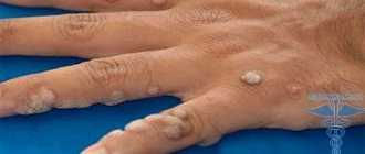
What are there
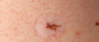
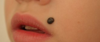
Moles differ from each other in size, shape and color.
By size they are distinguished:
- small (up to 15 mm);
- medium (1.5 – 10 cm);
- large (more than 10 cm);
- giant (moles that can occupy parts of the body - face, limbs, chest, etc.).
By form:
- convex;
- flat;
- moles “on a leg” (hanging);
- hair;
- warty;
- smooth.
By color:
- black;
- brown;
- blue;
- red;
- bodily.
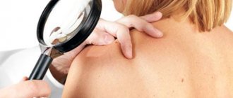
Symptoms that may bother you
There are, of course, post-operative symptoms that may be of particular concern to the patient. For example, what if a lump appears after a mole is removed? There is only one answer - again, turn to professionals for help. They must see this seal, feel it and then draw certain conclusions. Most often, such lumps are forming scar tissue, although it may be something more serious. In short, you should definitely contact a doctor for an examination if you have any questions, complaints or other reasons for concern. Only a specialist can actually determine whether these are serious reasons or imaginary ones.
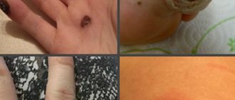
Stains
The types of spots that can form at the site of removed moles are different.
Black ones are formed upon completion of the operation, and the selected method does not play a role. Spots of this type are crusts, protect the wound from external influences and fall off on their own when the wound is completely healed.
Pink - represent young, delicate skin that forms under the crust, acquiring a natural color as all processes in the tissues normalize.
Red spot - appears when the crust is removed prematurely. The color of the spots indicates infection of the wound, so there is a high probability of complications.
Pigmented - caused by uncontrolled exposure to ultraviolet radiation on the area from which the nevus was removed. Pigmentation is a natural result of non-compliance with instructions for caring for newly formed skin.
Brown - indicate incomplete removal of the mole. Such a spot requires professional consultation to adjust the treatment technique or prescribe a repeat operation to excise the formation.
Burgundy (purple) - signal infection of the wound, inflammation. Spots of this type are often painted in darker shades, which indicates the beginning of the process of malignant degeneration of an incompletely removed nevus. After detecting such traces, it is not recommended to delay visiting a doctor.
White spots usually form if a mole has been removed with a laser. These spots disappear within six months from the moment of medical intervention, but sometimes remain for life.
If any deviations occur or you suspect the beginning of their development, you must immediately visit a doctor, since preventing further progression of pathological processes depends on the timeliness of the measures taken.
A lump appeared in place after the removal of a mole: what to do?
After removing a mole with a laser or as a result of surgery, one of the decisive periods begins, on the normal course of which the health of the entire body depends. Removal of a nevus is necessarily accompanied by a long rehabilitation period, during which it is necessary to properly handle, care for and monitor all changes at the site of removal.
If this area of the skin is treated correctly, not overheated in the sun and minimizes the influence of chemical and physical factors on it, it will slowly heal and there will be no trace left of the intervention. However, not everything always goes well and often after such operations unpleasant consequences arise, such as:
- A scar appeared at the site of removal. The situation is typical and does not pose any threat. There is no need to do anything, the scar is a natural phenomenon that will disappear on its own after some time. There's no need to worry.
- Redness after mole removal is an adequate reaction of the body to surgery. But if the redness around the mole does not go away over a long period of time, it is important to consult a specialist.
- A lump appeared at the site of the removed mole. If any formations appear, it is recommended to see a doctor immediately. Often, compaction under a mole signals the initial stage of melanoma (skin cancer) and the development of malignant tumors. They may be benign, but it is better to consult a doctor immediately in order to eliminate all health risks.
On average, the wound healing process takes 2-3 weeks.
During this period, it is important to carefully observe all changes in the skin at the site of removal, wipe the wound with products prescribed by specialists, and if it is noticed that something new has formed, immediately consult a doctor. If healing occurs normally, a small scar will remain after a couple of weeks, which will soon disappear.