There is no such person who does not have moles on his body. Some have one, others have many. The sizes of nevi can also vary. Almost everyone knows that touching or removing them is dangerous, as it can provoke the development of cancer. Not all nevi transform into cancer, so it is important to know how to check a mole for cancer. You can do this yourself, or you can go to a medical institution for an examination. But moles located in the armpits, chest, waist or head should not be subjected to injury or constant friction. If an injury does occur, it is recommended to consult a dermatologist for advice. After diagnosis, he will prescribe appropriate treatment.
Indications for checking moles
Here are the main reasons to visit a dermatologist more often:
- change in the mole itself: its color, size, volume;
- skin injury in the area of the mole,
- unpleasant sensations: soreness, itching;
- ultraviolet intolerance,
- frequent and long exposure to the sun without protection,
- presence of melanoma in the family history.
What kind of doctor deals with moles?
If you find even one of the above alarming parameters, you should consult a doctor. A logical question: which doctor checks moles?
The first doctor who should conduct an examination and collect a primary medical history is a therapist. If he considers that there is or may be cause for concern, he will give a referral to an appointment with a dermatologist or oncodermatologist.
Types of birthmarks
The main types of moles include:
- Vascular. They appear due to too rapid growth of skin capillaries.
- Pigmented. They appear due to the fact that the body produces too much melanin pigment.
- Congenital. The baby is born with them.
- Acquired spots, which depend on the patient’s genetics, skin type, and sun exposure.
Another classification:
In addition, there are nevi, the entire plane of which is located on the skin, and hanging ones, that is, on a stalk.
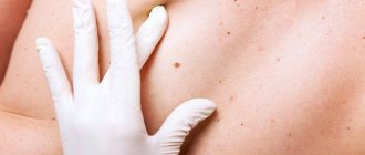
Checking moles
At the appointment, the dermatologist will conduct a more detailed medical history (patient history and family history) and a detailed examination. The process of checking moles is painless, the methods used are as follows:
Optical dermatoscopy. The point is to examine the skin with a special device - a dermatoscope - a special medical microscope with a magnification of 10 times or more, which records the structure and boundaries of the mole.
Typically, such a microscope is combined with a special camera or connected to a computer and displays the image on a monitor.
There is no need to prepare for the examination, although if an area of skin on the face is examined, it will be necessary to remove makeup first. The examination is absolutely painless and will take a few minutes.
Read here: The meaning of moles - research, the need for removal and the mechanism for assessing a mole (100 photos)
Epiluminescent dermatoscopy is a non-invasive (painless) method for examining nevi, which uses special polarizing lighting. It illuminates the mole from the inside, providing the doctor with a more informative picture.
Red flags
When assessing the condition of nevi, it is important not only their current condition, but the dynamics of the manifestation of changes, which the patient himself can tell about, is no less significant.
What should you pay attention to when checking moles?
The verification algorithm is called ACCORD after the first letters of the names of the key factors being assessed. This is what each letter encrypts.
- A - presence/absence of nevus asymmetry (normally, the mole should be symmetrical),
- K—edge evaluation. In normal nevi, the edges are clearly defined, smooth, not torn,
- K - presence/absence of bloody discharge from the nevus (normal, there should be no discharge from moles),
- O - color, degree of pigmentation of the nevus. Ideally, it should be homogeneous and not change throughout life.
- R - height, size. Normally, moles can grow throughout life, but this growth is very slow and almost unnoticeable.
- D—dynamics of change. Did cracks appear, did bloody discharge begin, hairs fell out, did itching or burning appear, etc.
Based on the assessment of each of these parameters, as well as assessing the condition of moles using a dermatoscope, taking into account medical history, the dermatologist makes a diagnosis, prescribes treatment or recommends consultation with a related specialist - an oncologist or a surgeon.
Type of malignant neoplasms
The skin is one of the most complex structures of the human body. Under the influence of various factors, various malignant tumors can form on the surface of the skin. According to statistics, the two most common types of cancer are carcinoma and melanoma. Each of these pathologies has its own key features and forms of development.
Carcinoma has two distinct forms, called basal cell and squamous cell. The reason for the appearance of this pathology is prolonged exposure to sunlight on the skin. According to experts, this disease is most often observed in patients whose age exceeds forty years. Carcinoma is considered to be a curable disease, provided that the pathology is detected in a timely manner.
Basal cell carcinoma is a disease better known by the abbreviation basal cell carcinoma . This type of cancer is considered one of the most common. Tumor formation occurs under the influence of atypical dermal cells. Basalioma has the appearance of nodules with a dense structure.
Over time, benign neoplasms can change, grow, change color, thereby becoming substandard
Most often, neoplasms appear at the site of accumulation of individual branches of the vascular system. These areas of the body include the upper torso, neck and face. Basal cell carcinoma does not metastasize to healthy tissue. The development of the disease is accompanied by inflammatory processes and the formation of ulcers on the affected tissues.
Squamous cell carcinoma affects various parts of the body, but most often cancerous tumors form in the mucous membranes of the mouth, face and extremities. Most often, this type of malignant tumors forms at the site of damage to the integrity of the skin, including burn injuries. A small red spot appears on the affected area of the body. At further stages of oncology development, hard nodules form on the surface of the spot, which cause pain when pressed.
Squamous cell carcinoma spreads deep under the skin, attacking healthy cells. This type of disease is formed against the background of the activity of certain cells that are located deep under the epidermis.
Melanoma is the most aggressive type of cancer . Let's find out what a malignant mole of this type looks like. This type of neoplasm looks like a small nodule that rises above the surface of the skin. Less commonly, melanoma is flat in appearance and literally pressed into the skin. The tumor has uneven borders and color, which varies from blood scarlet to dark brown. Sometimes, certain areas of the growth may not have color.
The degeneration of education is accompanied by certain modifications, which are easy to determine visually
The development of a tumor leads to the fact that cancer affects the lymph nodes and individual elements of the vascular system. The disease is formed under the influence of pathological processes occurring in the cells responsible for the pigment. If pathology is detected early, melanoma can be cured. However, this type of cancer is considered to be the most dangerous, since cancer metastases spread at high speed. In order to make an accurate diagnosis, a specialist will need data obtained through laboratory testing.
Where to get checked
A district clinic or a specialized dispensary is where moles are usually checked for oncology. In a cosmetology office, where a patient may ask to remove a mole that is bothering him, the cosmetologist, after conducting a visual examination, may in some cases recommend contacting a dermatologist-oncologist to obtain histological confirmation of the benign quality of the formation.
How to determine the oncogenicity of a tumor
Let's look at the main signs of malignant moles. First of all, various changes on the surface of the growth indicate the degeneration of birthmarks. Peeling, increase in diameter and change in structure are the main symptoms of nevus transformation. Symptoms such as itching, burning and tingling sensation in the area of moles are also important. Less commonly, various allergic reactions occur in those parts of the body that are adjacent to the growths.
Hair loss, color changes and the formation of papillomas are one of the clear signs of metamorphosis. Subsequently, new small-sized moles are added to the above-described symptoms, located close to the main growth. The appearance of small wounds and hemorrhages is also a negative symptom.
Skin cancer is a deadly disease!
Above are photos of moles that need to be removed in order to prevent the development of cancer. Each of the symptoms indicated in the list may be the beginning of pathological processes that have a destructive effect on the body. That is why it is very important to seek qualified help in a timely manner.