Intradermal skin nevus: types, symptoms and treatment, photo. Intradermal nevus is determined by the benign etiology of its manifestation on the dermis in the form of a birthmark, often congenital in nature, but can appear during puberty or during menopause.
According to doctors, a pigmented nevus inside the dermis does not degenerate into an oncogenic nevus, but in 16% it can cause the formation of melanoma.
- How to recognize a nevus inside the dermis
- Types of nevi
- Clinical symptoms
- Causes of neoplasms
- Treatment of nevi
- Alternative Methods
- Prevention
How to recognize a nevus inside the dermis
The intradermal nevus differs from other benign formations - anematic and sebaceous nevus, hemengioma, teratoma:
- soft surface that can be felt when pressed;
- uniform color that will not change even over time;
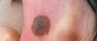
- clear boundaries with bright and smooth edges, as seen in the photo;
- absence of inflammation under and around the mole.
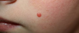
Initially, the nevus resembles a light elastic polypule, or a nodule on a broad base 2-4 mm in diameter or more.
The surface of the nevus is covered with brown spots and dilated capillaries.
Melanocytic nevus in pregnant women
The skin is both a hormone-producing and hormone-dependent organ. The regulator of the activity of this integument is the neurohumoral system. There are studies in the medical literature that indicate an increased risk of degeneration of melanocytic formations during pregnancy. This occurs due to significant changes in the functioning of the hormonal system. It would seem that this statement has a correct justification. However, it has not been confirmed by research. There have been no studies comparing the growth factors of birthmarks and melanoma cells with local hormone saturation.
Types
Intradermal nevi are of the following types:
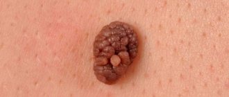
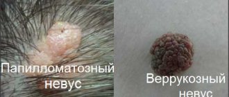
Papillomatous or warty
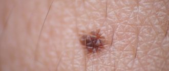
Since the intradermal nevus subsequently changes, as can be seen in the photo, it takes the form of a papilloma with a stalk and a warty surface (in the form of a blackberry), increasing in size.
In older people, papillomamatous nevus does not evolve.
Pigmented or melanocytic
This type is prone to malignancy. A melanocytic nevus can transform into melanoma, which spreads over the surface of the skin.
May be congenital or acquired during puberty.
New fragments appear throughout life.
Noncellular
Noncellular nevus. In the form of a small convex oval, as seen in the photo. Often appears on prominent areas of the skin of the neck and face.
Types of melanocytic nevi
When clarifying the question of what an intradermal melanocytic nevus is, the following factor must be taken into account. Birthmarks are new growths on the skin surface. However, they have a “conditional” relationship directly to the skin. Even during the period of embryonic development, nevus precursor cells can be “pinched off” from the neural tube, which “migrate” into the skin. Because of this, melanoma developing from melanocytic moles should be considered not a cancer of the skin, but a cancer of the nerve ending system.
This formation is a malformation of the skin. In this case, melanocytes, “derivatives” of the cellular structure of the neural crest, “split off” from the neural tube and end up in the structure of the skin. During a person's life, under the influence of ultraviolet radiation, estrogen or other factors, their nesting reproduction is stimulated.
Acquired melanocytic formations usually develop after the child turns 3 years old.
The number of moles on the body reaches a maximum in 16-year-old adolescents. Then, throughout life, the number of moles usually remains constant until age 35. After reaching this age, the spots atrophy and they gradually disappear.
Congenital melanocytic nevus
This type of mole comes in different sizes. A giant representative of these pigment formations sometimes “occupies” a significant part of the skin. Often, coarse hair grows on it, resembling animal fur. Previously, it was believed that a giant congenital melanocytic nevus is more often the cause of the development of a malignant tumor compared to small moles. However, researchers have found that the process of mengalization does not depend on the size of the tumor.
The sizes of such neoplasms are:
- small – do not exceed 15 mm;
- medium – up to 100 mm;
- gigantic (large).
The diameter of the last of them is more than 10 cm. At the same time, congenital formations also become larger as the child grows. The least dangerous small moles. Medium and large spots more often degenerate into a malignant neoplasm. The reasons for the appearance of large congenital birthmarks have not yet been fully elucidated. Parents of babies with congenital giant nevi should remember that it is imperative to consult a specialist.
The medical names of birthmarks are not always clear to the average person. However, this terminology is based on the following logical conclusions. The human skin includes the following layers:
- epidermis - this layer is located directly on the surface;
- dermis - is the middle layer (and the thickest);
- hypodermis – located most deeply.
Clusters of melanocytes can be located in any of these places. Depending on how deep the cells are located, the following types of pigment spots are distinguished:
- Epidermal. It “settles” in the epidermis, which is the top layer of the integument.
- Intradermal. Melanocytes accumulate in the thickest layer of the skin.
- Borderline. An increase in melanin concentration is observed between the epidermis and dermis.
- Hypodermal. The pigment is located in the hypodermis. This type of mole does not appear outwardly. Sometimes melanocytes move closer to the skin surface, after which the neoplasm becomes visible.
Intradermal melanocytic nevus
Interdermal (in other words, intradermal) nevus can usually be “found” in an adult. Doctors make long and incomprehensible diagnoses for the average person. These include, for example, papillomatous intradermal melanocytic nevus of the skin. The word “pigment” is added to such a long name.
This definition contains several concepts: the pigment melanin accumulates in melanocytes (which produce it), located deep in the layers of the skin. Outwardly it looks like a tubercle “rising” above the skin. A synonym for such a long expression is the concept of intradermal melanocytic neoplasm. If it is flesh-colored and is also located on a stalk, then the word “papillomatous” is added to the name. In medicine, there are other names for such a nevus - warty or hyperkeratonic. However, only a doctor can say for sure whether it is a papilloma, a nevus or a degenerated melanoma.
Borderline melanocytic nevus
When determining this type of formation, it is taken into account that nevus cells are located in a zone that is borderline. They are found between the outer layer (epidermis) and the deeper layer - the dermis. Most birthmarks located on the genitals, as well as the soles or palms, are classified as borderline nevi. They often appear on the body as a spot, sometimes a nodule that has an oval or round shape. Size - from a few millimeters to 1 centimeter (rarely - up to 5 cm).
A complex melanocytic nevus is usually formed when some melanocytes move into the next layer - the dermis. At the same time, some of them remain in the upper layer.
Clinical symptoms
- Papillomatous (warty) intradermal nevus can grow up to 1-1.5 cm and change in color to maroon, brown and black. Papillomatous nevus under the hair on the head is more common. They may grow short, dark, coarse hair. Moles grow and make it difficult to comb your hair. All the time, a person runs the risk of injuring (tearing or catching) the formation due to constant mechanical impact. Therefore, it is recommended to remove them.
- Melanocytic pigmented intradermal nevus has clear boundaries and a bright color, preserved for a long time due to its contents. In it, with a small size (2-5 mm), melanin cells accumulate in large quantities. During life, the spots change their shape, the surface can be smooth or bumpy, more often rough. Place of dislocation - areas of skin on the neck, under the breast or in the groin.
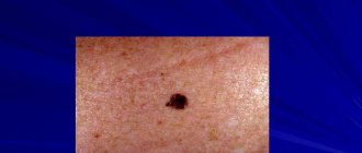
- Dysplastic melanocytic nevus can be of uneven color: from pinkish and beige to blue and black with a pronounced zone of hyperemia at the base. May have an irregular shape and unclear boundaries. Many moles cover the lower limbs, back, sternum, scalp, genitals and buttocks.
- Non-cellular intradermal nevus spoils the aesthetic appearance if it appears on the face or in the neck area. Therefore, it is recommended to treat or remove them. Most often they form during puberty in adolescents.
Diagnostics
Ordinary moles require diagnostics only when it is necessary to confirm their benignity. The first stage of the examination is a medical examination and dermatoscopy. It is necessary to differentiate intradermal nevi from dermatofibroma, vulgar warts, juvenile xanthoma, and cystic basal cell carcinoma.
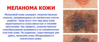
It is extremely important to exclude a malignant pigmented tumor – melanoma. It is distinguished from intradermal nevus by a number of clinical and morphological aspects:
- Fast growth.
- Large sizes.
- Wrong boundaries.
- Uneven coloring.
- Asymmetry.
Cytological diagnosis of a tumor (imprint smears) is not effective enough, so the only sure way to exclude malignant growth is a biopsy of the tumor. It provides the most valuable information for the clinician.
Of great importance in the diagnosis of intradermal nevi is their differential diagnosis with melanoma.
Causes of neoplasms
Why an intradermal nevus forms on the skin is still not clear to medicine.
According to doctors, melanocytes or nevus cells are formed in utero.
They affect the formation and development of nerve nodes and internal organs of the fetus.
Melanoblasts produce cells containing melanin, which give the baby's skin its color or hue.
And wandering immature nevus cells appear as spots on some deep areas of the skin.
That's why they are called intradermal.
Genetic factor in the formation of nevi
Heredity in the formation of birthmarks is of great importance; therefore, their manifestation is noted not only at birth, but also at 12 or even after 30 years.
The transformation of nevus cells occurs in stages:
- in infancy, intradermal moles are difficult to notice due to their location under the epithelium;
- when changing and moving into the layers of the skin, nevus cells find themselves between the dermis and epithelium;
- as the child grows, birthmarks with clear external parameters form;
- then the melanoblasts find themselves in the dermal layer, when they pass to the final stage of formation, the mole becomes noticeable;
- as a result of transformation, the mole takes its final shape: convex on a wide base or rounded on a stalk;
- the final stage is characterized by cessation of melanin production or discoloration of nevus cells.
Melanocytic nevus appears in women and men, often of a sporadic nature (single, random, non-familial).
There are cases of genetic inheritance of an autosomal dominant type.
With dysplastic nevus syndrome, the formation transforms into melanoma, especially if it is present in close relatives.
In this case, sun rays are not the cause of transformation into melanoma, but gene mutations are important.
Clones of mutational melanocytes are active in direct sunlight. Also, a nevus can transform with long-term use of immunosuppressants.
Why do they appear?
A specific reason for the development of intradermal nevi on the surface of human skin has not yet been established. There is only a version or assumption that, according to scientists, can explain the process of development of nevi.
Even during the development process, melanoblasts are formed in the mother’s abdomen—the so-called nevus cells, which play an important role in the development of some of the child’s internal organs, as well as its nerve nodes. In addition, melanin cells, which give the skin of the unborn child a certain color, are formed precisely inside melanoblasts. Birthmarks on the body of a newborn occur due to the accumulation of a certain number of immature nevus cells. Due to the fact that such nevi form in the deep layers of the epidermis, they are called intradermal.
Intradermal nevus treatment
Medical institutions examine the structure of nevi in laboratories under microscopes to determine their good quality and the number of nuclei in the cells.
If clinical signs of degeneration of a melanocytic dysplastic nevus into a malignant formation are noticeable, it is surgically removed.
If several degenerated nevi are removed, the patient is registered with a doctor and monitored throughout his life.
Removal of dysplastic nevi is carried out by capturing 2-3 mm of healthy skin.
This is done to prevent relapse of the disease and repeated re-excision.
Such patients are frequently and carefully examined. If surgical removal is not possible, applications of 5-fluorouracil (5%) or Trethionine are used.
It is especially important to visit a doctor for examination during hormonal transformations of the body - during pregnancy, adolescence, when using immunosuppressants, estrogens and oral contraceptives.
Treatment and its directions
Treatment of benign neoplasms is necessary if they represent a noticeable cosmetic defect. In other cases, the appropriateness of the intervention is determined by the doctor on an individual basis. Removal is necessary if the mole is constantly exposed to injury. But such manipulation must be carried out by a qualified specialist. An inept approach to surgery can provoke the degeneration of benign tissues into malignant ones.
Some moles are best monitored. If the outline of the neoplasm gradually changes, its color, or it begins to itch, the doctor raises the question of removal. To do this, he can recommend several methods:
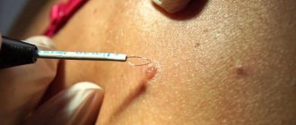
- Laser therapy. The procedure does not last long and is painless. With its help, you can remove tumors on any part of the body. There is no scar left if we are talking about a small area.
- Surgical removal. The cost of this method is the most affordable, but requires special equipment. After such manipulation, scars remain. It can be carried out only after pain relief.
- Treatment with liquid nitrogen. This method is called cryodestruction. This manipulation involves influencing the tissue of the mole by completely freezing it. There are no scars left after such a procedure, but complete excision of the pathological area is not guaranteed.
- Electrocoagulation. It is a method of applying electric current to a nevus.
Experts recommend avoiding traditional recipes for removing moles. Independent influence on neoplasms can lead to the most unpredictable results.
Under no circumstances should you cut out a mole yourself using scissors or a knife. This approach is so dangerous that without subsequent treatment of injured tissue, one can expect the formation of melanoma in 100% of cases.
Alternative Methods
For the treatment of intradermal moles the following is also used:
- laser therapy;
- cryodestruction;
- electrocoagulation;
- radiosurgery.
Benefits of laser removal
Laser surgery is considered the most effective because it can remove large birthmarks larger than 3 cm in size, as well as skin melanoma.
- absence of bleeding due to cauterization of small vessels;
- laser wound disinfection;
- no postoperative stitches are needed;
- minimum rehabilitation period;
- no scars.
Main characteristics
Small neoplasms on the skin represent a concentration of melanoblasts. A nevus can be recognized by its soft surface and uniform color, which does not change over time. The spot has clear, even boundaries, without inflammatory processes. In appearance it resembles a round, voluminous wart or “blackberry” on a thin stalk. On the body, a mole develops either one or several in different places.
The color of the spot varies from flesh to brown and dark burgundy, and the size ranges from 2 mm to 2 cm. If you look closely at the surface, it is easy to distinguish a thin network of capillaries and small inclusions. Rarely, pigmentation appears on the limbs, abdomen and back; often the formation occurs:
- in the armpit area;
- at the neck;
- in the folds of the groin;
- under the breasts of women.
No clear reasons for the growth have yet been discovered. There is a version that tries to rationally explain the processes of emergence. Nevus cells appear in a child during intrauterine development and play a role in the formation of internal organs.
Melanoblasts give the skin a certain shade, and birthmarks are immature elements of the epidermis. For some people, moles become noticeable after age 12, while for others, after age 30.
In childhood, the nevus is almost invisible, but as it grows, the cells move into the deeper layers of the skin. Over time, the spot develops a clear boundary and size. The convex neoplasm attaches to a thin stalk, and the melanoblasts stop producing pigment. The defects do not change for several years, but then actively grow. Often the reasons for “awakening” include:
- chronic venereal diseases;
- the mother being in unfavorable conditions;
- taking hormonal medications;
- bad habits.
Prevention
Since there is no way to prevent the appearance of an intradermal nevus, it is necessary to exclude its degeneration into melanoma.
For this:
- areas of skin with a mole are protected from direct ultraviolet radiation, especially on the beach;
- do not visit the solarium if you have large or multiple moles;
- if neoplasms appear, you should consult a doctor;
- After surgery, you should not ignore the examinations planned by your doctor.
Dangerous symptoms
Birthmarks range in size from one millimeter to extensive skin invasion. Flat nevi are almost invisible on the surface, and “blackberries” acquire a dark, rich color. Any changes in shape and color are a reason to consult a dermatovenerologist.
In the early stages of cancer, moles increase in size. Additional nodules and spots appear near or along the edges, and pigmentation is modified. In the later stages, bleeding begins, and metastases penetrate the lymphatic system and important organs. There are five signs that make it easy to notice dangerous changes:
- Itching. The skin in the area of the spot itches unnaturally.
- Blood. Discharge appears not only due to injuries, but also for no apparent reason.
- It is growing. The mole increases in size over the course of several weeks.
- Color. If the intradermal papillomatous melanocytic nevus changes color, this warns of a problem. Uneven coloring or a radical change in shade requires the attention of a specialist.
- Pigment spot. The skin around the lump darkens or lightens.
If you ignore the appearance of symptoms, the moles will degenerate into melanoma. A malignant tumor is difficult and takes a long time to treat, so doctors advise not to delay visiting a doctor. The doctor will examine the structure of the affected areas using a dermatoscope and prescribe more accurate histological studies.