What is actinic keratosis?
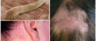
Actinic keratosis (AK, also known as solar keratosis, actinic keratosis) is a lesion that forms on sun-damaged skin. The lesions appear as small scaly patches on parts of the body such as the face, ears, bald crown of the head, arms and hands that have been exposed to the sun for a long time.
Actinic keratosis is a precancerous condition that affects only the top layer of skin (epidermis). The sun's ultraviolet (UV) rays damage the skin, and if left untreated, 15% of AKs can develop into a form of skin cancer called squamous cell carcinoma (SCC). When AK is detected early, it responds well to treatment.
Solar keratoses are more common in people with fair skin. The disorder affects 15% of people in North America and Europe.
Australia has the highest AK rate in the world. In Russia, the incidence rate is much lower.
Preventive actions
In order to prevent the development of actinic keratosis or to avoid its degeneration into a malignant formation, people need to follow simple preventive measures, especially over the age of 50:
sunscreens with SPF (ultraviolet protection factor) 30 or more. Women can be recommended to use any cosmetics applied to their hands and face with the same SPF value. These cosmetics should be applied 30 minutes before going out into the sun and reapplied every 2-3 hours of sun exposure. This is especially necessary if a person is bathing or has excessive sweating.
Use it all the time, especially in summer.- As you age, you need to stop visiting the solarium, as well as using various creams to improve your tan. Only spray tanning products are safe.
- Wear wide-brimmed hats, loose, long trousers made of linen or light cotton, long sleeves, and caps with a large visor that shade not only the face, but also the ears.
- Avoid tanning, especially sunburn. There is no need to stay in the sun for a long time without skin protection. Please note that cloud cover only absorbs a small portion of UV rays. The most dangerous time for being in the sun is from 12 am to 3 pm. It must be remembered that in winter you can receive a large dose of ultraviolet radiation, especially since ice and clean snow reflect these harmful rays. Therefore, even in winter, when walking in the sun, you need to remember about your own protection.
Causes and risk factors
Ultraviolet light from the sun and tanning lamps or UV lamps at home are the main causes of actinic keratosis. Ultraviolet light causes changes in the genetic material of skin cells. Changes in some genes can lead to abnormal, rapid cell growth and the formation of lesions.
Risk factors include:
- age 40 and older - keratoses can occur at any age, but the risk increases with the accumulation of solar radiation;
- immunosuppression - the body's ability to recognize and fight abnormal cells is reduced in people with weak immune systems (for example, as a result of taking organ transplant drugs, chemotherapy or AIDS), so cells with gene changes grow and form lesions;
- male gender - men, as a rule, work outside more often and accumulate more sunlight than women;
- fair-skinned people—also people with red or blond hair and those who are prone to burning rather than tanning are more likely to develop actinic keratosis;
- Sun exposure – People who work outdoors or are exposed to more sun are at increased risk of developing AK.
Features of the disease
Actinic keratosis develops due to constant or prolonged exposure to sunlight with a wavelength of 270-330 nm. Pathological changes appear over a long period of time; no changes may occur for 15–25 years. As a result, the disease is most often diagnosed in old age - 50-60 years, and is very rarely observed in young people.
Ultraviolet radiation on skin cells changes genetic material over time. As a result, atypical cells begin to appear - anaplastic, that is, undifferentiated, which do not have their clearly defined function. Anaplastic cells replace the normal epidermis, this disrupts the process of keratinization of the skin, which becomes rough and tough, this, in turn, speeds up the process of replacing healthy cells with atypical ones.
When conditions are favorable, anaplastic cells begin to penetrate the basement membrane, which separates the epidermis from the dermis. In this case, there is a possibility that akinetic keratosis develops into carcinoma. The probability of this happening is approximately 0.2%.
Classification of actinic keratosis
Actinic keratosis is classified solely by pathomorphological changes in the layers of the skin. Based on the localization of the active process in the epidermis and dermis, it is customary to distinguish typical variants of this disease:
- Hypertrophic actinic keratosis , when atypical large-nucleated cells appear in the epidermis, producing light and dark keratin. It is the alternation of layers of keratin that is a diagnostic sign of actinic keratosis.
- Pigmentary actinic keratosis is the accumulation of a large number of melanin cells, which stains the localization site dark brown in the basal layer of the epidermis.
- Lichenoid actinic keratosis is characterized by active processes at the border of the basal layer of the epidermis and the upper layers of the dermis, where lymphocytic infiltrates from dermal cells are formed on the basal layer of the epidermis “eaten away” by keratosis.
- Proliferative actinic keratosis occurs against the background of elastosis (colloid degeneration of the deep layers of the dermis) and is associated with the growth of epidermal cells into the skin and the formation of foci of hyperkeratosis.
- Atrophic actinic keratosis is localized in the upper layers of the dermis, thinning and destroying them locally through the formation of specific “lacunae” and cracks.
- A distinctive feature of acantholytic actinic keratosis is elastosis with the formation of epithelial-connective tissue foci deep in the dermis above existing “lacunae” and “cracks”. These local tumor-like formations grow towards the surface of the skin.
- Bowenoid (bovenoid) variant of actinic keratosis is the initial stage of cancer, characterized by an accumulation of dysplastic atypical cells in both the epidermis and the upper layers of the dermis. Atypical cells are in the so-called “dynamic equilibrium”: as many atypical cells appear, the same number die.
Moisture is your best friend
Since people with keratosis pilaris tend to have dry skin, and retinols and acids dry it out even more, it's important to combat this dryness with plenty of moisture. Contrary to popular belief, moisturizers do not technically hydrate the skin directly, but they do block water from entering the skin. (Exfoliating before applying moisturizer can help your product penetrate your skin better, and luckily you're already doing this if you follow rule two!)
It's always a good idea to drink plenty of water. And some people note that avoiding gluten and dairy products can help cope with keratoses. Unfortunately, there is not enough research yet to support this theory. However, doctors note that people with eczema may be more sensitive to certain foods, so keeping a food diary can't hurt.
Irritation after shaving: how to avoid and how to get rid of it
Symptoms and complications
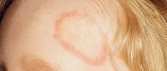
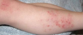
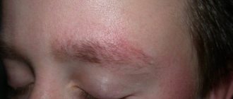
Actinic keratosis lesions appear as small, scaly patches that may appear flat or slightly raised. Color can range from skin tone to reddish brown. The spots range in width from 3 to 10 mm and can gradually increase in size (see photo above). The skin around the affected areas may show other signs of sun damage, such as dark spots, damaged blood vessels, and a yellowish tint.
Over time, the lesions caused by actinic keratosis may become thicker and harder. Their color may change from red to brown. Sometimes a cone-shaped growth occurs above the surface of the skin at the site of the lesion. This phenomenon is called cutaneous horn.
The most common location for actinic keratosis is the face, but it can occur on any part of the body that has been exposed to the sun for long periods of time, such as the arms, legs, and back of the arms. For example, sunbathers may develop actinic keratoses in other parts of the body.
The most significant complication of AK is skin cancer. Once an actinic keratosis lesion progresses and penetrates the lower layer of skin (dermis), it is classified as squamous cell carcinoma, a form of skin cancer. If left untreated, the cancer may spread to other parts of the body.
Lesions that are redder, larger and harder are more likely to develop into skin cancer.
Exfoliate your skin
Since keratosis pilaris is a genetic disease, there is no cure for it. But there are ways to cope with flare-ups and soothe your skin. The key to eliminating bumps is exfoliation, but it's vital to use chemical exfoliants rather than physical ones.
Look for treatments that will break down the skin's surface barrier. This means you need to choose acids and other products that help the skin exfoliate and eliminate rough skin cells that have formed around the hair follicles.
Lactic acid, urea, salicylic acid and retinoids work best. However, it's possible to overdose—you'll know you've overdone it when your skin becomes red and irritated. This is why it's best to start slowly and only use one acid at a time.
If you're new to the wild world of retinol, it's important to start slow. For keratoses on the face, dermatologists suggest starting with a low-percentage retinoid to avoid drying out the skin. Some people prefer to take breaks between retinol uses; others prefer the sandwich method, or layering a retinoid product between two applications of a moisturizing product. In any case, it is better to discuss this with a dermatologist.
Establishing diagnosis
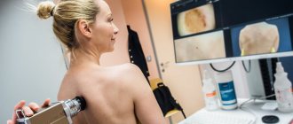
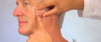
Actinic keratoses may look similar to other skin conditions such as psoriasis or skin cancer. A doctor usually discovers this condition during a physical examination, but the most accurate way to make a diagnosis is to perform a skin biopsy.
Hardware treatment methods
The basis of therapy for this disease is hardware-assisted techniques. The essence of treatment is to remove pathological foci. For this purpose, modern medicine suggests using one of several procedures suggested below.
- Cryotherapy (cauterization with liquid nitrogen). Recommended for large affected areas. After freezing, the flaky elements are removed, and the skin underneath becomes clean. One of the side effects of the procedure is skin hyperpigmentation.
- Laser therapy. Treatment of the pigmented form of solar keratosis is most often carried out using a laser. This is a relatively painless and at the same time effective way of influencing pathology. Under the influence of high temperatures, the plaque literally evaporates.
- Diathermocoagulation. During the procedure, the plaque is cleared using an electric current. The use of anesthesia is mandatory.
- Photodynamic therapy. The doctor applies a special cream to the keratotic areas, which increases the sensitivity of the dermis to the action of the applied waves. The lesion is then treated with a light beam. The procedure is well tolerated and is recommended for older people.
If during the diagnosis the doctor confirmed the benign nature of the process, treatment of solar keratosis is allowed in a beauty salon. A specialist can perform chemical peeling and dermabrasion. During these procedures, the upper layers of the epidermis are removed using chemicals and brushes. As a result, the patient leaves the cosmetologist’s office with renewed and healthy skin.
Treatment and prevention
There are several treatment options for actinic keratosis. Which one is best will depend on factors such as the size and location of the lesions, the number of lesions, and the person's overall health.
Cryotherapy - This method uses a very cold substance, liquid nitrogen, to freeze and kill the skin cells that make up the actinic keratosis lesion. Liquid nitrogen is applied as a spray. This treatment method is best for small numbers of lesions. Topical medications (see below) may be used prior to cryotherapy to improve results.
Excision is the surgical removal of a lesion using a sharp blade.
Electrodesiccation and curettage —the cells are dried with an electric current and then cleared using a curette (sharp instrument). These procedures require local anesthesia. Like cryotherapy, it is not suitable for large numbers of lesions.
Topical medications (medicines that are applied to the skin) may be used to treat actinic keratosis and superficial basal cell carcinoma. Topical medications include 5-fluorouracil* (also known as fluorouracil or 5-FU) and imiquimod.
- 5-fluorouracil belongs It acts by preventing the growth of cancer cells. The cream is applied to the skin in a thin layer and then spread. Treatment is repeated daily for several weeks. This drug is good for treating large areas of skin. 5-fluorouracil is contraindicated in pregnant women.
- Imiquimod is another topical drug for the treatment of actinic keratoses. It belongs to a group of drugs called immune response modifiers. This type of medicine works by stimulating the immune system to produce substances that fight cancer. The drug is applied to the skin twice a week for 16 weeks. This remedy is well suited for treating lesions on large areas of skin.
- Ingenol mebutate (picato gel) is another excellent drug for treating keratoses. It helps fight disease by killing skin cells and causing inflammation where it is applied. The gel is applied to the skin once a day for 2-3 days, depending on the location of the actinic keratosis.
Traditional medicine recipes
If surgical removal is not possible due to health reasons, preference is given to medicinal creams containing fluorouracil (this compound helps dissolve excessively keratinized cells), ointments with an anti-inflammatory effect, and a product called Imiquimod. Medicines are used in repeated courses. This therapy lasts a long time. Sometimes it leads to increased symptoms of the disease (inflammation worsens, burning and itching become much more pronounced). Experts suggest eliminating the ailment using traditional medicine.
Best suited for these purposes:
- applications made from grated raw potatoes;
- compresses with young aloe leaves;
- lotions with a decoction made from onion peels.
The effectiveness of such products has not been scientifically proven, but patient reviews indicate that they can be used to try to combat the manifestations of actinic keratosis.
Forecast
Actinic keratosis is a marker of severe changes and damage to the skin resulting from exposure to ultraviolet light. These changes put a person with actinic keratosis at high risk of developing skin cancer.
People who develop actinic keratosis sooner or later have a better chance of eliminating the risk of developing cancer. However, without treatment, actinic keratosis can develop into skin cancer.
After treatment for actinic keratosis, a person will likely need to schedule yearly visits with a dermatologist to check for recurrence and be screened for other signs of skin cancer.
Diagnostics
The diagnosis of actinic keratosis is made through a thorough physical examination based on a combination of visual observation and touch. If the lesion is large in diameter, thick or bleeding, a biopsy is ordered to rule out malignancy, especially if the patient has a history of skin cancer.
In addition, actinic keratoses may be mistaken for other skin lesions such as seborrheic keratoses, basal cell carcinoma, lichenoid keratoses, viral warts, inflammatory dermatoses, or melanomas.
The biopsy procedure involves taking skin samples from the affected area. It will be performed by a therapist using local anesthesia. The sample is sent to a laboratory where it is analyzed under a microscope. Common methods for obtaining material for the study of actinic keratoses:
- Scarification biopsy: extends to the dermis level.
- Punch biopsy: extends to subcutaneous fat.
On this topic
- Keratosis
Why is solar keratosis dangerous?
- Olga Aleksandrovna Kalinina
- July 13, 2021
Many cases of actinic keratosis are managed by a general practitioner. A referral to a dermatologist is made when:
- the diagnosis is unclear;
- skin lesions are severe or widespread ;
- symptoms do not respond to treatment;
- the patient is taking immunosuppressive drugs;
- there is a high risk associated with malignancy.
The dermatologist also uses other methods for diagnosing actinic keratosis, for example, fluorescence with a photosensitizing agent.
Any AK lesion can have one of three outcomes. Enter spontaneous remission, remain stable, without further progression, transform into invasive squamous cell carcinoma, which rarely metastasizes (but the risk should not be underestimated).
Clinical picture
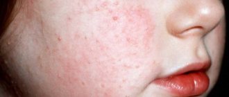
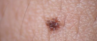
A typical manifestation of the disease is the formation of multiple dense nodules on the skin of various parts of the body (on the face, sides of the neck, upper and lower extremities, back). At the beginning of the process, a focus of hyperpigmentation appears on slightly atrophic or unchanged skin, which gradually rises above the surface, turning into a rounded papule. The size of the nodule varies - from a few millimeters to several centimeters. Color varies from normal skin to various shades of red or brown. The nodules are painless and not accompanied by itching.
Solar keratosis
The following clinical forms of solar keratosis are distinguished:
- The erythematous form begins with the appearance on the skin of round or oval spots covered with horny scales. The lesions are clearly demarcated from the surrounding tissue, and a focus of hyperemia forms around them. When injured, keratomas bleed heavily due to the large accumulation of capillaries in them.
- Keratotic papular, or hypertrophic form. Dense gray or dirty brown horny masses grow on the spots of hyperpigmentation. When these masses are removed, an eroded surface with small cracks remains.
- Warty form. With this form of solar keratosis, the nodules resemble vulgar warts in appearance.
- Horny form, or “cutaneous horn”. Keratomas are dense, rise above the surface of the skin, and resemble the shape of animal horns. Tumors are located mainly in the area of the ears and on the face.
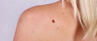
- The pigmented form is manifested by the appearance of brownish spots of various shapes on the skin. The lesions spread over the skin of the back and on the back of the hands.
- Spreading pigment form. With this type of keratosis, a large number of nodules appear in the facial area, varying in size and color. The surface of the lesions is covered with scales or smooth. This form is dangerous due to the high risk of transformation into a malignant neoplasm.
- Proliferative form. An oval plaque with a flaky surface appears on the skin. The boundaries of the lesion are unclear, the plaque gradually grows and can increase to 3-4 cm in diameter.
Keratosis can appear on the mucous membrane of the eye (conjunctiva) and the red border of the lips. When localized on the eye, the keratome looks like a dense wedge-shaped thickening in the area of the limbus - the place where the cornea transitions into the protein membrane (sclera). The red border of the lips affected by the disease thickens, begins to peel off, and becomes covered with small cracks. When erosions appear, we can talk about the degeneration of the process into squamous cell carcinoma.
Etiology and pathogenesis
One of the significant factors in the development of keratosis is constant exposure to ultraviolet radiation . A special role is played by ultraviolet light B (UVB, 290–320 nm) and A (UVA, 320–400 nm) - it induces p53-dependent apoptosis of epidermal keratinocytes. These apoptotic keratinocytes are often called “sunburn cells” (SBCs) and can be detected by histological examination of epidermis that has been overexposed to sunlight or tanning bed lamps.
In the development of keratosis, the UV-induced mutation of the tumor suppressor gene TP53, which is related to the above-mentioned p53 protein, is of great importance. Basal keratinocytes with mutant TP53 do not undergo apoptosis, which allows them to pass on genetic abnormalities to new cells. Similar changes are found in squamous cell carcinoma and other malignant tumors.
Actinic keratoses may be promoted by a general suppression of the body's immune response. The risk group includes patients who have undergone organ or tissue transplantation and are taking immunosuppressants.
Some medications can act as photosensitizers, increasing skin sensitivity to ultraviolet light and promoting the development of keratoses.
Microscopy of keratotic lesions usually reveals dysplastic changes in the basal layer of the epidermis, accompanied by parakeratosis and solar elastosis of varying severity. An increase in the size and pleomorphism of nuclei, hyperchromatosis, an increase in mitotic activity or an atypical nature of mitoses, and cytoplasmic pallor are recorded.
Dysplastic epidermal growths may extend deep into the papillary dermis and in some cases are difficult to distinguish from superficial invasive squamous cell carcinoma of the skin. Dysplastic epidermal changes, as a rule, do not affect skin appendages (sebaceous and sweat glands, hair, nails) ( Fig. 1 ).
Rice. 1. Histological features of keratosis (www.medscape.com)
In the epidermis, moderate hyperkeratosis is observed with dysplasia of basal keratinocytes and the formation of small outgrowths extending into the papillary dermis. The presence of solar elastosis is noticeable. Dysplastic changes primarily affect the interfollicular epidermis.