Primary period of syphilis
At the beginning of the disease, the rash is represented by only one element.
It appears on the genitals, less often in other places (on the lips, in the mouth, under the nails, in the anus).
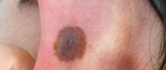
The only element of the rash is a painless erosion.
Sometimes it transforms into an ulcer, which is characterized by a greater depth of damage.
Item Features:
- Red color
- gray coating on top
- regular round shape
- no pain
- there is no growth either in depth or along the periphery
Erosion disappears on its own after 1-2 months.
If a person receives treatment, it regresses much faster.
Rash in the secondary period of syphilis
Most types of rash with syphilitic infection appear in the secondary period.
At this time, the disease is characterized by a wave-like course.
Symptoms from the skin and mucous membranes predominate.
There is usually no significant damage to internal organs.
The pathology either worsens or goes into a latent course.
Initially, multiple rashes appear on the body with syphilis, located symmetrically.
This is fresh syphilis.
There is usually a lot of rash.
It is located everywhere.
When examining such patients, it is still possible to detect the place where primary syphiloma was recently located.
It usually presents as a scar or pigmentation.
Regardless of whether a person receives specific therapy, the elements of the rash gradually regress.
Syphilis occurs in a latent form for a long time.
It recurs in 25% of patients.
In two cases out of three, this occurs no later than 6 months after the disappearance of the skin rash.
Less often - after 1-2 years.
If more than 5 years have passed, but there is no clinical relapse, then with a high probability it will no longer occur.
In the malignant course of syphilitic infection (mainly in HIV-infected people), there is practically no period of remission.
Exacerbations replace each other continuously.
Each subsequent one has more and more pronounced clinical manifestations.
Typically, the larger the rash and the more often it appears, the higher the risk of damage to internal organs, and the faster the syphilitic infection will progress to the tertiary stage.
Features of clinical relapses:
- less rash
- is not located throughout the body, but in separate areas
- no symmetry
The most diverse morphological elements appear.
There are secondary syphilides: papular, vesicular, macular or pustular.
Within each type there are many different rashes.
They differ in the location of morphological elements, grouping, and depth of tissue damage.
Different types of rash are determined by many factors:
- localization
- gender and age
- reactivity of the body
- concomitant infections
- aggravating circumstances (somatic illnesses, alcoholism)
- drugs used
General features of any rash with syphilis in the secondary period:
- elements do not appear suddenly, but gradually
- the patient does not complain of anything (it is extremely rare with syphilis that the rash itches or hurts);
- can appear on any part of the body
- both true and false polymorphism are observed (different elements, as well as different stages of development of the same elements at the time of examination)
- the rash is always red (the exception is fresh rashes, which may be pink)
- the elements that appear do not enlarge or merge (except in cases of local treatment or location in skin folds)
- independent and traceless disappearance, even if no treatment is carried out;
- resolution starts from the center and goes to the periphery
- no scars unless ulceration of the element occurs
- there is no increase in body temperature - the difference between syphilis and most other infections that lead to the appearance of a rash
- rapid disappearance of rashes under the influence of antibiotics
Differential diagnosis of papules for skin pathologies other than syphilis
Parapsoriasis or Broca's disease
Parapsoriasis is a rare, non-contagious skin disease that belongs to lymphoproliferative dermatoses.
It occurs predominantly in adult men of middle age.
Today there is no precise definition of this disorder.
Due to its variable clinical presentation and lack of specific histopathology findings.
However, the lesions usually appear as pink and scaly patches.
They vary in size from 1 to 5 cm.
The rashes are often chronic and resistant to medication.
There are two types of parapsoriasis: small-globular parapsoriasis (in English terminology SPP) and plaque parapsoriasis (LPP).
Of the two types, LPP is considered a premalignant dermatosis that can develop into T-cell lymphomas.
Whereas SPP is a chronic benign condition.
Again, because this disorder is difficult to diagnose, there are no specific epidemiological statistics.
In 10% of those who have plaque-type parapsoriasis, the disease progresses to a malignant form.
The cause of parapsoriasis is unknown; research on this topic is still underway.
It is conventionally accepted that parapsoriasis develops against the background of infectious processes and immune disorders.
Fine-grained psoriasis is usually asymptomatic.
There is no deterioration in general health, but rashes develop, ranging from pink to yellow-brown.
Round or oval spots (macula) ranging in size from 2 to 5 cm.
Maculae are mainly located on the abdomen, sides, back or lower extremities.
When examined under a microscope, there are very nonspecific data: mild spongiosis, parakeratosis, exocytosis of small lymphocytes.
There is also a variant of the disease called digital dermatosis.
In this case, the rashes look like elongated spots rather than round ones.
To make a definitive diagnosis, an epidermal biopsy is recommended.
It is advisable to perform several procedures on different areas of the skin.
Treatment begins with moderate-to-high potency corticosteroids for 8 to 12 weeks.
Wilson's ringworm
Lichen planus is an acquired, chronic, immune system-mediated disease.
Appears as polygonal, purple, itchy, papules or plaques on the skin and similar lesions on mucous membranes, such as the oral cavity.
Lichen planus is thought to be an immunologically mediated disorder.
This appears to be a response of CD8 cells to antigens in the basal cell layer and dermoepidermal junction.
Mainly in keratinocyte cells.
The reason for this phenomenon is still unknown.
There is only some speculation about the effects of certain viruses (eg, hepatitis C and hepatitis B) or drug factors.
Characteristic skin lesions of the lichen plan are polygonal, purple, itchy, flat papules.
They can stick together to form plaques.
Lesions may be close together or widely separated from each other.
They may have white spots or lines called Wickham's furrows, and they may also be hyperpigmented.
The lesions are usually distributed symmetrically on the upper/lower extremities, especially around the elbows and wrists.
However, they can be found anywhere on the body, including the glans penis and vulva.
Papules tend to disappear after some time, leaving signs of hyperpigmentation of the skin.
Psoriasis vulgaris (vulgar psoriasis)
Psoriasis vulgaris or scaly lichen is the most common form of psoriatic skin lesions.
Occurs in almost 80% of patients.
The disease is characterized by thickening of the epidermis, development of an inflammatory reaction on its surface, redness and silvery-white scales.
Skin lesions may be accompanied by itching, burning and pain on palpation.
Typical, inflammatory, erythematous, pruritic, and silvery-scaly patches of skin occur in psoriasis vulgaris on the extensor sides of the elbows and knees.
Also on the scalp, behind the ears.
However, other parts of the body may also be affected: the skin of the genitals, perineum, mammary glands, palms of the hands.
In turn, scaly lichen is divided into two subtypes:
- Type I: The disease usually occurs in patients under 40 years of age and is therefore called the early type. Other family members may also suffer from this form of psoriasis. Often, patients’ blood contains genetic markers of susceptibility to the disease.
- Type II. Called late type, it develops after 40 years. There is usually no family predisposition; DNA markers in the patient’s blood are significantly lower than in the first subtype. This type of psoriasis is milder and responds more effectively to treatment.
Miliary papular syphilide
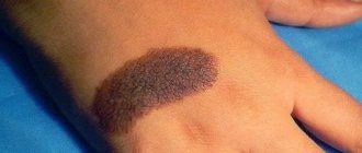
Rapulosa milliaris nigra is a benign skin disease.
Often affects patients with secondary syphilis.
The symptom is accompanied by the appearance of soft, dermal papules on the skin.
The color is practically no different from the natural shade of the skin.
Often localized in the area of hair follicles, on the face, around the eyes, and back.
Papules are harmless, but may be accompanied by severe itching and irritation.
The disease is quite rare and tends to occur in patients with dark skin, more often in African Americans.
The pathology is quite difficult to respond to drug therapy and practically does not respond to medications.
There are several methods for removing and treating rapulosa milliaris: curettage, electrocoagulation, electrosurgery, cryotherapy and electrotherapy.
Each treatment carries a risk of scar tissue formation, decreased pigmentation, and keloid formation.
It is important to differentiate miliary syphilide from lichen scrofulous.
In this case, the rashes are grouped and are papules in the form of perifollicular nodules that affect children or adults with tuberculosis infection.
Yellowish-reddish-brown papules usually develop on the back, chest, and abdomen.
Not accompanied by the formation of scar tissue.
The pathology can be found under synonyms - Lichen Scrofulosorum, Tuberculosis Cutis Lichenoides.
Spotted syphilide
The most common option.
It is observed in all patients without exception.
Most often - at the beginning of the secondary period.
Much less often - in case of relapse.
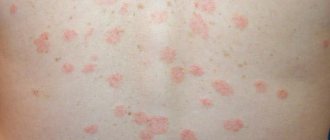
The most common element is roseola.
This is a small spot that starts out pink and then turns red.
Completely disappears when pressed.
The dimensions of one element can reach one and a half centimeters, although they are usually less than 1 cm.
The first spots may appear against the background of fever.
But then the body temperature returns to normal.
Features of roseola fresh syphilis:
- small sizes
- a large number of
- symmetrical arrangement
- pink color
- localization on the stomach and sides
Features of roseola recurrent syphilis:
- large sizes
- small number of elements
- asymmetry
- tendency to group with the formation of various figures (most often rings)
- bluish tint
Special types of roseola:
- raised - resembles a blister
- follicular - has a granular surface
- drain - formed by several adjacent elements
Treatment of syphilitic rash
Treatment of syphilitic rashes during all periods of the disease is not carried out separately, since it is only a separate symptom of the disease. Along with it, the entire body is affected and complex therapy for the disease is required.
To treat the disease, antibacterial agents from the penicillin series are used; if they are intolerant, other antibiotics from the group of macrolides, tetracyclines and cephalosporins are prescribed. The schematic prescription of drugs will depend on the stage of the disease and the general condition of the patient.
Treponema pallidum is most sensitive to penicillin-containing drugs that are administered by injection.
Table No. 2. Treatment at all stages of syphilis:
| Stage of syphilis | Features of treatment |
| First | Therapy at the first stage is carried out using water-soluble penicillins. Drugs in this group are most active against the causative agent of syphilis. Treatment is carried out in a hospital setting, since the drugs require frequent injections to maintain the required therapeutic concentration. The penicillin group is the drugs of choice, not only because of its high efficiency, but also because of its availability. The price of medicines is much lower than similar ones. Medicines:
|
| Second | Treatment of the second stage does not differ significantly from the treatment of primary syphilis; penicillin drugs are also prescribed. If they are intolerant, an alternative replacement is selected from the group of macrolides, tetracyclines and cephalosporins. Replacement drugs:
|
| Third | Therapy of the third stage is complicated by pathological lesions of the body, so treatment is prescribed based on the degree of damage to the body and the patient’s condition. As with the first two stages, the main thing is to take antibacterial drugs, but according to a specific regimen prescribed by the doctor. Further, complex treatment includes taking bismuth-based drugs and medications for the symptomatic treatment of damaged organs. |
Important. The instructions for bismuth preparations prohibit the use of the drug by patients with renal and liver failure.
The video in this article is about skin lesions caused by secondary syphilis.
Damage to the palms and soles due to syphilis
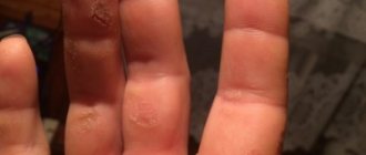
Papules often appear on the palms and soles.
There are the following types of syphilide:
- lenticular - multi-colored papules (yellow, pink, purple), which look like spots, but the infiltrate is determined by palpation;
- annular - in the form of rings and other figures;
- horny - covered with dense layers, reminiscent of callus;
- wide - large multiple plaques with irregular contours and horny layers (usually appears in places of constant skin irritation).
Vesicular syphilide
It is also called herpetiformis because the elements formed resemble those of a herpetic infection.
It is a rare form of the disease.
Indicates an unfavorable course.
Small papules appear.
They then transform into bubbles.
They are grouped into plaques several centimeters in diameter.
There are no subjective sensations.
In the form of blisters, the rash lasts only 1-2 days, and sometimes for several hours.
It quickly opens with the formation of multiple erosive and ulcerative elements.
After this comes the cortical stage, which lasts several weeks.
Gradually the infiltrate resolves and the crusts come off.
After resolution of the process, small multiple scars remain.
Tertiary period of syphilis
Signs of damage to internal organs, the brain and spinal cord dominate.
Elements of the rash in this period are presented:
- tubercles
- gums
- late roseola
Items appear slowly.
There are usually no subjective sensations.
Gummas develop in the subcutaneous fat layer, and tubercles develop in the dermis.
They are located in isolation and do not merge.
They may dissolve or rupture to form ulcers.
Whatever the resolution, these skin lesions are deep and leave scars.
The number of tubercles is usually greater than gummas, which are single.
What is a papule?
The papular morphological element of the skin is a cellular infiltrate located in the upper layers of the dermis.
Its features:
- round or oval shape
- rises above unchanged skin
- has clear boundaries
- Red color
- dense consistency
Syphilitic papules in the vast majority of cases are located separately and do not merge.
Once they appear, they no longer increase in size.
The exception is papules located in the folds of the skin.
They grow, and merge, and differ in appearance from everyone else.
There are different variants of papular elements.
Basically, additional varieties appear as a result of constant irritation, high humidity, attempts at self-medication, etc.
Ulcers, vegetations, and hypertrophic changes may appear in these places.
If the papules are not touched, and they are not located on damp skin or in folds, then they rarely become complicated.
They usually last no more than 2 months.
Then they begin to peel off, change color and disappear.
Hyperpigmented spots may remain at the site of the formations for some time.
Next, we will consider individual types of papular syphilides.
