Dermatovenerologist
Khasanova
Alina Rashidovna
8 years of experience
Make an appointment
One of the forms of vasculitis that develops as a result of an allergic reaction of the body is erythema nodosum. This is the name for inflammation of the blood vessels that penetrate the epidermis, as a result of which painful pink or bluish nodules form in the dermis and subcutaneous fatty tissue. Vascular damage is local in nature and is limited mainly to the lower extremities.
Causes of erythema nodosum
Erythema nodosum is a polyetiological syndrome that is found in the practice of many specialists and has two clinical forms: idiopathic and symptomatic.
Some researchers express an opinion about the viral origin of idiopathic erythema nodosum, others believe that this disease, especially in children, is an allergic manifestation in most cases of tuberculosis infection and is observed mainly in people who have had or are suffering from tuberculosis in its various manifestations. In most of these patients, the Mantoux reaction is sharply positive, with exudative phenomena, lymphangitis and general symptoms.
Acute erythema nodosum can develop during or after infectious diseases caused by:
- streptococci,
- viruses,
- rickettsia,
- salmonella,
- chlamydia,
- Yersinia, etc.
The pathogenesis of erythema in such cases is not entirely clear - an allergic reaction, septic granuloma, or a reaction to bacterial toxins. It is possible to develop erythema nodosum against the background of:
- taking medications (sulfonamides, antibiotics, iodine preparations, oral contraceptives),
- sarcoidosis,
- Behçet's disease,
- ulcerative colitis,
- Löfgren's syndrome,
- rheumatism,
- vaccinations and the like.
There are familial cases of erythema nodosum associated with a hereditary predisposition to sensitization of the body by infectious or other agents.
Some researchers classify erythema nodosum as a deep vasculitis.
In 40% of cases, the cause of erythema nodosum remains unknown.
The development of changes in the skin with erythema may be preceded by prodromal phenomena in the form of:
- ailments,
- increase in body temperature,
- arthralgia,
- myalgia,
- catarrhal phenomena.
They are observed 3-5, less often 7-10 days before the appearance of nodes.
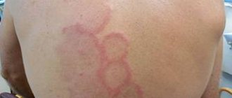
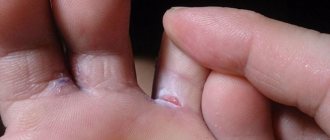
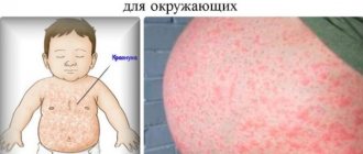
The clinic is characterized by the appearance on the front and lateral surfaces of the legs of painful, dense, acutely inflammatory (looking like erythema), bright red nodes (from 2 to 50), the size of a pea to a walnut, located deep in the subcutaneous tissue focally and symmetrically. Sometimes they can merge, their contours are unclear, which is associated with swelling of the surrounding tissues. In some cases, nodes may appear on the back of the legs, forearms, thighs, buttocks, torso, and even more rarely on the feet, hands, and face. Sometimes a macular, papular, urticarial or hemorrhagic rash or a rash characteristic of erythema multiforme may be observed next to the nodes. After a few days, the nodes acquire a bluish, then yellow-green color, reminiscent of the color changes of a bruise, their pain decreases, and complete regression is observed after 2-3, less often 4-6 weeks, leaving temporary pigmentation. These clinical manifestations during the period of their occurrence are also accompanied by fever, disturbances in general condition, pain in the joints and bones. Some patients may have gastrointestinal disorders and neurological symptoms. Leukocytosis or leukopenia and accelerated ESR are observed in the blood.
Histological changes in the tissue of nodes can be of three types:
- tuberculoid structure of the cellular inflammatory infiltrate in the subcutaneous tissue with giant Langerhans cells without caseous decay;
- nested placement of cellular infiltrate with giant Langerhans cells without a clear tuberculoid structure;
- nonspecific inflammatory changes in the subcutaneous tissue.
In addition, there is also chronic erythema nodosum, which is not accompanied by acute inflammatory phenomena; the nodes are mildly painful, remain unchanged for months and resolve without decay, leaving no lasting trace. In such patients, foci of focal infection are often detected.
It is believed that the clinical variants of erythema nodosum should also include migratory erythema nodosum, described by Beferstedt in 1954. In this case, the provoking factors are pregnancy (up to 40%), especially in the first trimester, streptococcal infections are in second place, and sarcoidosis is in third place. The number of nodes is formed from 1 to 8, often asymmetrical placement, although a symmetrical version can also be observed. The nodes are susceptible to migration and relapse, the average duration of the disease is 4-5 months, and they regress without leaving a trace.
Symptoms
A typical manifestation of erythema nodosum is dense nodes located in the lower dermis or subcutaneous tissue. The diameter of the nodes varies from 5 mm to 5 cm. The skin over them is smooth and colored red. Elements of erythema nodosum rise somewhat above the general level of the skin, their boundaries are blurred due to swelling of the surrounding tissues. Having quickly grown to a certain size, the nodes stop growing. Pain syndrome in patients with erythema nodosum can have varying severity and is noted not only during palpation of the nodes, but also spontaneously. There is no itching. After 3-5 days, the resolution of the nodes begins, which is manifested by their compaction and is not accompanied by disintegration. Characteristic of erythema nodosum is a change in skin color over the nodes, which resembles the process of resolution of a bruise. Initially red, it becomes brown, and then bluish, greenish and yellow. The most typical location of nodes in erythema nodosum is the anterior surface of the legs. More often, symmetry of the lesion is observed, but unilateral or single rashes are possible. Elements of erythema nodosum can occur wherever there is subcutaneous fatty tissue: on the thighs, calves, buttocks, forearms, face and even the episclera of the eyeball. In most cases, erythema nodosum has an acute onset and is accompanied by fever, anorexia, general malaise, and chills. Approximately 2/3 of patients experience arthropathy: joint pain (arthralgia), pain when palpating, stiffness in the morning. In 1/3 of patients with erythema nodosum, subjective symptoms are accompanied by objective signs of inflammation in the joint (arthritis): swelling and redness of the skin in the joint area, increased local temperature, and the presence of intra-articular effusion. Articular syndrome with erythema nodosum is characterized by symmetrical damage to large joints. Swelling of the small joints of the feet and hands is possible. General symptoms and arthropathy may precede the appearance of skin elements by several days. As a rule, complete resolution of erythema nodosum nodes occurs within 2-3 weeks. In their place, temporary hyperpigmentation and peeling may occur. Along with the skin symptoms, the joint syndrome also disappears. In total, the acute form of erythema nodosum lasts about 1 month. Much less often, erythema nodosum has a persistently relapsing chronic course. Exacerbations of the disease are manifested by the appearance of a small number of single bluish-pink nodules of dense consistency, which persist for several months. Skin manifestations may be accompanied by chronic arthropathy that occurs without joint deformation.
How to treat erythema nodosum?
Treatment of erythema nodosum should begin with identifying its cause, and it largely consists of eliminating this cause. However, this is not always possible. In cases where erythema nodosum is associated with infectious factors, antibiotics are indicated - depending on the nature of these factors, rifampicin, streptomycin, penicillin, tetracycline and the like. The drugs are taken in usual doses for 1-2 weeks. The effect is significantly enhanced when antibiotics are combined with small doses of corticosteroids - 15-20 mg of prednisolone once a day after breakfast. Systemic glucocorticoids are effective in the treatment of erythema nodosum , but they should be used as a last resort, since they usually affect the course of the underlying disease.
Anti-inflammatory drugs are also used - acetylsalicylic acid, indomethacin (methindol), butadione, ibuprofen and others. Potassium iodide is quite effective in daily doses of 300-900 mg for 2-4 weeks. In cases of a clear connection between exacerbations of erythema nodosum and menstruation, oral contraceptives are indicated for 3-6 cycles. The use of such drugs for medicinal purposes is permissible after consultation with a gynecologist.
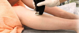
Of the physiotherapeutic methods, the following have a good effect on erythema nodosum:
- UV in erythemal doses,
- magnetotherapy,
- laser therapy,
- phonophoresis with hydrocortisone on the area of inflammatory nodes or affected joints.
Prevention
There are no clearly effective measures that will completely protect against the occurrence of such pathology. Since different forms of the disease are provoked by various factors, it is impossible to exclude them all. However, you can reduce the risk of erythema by strengthening the body's defenses. In general, prevention of this disease comes down to strengthening the immune system, and in the presence of allergic reactions, minimizing contact with allergens.
- It is necessary to follow a sleep and rest schedule and avoid stress. One of the indirect causes that causes dermatological diseases is chronic stress.
- You should lead a moderately active lifestyle. Regular jogging or physical exercise, cardio exercise is a good way to strengthen the walls of blood vessels. Such measures reduce the risk of rashes.
- A balanced diet with a predominance of protein and plant foods. It is necessary to minimize the consumption of fatty or spicy foods, the amount of sugar, and products with chemical dyes or preservatives. They can provoke allergic reactions.
- If your immune system is weakened or after an illness, you should take courses of multivitamins and mineral preparations. They should be selected by a therapist.
- Staying in the open sun should be limited to morning and evening hours. Before sunbathing, be sure to use sunscreen cosmetics.
- To reduce the risk of erythema, you should give up bad habits. Smoking and regular consumption of alcoholic beverages often lead to the appearance of erythema.
What diseases can it be associated with?
Erythema nodosum is not so much an independent disease as a symptom of other disorders in the body. There are many points of view on the origin of erythema nodosum, the main ones are as follows:
- against the background of infectious diseases caused by: streptococci, the manifestations of which are tonsillitis, scarlet fever, acute pharyngitis, streptoderma, erysipelas, otitis media, cystitis, rheumatoid arthritis,
- viruses,
- rickettsia,
- salmonella,
- chlamydia,
- yersinia,
- Mycobacterium tuberculosis, etc.;
- sarcoidosis,
- ulcerative colitis,
Diagnostics
Blood test as a diagnostic method Only a specialist can correctly diagnose erythema nodosum, since it is necessary to distinguish the disease from other pathologies with similar symptoms, in particular joint diseases, in order to correctly prescribe treatment and prevent the disease from becoming chronic. To do this, they prescribe clinical tests of blood, urine and feces, diagnose tuberculosis, and take a culture from the nasopharynx to detect the presence of streptococcus. If the joints hurt, the patient must be referred to an appointment with a rheumatologist and for appropriate studies, for example, radiography, MRI. Depending on what triggered the disease, a consultation with various specialists may be prescribed, for example, a phlebologist, an ENT specialist, an infectious disease specialist, etc. An X-ray of the lungs is often prescribed to determine the presence of tuberculosis or sarcoidosis.
What medications are used to treat erythema nodosum?
Antibiotics:
- rifampicin - 10 mg/kg once a day or 15 mg/kg 2-3 times a week;
- streptomycin - dosage is individual, depending on the underlying disease;
- penicillin - dosage is individual, administered intramuscularly, intravenously, subcutaneously, endolumbarally;
- tetracycline - a single dose for adults is 250 mg every 6 hours.
Corticosteroids:
- prednisolone - 15-20 mg 1 time per day after breakfast.
Anti-inflammatory drugs:
- acetylsalicylic acid - 0.5-1 g per day (maximum up to 3 g), can be used 3 times a day;
- butadione - 0.2-0.4 g during or after meals 3-4 times a day;
- ibuprofen - the dosage is individual, depending on the underlying disease;
- indomethacin - 25 mg 2-3 times a day;
- potassium iodide - daily dose 300-900 mg for 2-4 weeks.
Treatment of erythema nodosum with traditional methods
Conservative treatment of erythema nodosum can be supplemented by the use of folk remedies. For oral administration, the following medicinal herbs and berries are used:
- lingonberry leaves,
- Melissa,
- mint,
- birch,
- yarrow,
- elder,
- hawthorn,
- rose hip,
- red rowan
Any herb listed above in the amount of 1 tbsp. should be infused in ½ liter of boiling water and taken 1/3 cup before meals.
Compresses and ointments based on nettle, mistletoe, and arnica are used locally:
- Grind 100 grams of dried arnica roots to a powder, combine with an equal amount of fresh pork fat and leave on low heat or in the oven for up to three hours; when the medicine has cooled, it will take the form of an ointment, which should then be used at night under gauze bandages;
- Use freshly squeezed nettle juice for compresses, moistening the same gauze bandage in it.
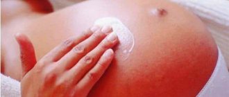
Treatment of erythema nodosum during pregnancy
Erythema nodosum often develops in pregnant women, in such cases it is idiopathic in nature. The exception is chronic diseases present at the time of pregnancy.
The factors causing erythema in a pregnant woman are usually:
- seasonal weather changes,
- hypothermia of the body,
- congestion in the lower extremities,
- hypertensive changes,
- restructuring of the endocrine-immune cascade.
An expectant mother should contact specialized specialists with a similar problem. Profile diagnostics and conservative treatment should be carried out exclusively under their supervision. The doctor will have a goal to reduce the severity of skin manifestations of the pathology and exclude the presence of infection and other serious causes of erythema.
It is usually recommended for a woman to:
- bed rest,
- non-steroidal anti-inflammatory drugs,
- antibacterial drugs, preferably in the second trimester and provided that their use is inevitable, and the existing infection is obviously more destructive than the medicine being taken - these are penicillins, cephalosporins and some macrolides.
For preventive purposes the following is used:
- sanitation of foci of focal infection,
- treatment of concomitant diseases,
- avoiding the use of drugs that provoke the disease.
Principles of diagnosis
Often patients have a question about which doctor to contact for a woman or a man who is faced with such changes in the body.
Diagnosis depends on the cause of the disease and is often complex. It takes place with the participation of several specialists.
At the initial stage you will need:
- Donate blood and urine for general and biochemical analysis to the laboratory.
- Get examined by a therapist, check your blood pressure, do an ECG.
- Submit biological material for analysis and make sure that there are no hidden infections in the body.
The key is to palpate the nodules for pain and mobility. If required, the doctor gives a referral for an ultrasound examination of the formations in order to clarify their nature of appearance and characteristics.
The diagnosis will be carried out under the supervision of a doctor, and its direction will be determined by the doctor. He selects the necessary procedures that can help make the patient the correct diagnosis and prescribe treatment.
Which doctors should you contact if you have erythema nodosum?
- Dermatologist
- Rheumatologist
The diagnosis used for erythema nodosum is not specific; it is aimed at identifying the underlying cause of the erythema. The methods used make it possible to differentiate erythema from other dermatological disorders. A blood test reveals neutrophilic leukocytosis and increased ESR.
Bacterial culture of stool and nasopharyngeal smear is carried out, tuberculin diagnostics, and a blood test for rheumatoid factor are appropriate.
To confirm the analysis, the nodule is subjected to biopsy and histological examination for the presence of inflammation.
In the process of identifying the etiological factor, the patient may be referred for consultation to:
- pulmonologist,
- infectious disease specialist
- otolaryngologist,
- vascular surgeon
- phlebologist.
Diagnostic methods that may be appropriate:
- rhinoscopy,
- pharyngoscopy,
- CT scan,
- radiography of the lungs,
- rheovasography of the lower extremities.
Differential diagnosis of erythema nodosum is carried out with indurative erythema in cutaneous tuberculosis, migratory thrombophlebitis, panniculitis, nodular vasculitis formed in syphilis.
Erythema nodosum is also differentiated from the following rarer diseases:
- Christian-Weber febrile nodular panniculitis. It is characterized by the presence of single or multiple, somewhat painful nodes in the subcutaneous tissue, often located asymmetrically on the legs, thighs, arms, and torso. At first, the nodes are dense, subsequently softer, the skin over them is initially somewhat hyperemic, and at a later stage remains unchanged. The nodes appear in paroxysms at intervals of several days or months and resolve, leaving a saucer-shaped recess, hyper- or depigmentation on the skin, does not suppurate. The onset is subacute with fever, impaired general condition, joint pain; with relapses, the general condition may not be affected. The duration of the disease is years and decades. Women aged 30-40 years are most often affected. Similar changes can occur in the retroperitoneal, perirenal tissue, and omentum, which indicates the systemic nature of the damage to the fatty tissue. Histologically, pseudoxanthoma cells are detected.
- Subacute migratory nodular hypodermatitis of Vilanova-Piñol. Women are more likely to get sick; the rash often occurs after a sore throat or flu. Asymmetrically, a deep nodular infiltrate the size of a palm appears on the anterior surface of the leg, slightly painful on palpation, with clear contours and a chronic course. Histologically, it differs from erythema nodosum in that it affects not large vessels, but the capillaries of the subcutaneous tissue. Hypodermatitis nodosa can sometimes develop as a reaction to a tuberculosis infection.
Kinds
In total, there are three types of erythema nodosum:
- Acute;
- Chronic;
- Migratory.
Erythema nodosum of the legs Most often it is acute erythema nodosum of the lower extremities. Nodules begin to appear on the legs, symmetrically on both legs. They are painful when pressed; in addition, the disease develops suddenly, the general condition worsens, fever may occur, and weakness may occur throughout the body. In addition, the nodules themselves turn red and swell. If inflammation occurs near the joint, then it is quite possible that it will be involved in the process, then the pain intensifies, the motor activity of the limb is impaired, and the patient’s general condition worsens. Over time, the nodes decrease, redness subsides, and yellowish spots remain on the skin that resemble bruises. Chronic erythema most often occurs in women over 45 years of age who have a history of various tumors and chronic infections in the body. Unlike acute erythema, the disease passes without deterioration in general health, the nodules are not externally visible, the skin does not become red or inflamed. Pain and obvious signs of inflammation may occur during an exacerbation of the disease. With erythema nodosum migrans, the symptoms are rather vague, the temperature does not rise much, and the person feels general weakness. In this case, the node appears alone, it does not increase much, and can migrate, leaving pale plaques.
Treatment of other diseases starting with letter - y
| Acne treatment |
| Treatment of kidney duplication |
| Treatment of polyarteritis nodosa |
| Treatment of nodular goiter |
| Treatment of ureaplasmosis |
| Treatment of ureterohydronephrosis |
| Treatment of urethritis |
The information is for educational purposes only. Do not self-medicate; For all questions regarding the definition of the disease and methods of its treatment, consult your doctor. EUROLAB is not responsible for the consequences caused by the use of information posted on the portal.