Pigmented nevus is the most common. In Latin, pigment means a relation to color. In this case, we use this term to indicate the presence of skin pigment cells - melanocytes, which give the skin a dark color. It is not at all necessary that a pigmented nevus will be colored. Intradermal nevus and Spitz nevus are pigmented. However, most often they are not dark in color. The term pigmented was coined to refer to color. In such moles there are foci with actively multiplying melanocytes (nevocytes). These cells are characterized as slightly damaged melanoblasts that have migrated from the neural crest into the epidermis. Melanocytes often cluster together to form nests. The natural development of pigmented nevi is not fully understood. The most common theory suggests that melanocytes gradually move into the deeper layer of skin, the dermis. All pigmented nevi can be divided into 3 main types: acquired, congenital, dermal. In turn, acquired nevi are divided into 3 main subtypes: borderline, complex, intradermal. The first three types are acquired nevi; on their basis, many other varieties are formed, such as dysplastic nevus or Setton. These varieties are named according to their external characteristics. In essence, they are only modified acquired nevi of one of the 3 main types. The Spitz nevus stands somewhat to the side; the growth of blood vessels radically changes its structure, and the original shape becomes incomprehensible. As the name implies, congenital pigmented nevus is already observed in newborns and appears during the embryonic period of development. Dermal pigmented nevi are so named because their melanocytes are located in the dense layer of skin called the dermis, most of them are found in Asians.
Pigmented nevus: what is it (photo)
The formation of a nevus occurs in the stratum corneum of the skin, where cells unite; sometimes such processes can be observed in the area of subcutaneous tissue.
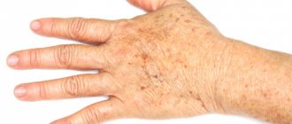
Moles can appear on any part of the skin - in the back, abdomen, face and even in the fundus of the eye.
Early age groups of children are susceptible to the occurrence of formation, and papules are most often found in people of the Caucasian race.
Symptoms
Apart from external manifestations in the form of spots, neither acquired nor congenital melanocytic nevus no longer has any clinical signs. Neoplasms are not accompanied by pain and burning, as well as other uncomfortable sensations. However, quite often you can notice single or multiple hairs on them, which are denser and darker than the rest of the body hair.
Separately, it is worth noting about those types of nevus that are prone to malignancy. When the process of malignancy occurs, often under the influence of ultraviolet radiation, symptoms may appear that should be the impetus for seeking qualified help.
Transformation to melanoma may be accompanied by:
- rapid growth of age spots;
- changes in the outlines of the neoplasm;
- the formation of a dark rim around the mole;
- color enhancement;
- inflammation, redness or swelling of the skin surrounding the nevus;
- secretion of ichor or other pathological fluids;
- a sharp increase in the number of melanocytic nevus;
- sensation of burning, itching and tingling;
- peeling of the skin in the problem area;
- moderate pain syndrome.
Trauma, chemical or thermal damage to the mole can lead to malignancy.
Congenital pigmented nevus
Congenital nevus can be large. This type of formation is grouped depending on its size - up to 1.5 cm; up to 10 cm; more than 10 cm.
It is the second group that carries high risks - congenital nevus up to 10 cm, since it can transform into a malignant tumor.
One of the subtypes of congenital formations is a giant pigmented nevus.
This formation has an uneven, often lumpy surface on which depressions form.
A giant pigmented nevus in children continues to grow along with the body and carries a direct threat of degeneration into a malignant neoplasm.
Prevention of mole degeneration
Normally, pigment formation does not threaten human life. But under certain conditions it can become malignant. To prevent this, you must adhere to basic preventive rules:
- Before going outside, apply sunscreen to your body.
- Avoid direct sunlight. Wear protective clothing if possible.
- Do not visit a solarium, do not pull out hair and do not use aggressive cosmetics.
- Try not to injure the mole.
- If there are any changes, consult your doctor.
Only an experienced specialist will be able to determine whether a pigmented nevus is dangerous. And he makes the decision to remove the formation after a complete examination of the patient for malignancy.
Acquired pigmented nevus
Birthmarks of this group are clearly distinguished by their histological characteristics:
- Borderline pigmented nevus - formation cells are located on the border between the stratum corneum and the surface of the dermis.
- Intradermal type - localized in the skin itself, often in the neck and face. It has the shape of a nodule and is yellowish in color, interspersed with brown. By the age of 30, this birthmark takes the form of a warty papule.
- A complex type of acquired formation is a combination of two histological elements at once. One nevus combines intradermal and borderline types of formation. Therefore, its treatment is most difficult. A complex nevus is located slightly above the surface of the dermis and may have a black color.
Stages of development
Melanocytic nevi of epidermal origin appear in childhood and reach their maximum number in adolescence. The appearance of new nevi in adults is possible, but not so common. In their development, they always go through several stages, which culminate in involution and fibrosis.
In children, borderline nevi are most often observed, in which nests of nevus cells are located above the basement membrane, at the border of the epidermis and dermis.
With age, nevus cells usually gradually penetrate into the papillary dermis and the nevus becomes complex or intradermal.
Intradermal nevus is the last stage of development of melanocytic nevus. Immersion into the dermis is complete. Here the nevus continues to grow or enters a dormant stage. Over time, it undergoes fibrosis. As nevus cells sink into the dermis, they lose the ability to synthesize melanin, and the noncellular nevus loses pigmentation. The fewer nests of nevus cells left in the epidermis, the lighter the color of the nevus.
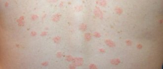
Dysplastic pigmented nevus
Pigmented nevus of the eye
Dysplastic nevus has the most dangerous distribution for humans. This formation can manifest itself in any age group.
Their difference from other subspecies is their inability to die off and disappear on their own with age.
It is dysplastic nevi that are the most dangerous, therefore, at the slightest deviation in the appearance of this papule or when painful sensations appear, it requires immediate medical intervention so as not to miss malignant consequences.
Based on their clinical manifestations, nevi are classified into the following groups:
- papillomomatous;
- warty;
- flat;
- knotty;
- hairy.
What complications can you encounter?
The main complication that can happen is the degeneration of a mole into a malignant tumor. But not all such formations can change like this. You need to closely monitor the development of melanoma-dangerous moles.
Despite this, under the influence of certain factors, a seemingly harmless mole can cause harm to the body. This often happens as a result of injury or spontaneous removal.
The beginning of the degeneration of a mole is detected by the following signs:
- education is rapidly increasing in size,
- the mole hurts or itches,
- its color changes,
- the surface layer changes,
- previously clear boundaries become blurred,
- the nevus is bleeding.
Malignancy can occur at any age, so if suspicious spots appear, you should immediately contact a specialist.
Diagnosis of pigmented nevus
Pigmented papule requires differentiated diagnostic studies, due to its similarity with other types of formations.
For example, a complex nevus is similar in appearance to senile keratoma or nodular melanoma.
Borderline nevus can be confused with lentigo. And the nevus, which is located inside the dermis, has similar symptoms to neurofibroma and the growth of the sebaceous glands.
Only a full range of histological studies will help distinguish between formations and establish the correct diagnosis.
Diagnostic procedures
If a mole appears, even if it is not accompanied by negative symptoms, it is recommended to consult a doctor.
A professional examination of a papillomatous mole is aimed at identifying the degree of danger of the neoplasm to health. The main task when making a diagnosis is considered to be the correct differentiation of papillomatous nevus from other skin neoplasms (papillomas, pigmented borderline nevus, etc.). If during the examination there is a suspicion of the presence of cancer cells, the patient is referred for consultation to a dermato-oncologist. Accurate diagnosis of the tumor helps to avoid the development of melanoma and other malignant skin tumors and, if necessary, begin timely treatment. The diagnosis is usually made by a dermatologist. He conducts a visual examination and, based on the preliminary diagnosis, prescribes additional confirmatory diagnostics:
- dermatoscopy;
- microscopic examination;
- biopsy.
Pigmented nevus removal and treatment
There is no drug treatment for this type of formation.
Treatment involves removal of the pigmented nevus when indicated.
In order to remove a formation, many factors are taken into account:
- location - mucous membrane, groin area, facial part, sole of the foot or scalp;
- shade - if the nevus is unevenly colored or has changed its color, then this situation requires treatment by removing the mole;
- shape - if the birthmark has unclear boundaries or growth that is intermittent in places, then this also indicates the need for surgical intervention.
Indications for nevus excision also include:
- education bleeds;
- painful sensations upon contact;
- the papule itches and peels off;
- the skin around the mole is swollen and red.
Surgical resection is prescribed when optical microscopy examination was unsatisfactory and indicated signs of malignancy.
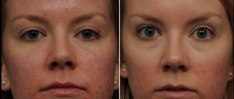
The aesthetic side of the removal issue can be resolved in beauty salons through the use of electrocoagulation.
If the mole is swollen or has redness around it, you should immediately consult a doctor.
Features of the view
Warty nevus is divided into subtypes, differentiation occurs:
- according to the drawing;
- distribution features:
- presence of inflammation.
https://www.youtube.com/watch?v=cgRiUHSebLg
Usually the plaques are located in one part of the body and are small in size. It is a dark brown or yellowish warty growth. Among them you can see soft ones with a dark velvety surface. The growth consists of several tubercles located very closely, with a flat top.
We invite you to familiarize yourself with Laser removal of nevus consequences photo
This is the name for the type that is located on one side of the body. It resembles linear garlands on the limbs or is located in bunches on the body. Spreads in areas along Blaschko lines, alternating with clean skin. It happens that a skin formation covers the human body, going down from head to toe.
The form extends to equal areas of the body, being located on both sides of the body, usually symmetrically. It refers to extended growths and causes significant aesthetic discomfort to a person. The elements also extend along Blaschko's lines; the growths can be of different lengths, but the species is characterized by an arrangement strictly up to the middle of the body. Sometimes several growths merge into one large element.
This type is characterized by the presence of inflammation. This type is always located on one side: usually observed in the lower half of the torso and limbs, often in the gluteal region. It is distinguished from the classic warty nevus by:
- more scales;
- it is accompanied by itching;
- redness.
It is often confused with lichen planus and linear lichen or manifestations of psoriasis, although these are completely different diseases. Sometimes a histological examination is required to clarify the diagnosis.
Pigmented nevus according to ICD 10
Pigmented nevus (melanocytic) in the Russian version of ICD-10 is called melanoform and is assigned codes from the D22 series.
- D 22.0 pigmented nevus of the lip;
- D 22.1 pigmented nevus of the eyelids or their adhesions;
- D 22.2 ear and external auditory canal;
- D 22.3 persons without specification;
- D 22.4 scalp and neck;
- D 22.5 body;
- D 22.6 arms and shoulder girdle;
- D 22.7 legs and hip area;
- D 22.9 without specifying the location.
Should I delete education data?
Everyone has probably asked a similar question. For some, these nevi are completely safe and the risk of their germination is minimal. For others (especially those who have a genetic predisposition), the risk of the spot degenerating into melanoma is extremely high, and a late decision on treatment can be fatal.
Therefore, the question of removing each nevus is purely individual. If you are ready to take the risk of surgery, it is best to remove it and forget about its existence. If you are ready to risk your health and not touch the nevus, that’s up to you, however, from a medical point of view, it is better to take care of your health prematurely and get rid of this small but very formidable formation, so as not to regret your decision later.
Included:
- morphological codes M872-M879 with tumor character code /0
- nevus:
- NOS
- cyan [blue]
- hair
- pigmentary
Anal region:
- the edges
- skin
Skin of the perianal area
Breast skin