Epidemiology of the disease
Hand fungus is ubiquitous. The source of infection is a sick person. Infection occurs in public places, family and workplaces. Fungi are transmitted both through close contact with a sick person and through infected objects: washcloth, towel, shoes, clothes, etc.
In the chronic course of foot rubrophytosis, the infection often spreads to the skin of the patient’s hands - first the working hand, and then to both hands.
The following factors contribute to the spread of infection:
- Minor injuries and abrasions of the skin of the hands, which occurs during prolonged rubbing of the skin of the palms with tools, washing, peeling and processing vegetables, etc.
- The skin of people whose hands are exposed to a damp environment for a long time is affected by fungi: when cleaning, cooking, doing construction work, etc.
- People with reduced body resistance are susceptible to the disease, which is observed in endocrine diseases, chronic infections, long-term somatic diseases, and immunodeficiencies.
- People with a genetic predisposition to fungal diseases are at risk.
Rice. 2. In the photo on the left is a view of the fungi Trichphyton rubrum under microscopy of a pure culture. On the hyphae there are pear-shaped single-celled spores (microconidia). The photo on the right shows colonies of pathogens, some varieties of which produce red pigment when growing on a nutrient medium.
Prevention measures
To reduce the likelihood of infection with pathogenic fungi, the following prevention methods must be followed:
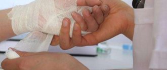
- when it is necessary to work with chemicals, it is recommended to use gloves;
- promptly treat all damage to the skin of the hands with special antibacterial agents;
- regularly wash your hands with soap after visiting public places;
- regularly saturate the body with vitamins;
- lead a healthy lifestyle, reduce alcohol consumption;
- Treat manicure sets with alcohol before use, and do not use other people’s manicure items.
Let's celebrate! When visiting nail salons, it is recommended to ensure that all tools are treated with an antiseptic in front of the client. Most cases of fungal infection occur during nail care procedures.
Infection of the skin of the hands with a fungus can lead to complex consequences that require long-term treatment. You should not fight the disease on your own, since each type of hand infection may require an individual approach to treating the problem.
Hand fungus caused by Trichophyton rubrum
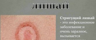
The cause of about 90% of all hand mycoses is the anthropophyll fungus Trichophyton rubrum (red trichophyton). In addition to the skin of the hands, pathogens affect the feet, nails and smooth skin, and extremely rarely hair. Foci of infection have a characteristic appearance. The skin lesion is erythemato-squamous in nature (erythema - redness, squamous - peeling). With a long course and the presence of predisposing factors, the disease progresses until the development of erythroderma. Mycosis lasts from several months to several years.
Pathogens most often penetrate the skin of the hands from lesions localized on the feet or, which is rare, when fungi are introduced from outside. With rubrophytosis of the feet, one hand (working) is first affected, then the second.
There are several clinical forms of hand mycosis:
- Squamous.
- Squamous-hyperkeratotic.
- Intertriginous (interdigital).
- Dyshidrotic.
Rice. 3. Fungus on the hands, svamozny form. Damage to the skin of one (working) hand occurs with rubrophytosis of the feet. Against the background of hyperemia, mealy peeling is clearly visible.
Rice. 4. Infection of Trichphyton rubrum on the feet and hands.
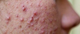
Squamous form of fungus on hands
The disease begins with dry skin and mild erythema (redness) of the palms, where floury peeling appears, especially pronounced in the skin furrows. Peeling of the skin may be minor due to frequent hand washing. In the area of the arch of the hand, the skin pattern intensifies. Mycosis is subjectively accompanied by itching.
Rice. 5. Fungus on the hands. The palmar and dorsum of the hand are affected.
Rice. 6. Floury peeling when palms are infected with Trichphyton rubrum fungi.
Squamous-hyperkeratotic form of fungus on the hands
Due to the long chronic course of the disease, areas of hyperkeratosis appear on the palms (especially in places of mechanical stress) against the background of floury peeling. Elasticity decreases and dryness of the skin increases, small cracks appear. The skin becomes reddish-bluish in color. Lamellar (pityriasis-like) or ring-shaped peeling appears, spreading to the palmar surface of the fingers; with frequent hand washing it becomes hardly noticeable.
Rice. 7. Squamous-hyperkeratotic form of fungus on the hands. Against the background of floury peeling, areas of hyperkeratosis and a crack on the finger are clearly visible.
Intertriginous (interdigital) form of fungus on the hands
In some cases, an interdigital form of mycosis occurs. The disease begins with slight redness and peeling of the interdigital folds. Without treatment, mycosis progresses, maceration and erosion appear, and cracks appear in the depths of the folds. Without adequate treatment, this form of mycosis can transform into a dyshidrotic form.
Rice. 8. Interdigital form of the disease.
Rice. 9. Rubrophytosis of the hands.
Rice. 10. Fungus on the fingers.
Dyshidrotic form of fungus on the hands
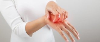
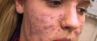
With this form of the disease, papules and blisters appear on the skin of the palms, and when opened, erosions form. When merging, more extensive lesions are formed with oozing inside and clear boundaries along the periphery. In some cases, allergic rashes appear on the palms, palmar surfaces of the fingers and forearms. Patients are concerned about attacks of itching, and pain when a bacterial infection occurs.
Often the infectious process spreads to the back of the hand, where foci of hyperemia appear with peeling in the center and an intermittent ridge along the periphery, consisting of vesicles, papules and crusts. Damage to the hands can be combined with fungal infection of the nails.
Rice. 11. Dyshidrotic form of fungus on the fingers and palms with rubrophytosis.
Rice. 12. Fungus on the fingers. The back of the hand is affected.
Rice. 13. Spread of infection from the palms to the back of the hand.
Rice. 14. The source of inflammation has clear boundaries. Along the periphery there is an intermittent raised ridge with papules, vesicles and scales.
Rice. 15. Spread of mycosis to the skin of the forearm.
What is the source of infection?
You can become infected with onychomycosis in the following ways:
- Living with a person suffering from mycosis. Use of his personal belongings: towels, slippers;
- Visiting water parks, saunas, swimming pools, showers;
- Using manicure tools that have not been disinfected.
However, the contact of pathogenic microorganisms on the nail does not always provoke the onset of the disease. At this stage, the immune system can cope with the infection. Accelerated development of the disease usually occurs against the background of the following factors:
- Warm climates that cause sweaty feet;
- Stressful conditions;
- Unhealthy diet, in which sweets and fatty foods predominate;
- Improper hygiene, rarely changing socks, irregular foot washing;
- The use of artificial nails, under which the infection develops especially actively;
- Weakened immune system;
- Taking antibiotics and oral contraceptives that can change normal acidity;
- Diabetes.
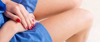
It is worth noting that onychomycosis on the hands occurs relatively rarely. This is a disease of housewives, since constant contact with household chemicals without gloves leads to the formation of microcracks on the skin. They are literally a gateway for infection.
If these circumstances are present, the person is most vulnerable to the onset of the disease. However, this does not mean that these factors lead to infection. They just allow the disease to actively develop.
Hand fungus caused by Trichophyton interdigitale
Trichophyton mentagrophytes var. interdigitale rarely affects the hands. The disease occurs with a pronounced inflammatory component and the development of allergic reactions in the form of a rash on the skin of the limbs, trunk and face. The disease begins acutely with damage to the interdigital folds, where hyperemia and maceration of the skin are noted. In appearance, the affected area resembles eczema.
Rice. 16. Fungi Trichophyton mentagrophytes var. interdigitale under a microscope (photo on the left). The mycelium is long, with spiral curls at the ends. The photo on the right shows pathogen colonies growing on a nutrient medium. The colonies are mealy, velvety, surrounded on the periphery by a rim consisting of young shoots.
Rice. 17. Fungus on the fingers when affected by Trichophyton interdigitale. The disease is acute, the inflammatory component is significantly expressed.
The most obvious signs of mycosis
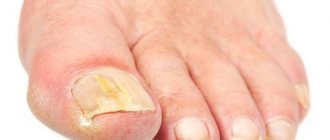
What does fingernail fungus look like? Let's look at its most obvious signs:
- The nail begins to change its color. What shade it takes depends on what pathogenic microorganisms caused the disease. The plate may turn yellow, blue, green. White and yellow tints indicate superficial mycosis. Green color indicates moldy fungus, black or purple indicates Candida microorganisms;
- The structure of the nail changes. It becomes thinner, thickening appears in some places. The plate increases due to the resulting inflammatory process;
- The plates delaminate and become more brittle;
- In the later stages, when microorganisms have already penetrated deep inside, the nail begins to move away from the bed. It may be completely detached.
Against the background of pathological processes, allergic reactions occur, since the infection is hostile to the body. A rash and redness may appear.
Hand skin candidiasis
Yeast-like fungi of the genus Candida most often affect the palms of women, and dermatophyte fungi in men. The disease affects people whose hands are exposed to a humid environment for a long time - housewives, people working in food factories, confectionery shops, when working with vegetables and fruits, etc. The disease in men is more often occupational.
Rice. 18. Candida albicans fungi under a microscope. Round pathogen cells and filaments of pseudomycelium are visible.
Damage to interdigital folds
The disease begins with the appearance of very small blisters on the skin of the contacting surfaces (usually 3 and 4 interdigital folds). Hyperemia of the transitional fold and maceration are noted. The rejected epidermis becomes white. A hyperemic, shiny, smooth surface is exposed underneath. The boundaries of the lesion are clear, surrounded at the periphery by a rim of exfoliating epithelium, rarely extending to the back of the hand. Subjectively, the patient experiences itching and burning.
Rice. 19. Damage to interdigital folds due to candidiasis.
Rice. 20. Candidiasis of interdigital folds.
Damage to the skin of the palms
This form of the disease is rare and has several forms of manifestation:
- In some cases, the disease occurs as dry lamellar dyshidrosis (superficial ring-shaped, garland-shaped or lamellar peeling is noted). The disease should be distinguished from dyshidrotic eczema.
- The vesicular-pustular form is characterized by the appearance of vesicles and pustules against the background of hyperemia and edema.
- Hyperkeratotic eczema is characterized by the appearance of wide skin furrows of a dirty brown color in areas of keratinized skin.
Rice. 21. Candidiasis of the palms. Dry lamellar dyshidrosis.
Candidiasis of the periungual fold
Prolonged stay in a humid environment and trauma contribute to the development of candidal inflammation of the periungual ridges. The disease begins with hyperemia and edema. The skin becomes thinner and becomes shiny. The nail skin disappears. When pressed, ichor, a crumbly white mass, or a drop of pus may be released. In some cases, the nail plates are affected. Patients are bothered by pain.
Rice. 22. Candida paronychia - inflammation of the periungual fold.
Rice. 23. Candidiasis of the hands (photo on the left) should be distinguished from eczema (photo on the right).
Rice. 24. Interdigital candidiasis (photo on the left) and skin lesions on the hands due to rubrophytosis (photo on the right).
Diagnostic methods
When the first symptoms of the disease appear, you should consult a dermatologist who will perform the following procedures for diagnosing the disease:
- familiarization with the patient’s symptoms and visual inspection of lesions;
- PCR analysis to determine the type of infection;
- scraping a sample from the affected area;
- general blood analysis.
Note! If necessary, additional tests may be prescribed to help accurately determine the type of infection and prescribe appropriate treatment.
Laboratory diagnosis of fungus on the hands
The diagnosis of mycosis of the hands is made on the basis of the clinical picture, confirmation of the fungal nature of the disease during microscopy and identification of pathogens when cultured on nutrient media. Skin flakes and scraps of macerated epidermis are used as diagnostic material for suspected hand fungus.
Rice. 25. View of mushrooms under a microscope.
Rice. 26. Species Trichphyton rubrum growing on nutrient media.
Signs and symptoms
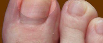
If a person has nail fungus, the following signs are noticeable:
- Part of the plate becomes white, yellow, brown or another color. You may first see a patch of discoloration at the tip of the nail. Without treatment, this discoloration can spread to cover the entire surface of the nail.
- Irregularities appear under the nail.
- The nail begins to lift, which leads to its fragility and painful condition of the finger.
- The nail becomes white, and the surface of the nail may become soft, dry, and powdery. The plate also thickens.
- The plates thicken and turn yellow or brown, often affecting the entire nail.
- The plate splits or collapses.
The initial stage of a fungal infection is usually painless. However, if treatment is ignored, the fungus can grow and contact with the nail becomes unpleasant.
If any changes in the nail become noticeable, you should immediately visit a dermatologist. As onychomycosis grows, it becomes increasingly difficult to clear the infection.
Differential diagnosis of fungus on the hands
Hand skin fungus should be distinguished from psoriasis, keratoderma, chronic eczema, diffuse and limited neurodermatitis, and allergic contact dermatitis.
Rice. 27. In the photo on the left is herpes, on the right is dyshidrosis.
Rice. 28. The photo shows dyshidrosis with symptoms of eczematization.
Rice. 29. In the photo on the left is Devergie’s disease, on the right is psoriasis.
Rice. 30. In the photo there is neurodermatitis. The skin on the back of the hand is affected.
How do we get infected?
There are several ways to get a fungal infection. They may be:
- from oneself if a person is infected with foot fungus;
- intra-family infection if someone is already sick at home;
- by shaking hands;
- the use of other people's unprocessed manicure accessories;
- going to baths, saunas or swimming pools;
- touching the handrails.
Factors predisposing to infection are:
- presence of cuts and abrasions;
- poor blood circulation;
- decreased immunity;
- excessive sweating of the palms;
- stressful situations;
- use of toxic drugs;
- changes in hormonal levels;
- the appearance of endocrine disorders;
- poor nutrition;
- failure to comply with personal hygiene rules.
It is very important to maintain your personal hygiene in order to avoid infection.
Treatment of fungus on hands
Treatment of fungus on the hands is aimed at combating inflammation and mycosis pathogens. Predisposing factors such as trauma to the palms and treatment of rubrophytosis of the feet should be eliminated. Treatment uses local and systemic antifungal drugs (oral).
Treatment of acute inflammation
For severe inflammation and itching, antihistamines and lotions with solutions of boric acid, potassium permanganate, resorcinol, methylene blue, brilliant green or fucorcin are prescribed.
Next, pastes are used: boron-naphthalan, ichthyol-naphthalan or ACD paste.
Multicomponent drugs containing an antifungal agent, corticosteroid and/or antibiotic have a good effect:
- Triderm cream and ointment, Pimafukort ointment, cream and lotion (contain antifungal, antibacterial and hormonal components).
- Mikozolon ointment and Candid-B cream (contain antifungal and hormonal components).
- Ointments Lorinden S, Dermozolon and Sinalar K (contain antimicrobial and hormonal components).
After the acute symptoms have subsided, treatment with topical antifungal drugs begins.
Treatment of interdigital and dyshidrotic forms of fungus on the hands
When treating these forms of mycosis, combination drugs are used, which include an antifungal component and a corticosteroid: Triderm cream and ointment, Pimafucort ointment, cream and lotion.
It must be remembered that when using corticosteroid ointments, foci of inflammation subside quickly, and the fungal infection takes on an erased course!
Treatment of squamous fungus on the hands
When treating the squamous form of mycosis of the hands, antifungal drugs are used in the form of ointment, cream, spray and solution: Isoconazole, Ketoconazole, Clotrimazole, Terbinafine, Miconazole, Mikozoral, Kanizol, Mikozol, Mikospor, Mifungar, Lamisil, Batrafen, Travogen, Mikoseptin, etc.
Treatment of the initial stage of onychomycosis
If nail fungus is detected at the beginning of its development, then getting rid of it is quite simple. But self-treatment is strictly prohibited, since the type of pathogen plays a huge role.
The doctor initially refers the patient for diagnostics. After receiving the results, adequate therapy is prescribed, corresponding to the type of fungal infection.
In the initial stages, the use of local drugs (gels, varnishes, creams, ointments, liquid solutions) is sometimes sufficient. But in some cases, the dermatologist prescribes a tablet form of medication, which helps destroy the pathogen at the cellular level.
Drug therapy
Application of varnishes
Therapeutic varnishes are innovative preparations that do not require frequent application of the product to the surface of the nail (convenient if it is not possible to perform procedures during the day). Before using them, you need to get rid of the dry parts of the plate. To do this, special files are placed in the set with varnish. Varnishes can be transparent or colored.
List of antifungal varnishes:
- Demicten is applied daily;
- Batrafen is used according to the following scheme: the first 30 days once every two days, the second month twice a week, the third - once a month;
- Loceryl is used 2 times a week;
- Mikozan does not dry out, so you need to apply it 3 times a day;
The active ingredients of each varnish have a healing effect over a specified period of time.
Using sprays
The spray is a liquid solution that is sprayed onto the nails and skin. Popular antimycotic drugs of this form:
- Terbix;
- Lamisil;
- Terbinafine;
- Mycostop.
Ointments and creams
Creamy mixtures are applied to the affected areas 1 to 3 times a day. The most popular means:
- Lamisil;
- Exoderil;
- Terbinafine;
- Mycozoral;
- Zalain;
- Ketoconazole;
- Fundizol;
- Clotrimazole;
- Nizoral;
- Mifungar;
- Nitrofungin;
- Itraconazole
Pills
Systemic treatment is prescribed if there is no positive effect after using external agents. In the initial stages, tablets are rarely used.
Drug groups:
- based on triazoles (fluconazoles and intraconazoles): Irunin, Medoflucon, Rumikoz, Orungal, Diflucan, Diflazon;
- based on allylamines: Exifin, Fungoterbin, Terbinafine, Exiter, Termicon, Terbizil, Terbinox, Tebikur, Onychon, Lamican;
- based on imidazoles: Oronazole, Fungistab, Ketoconazole, Fungikok, Fungavis, Nizoral, Ketoconazole.
Folk remedies
Traditional medicine is actively used to treat nail fungus at the initial stage. They are harmless to health as they contain substances of natural origin. Their cost is low, but do not forget that the uncontrolled use of such funds can lead to serious consequences.
Before using folk remedies, be sure to visit your treating dermatologist or mycologist. Only a doctor after an examination can prescribe this or that remedy.
Salt and soda
The most effective recipes:
- Take a liter of warm water, add a teaspoon each of soda and table salt, mix thoroughly and dip your fingernails into the solution. Keep for no more than 15 minutes. Do the procedure daily.
- Combine 1 liter of water with a tablespoon of salt and the same amount of lemon juice. Add lavender oil (2-3 drops). Take baths. Course duration is 10-12 days.
- Salt compress. Make a brine, soak a cotton pad in it and apply to the affected area for an hour. Apply the compress 2 times a day.
- Pour baking soda into a saucer and add a little water (you should get a paste). Apply baking soda to your nail for 15-20 minutes, then rinse with water.
Vinegar
The following recipes are popular:
- Baths with table vinegar (9%). You will need 20 ml of product per glass of hot water. Steam your nails for 15-20 minutes. The procedures are carried out every other day.
- You need to take 1 tsp. 70% vinegar. Add the same amount of vegetable oil and Dimethylphthalate plus one egg. Apply the mixture to your nails, cover with cling film, and put on cotton gloves on top. Keep for 1-3 hours. A total of 4 procedures need to be done, repeated every other day.
- Combine 2 tbsp. l. apple cider vinegar with 1 tbsp. l. olive oil. Apply the mixture daily until recovery.
Hydrogen peroxide, ammonia
The most effective recipes are:
- 2 tbsp. l. Pour 3% peroxide into 2 liters of hot water. You need to steam your fingernails for 15-20 minutes throughout the week.
- Steam your hands in a soda solution, rinse with water, and wipe dry. Soak cotton wool with hydrogen peroxide and apply to the nail plate for an hour. Place cling film and cloth on top. Do twice a day for 2 weeks.
- Take 1 tbsp. l. ammonia and 200 ml of water. Soak a cloth flap in the solution, wrap your nails with polyethylene and gauze. Keep it on all night. Repeat the procedure daily for a week.
Propolis
At the initial stage of nail fungus, the following recipes are recommended:
- Combine ethyl alcohol (100 ml) with a teaspoon of propolis. Apply the resulting mixture to the nail and under it. Wrap with cling film and cloth. You can keep it for several hours. A total of 3 procedures are performed.
- Buy propolis tincture (20%) at the pharmacy. Moisten a cotton swab and apply it to the fungus-affected area. You can remove the tampon after the solution has dried.
Coffee
The following recipes will help with onychomycosis:
- Brew stronger bean coffee and let cool to room temperature. Lower your fingers for 5-7 minutes. At the end of the procedure, apply antifungal cream.
- Prepare coffee in the same way as in the previous case, but put your hands in a hot bath. The procedure lasts 20 minutes.
Soap
The following options are valid:
- Lubricate your nails with brown laundry soap several times throughout the day.
- You can use tar soap. Generously lubricate the affected area, then immediately dip your nails in the saline solution, quickly remove and put on plastic gloves with cotton gloves on top. Keep it on all night.
Garlic, onion
The following recipes are effective:
- Rub your nail plates with a clove of garlic 2-3 times a day. Additionally, garlic can be dipped in salt.
- Grind the onions in a meat grinder or the finest grater. Place the mixture in cheesecloth, shaping it into a bag. Apply to your nails for half an hour.
- Chop the garlic, throw it into boiling water for 5 minutes (5-6 cloves per glass of water). Cool and take a bath for 15-20 minutes.
Herbal infusions
The following recipes will help you get rid of nail fungus at the initial stage:
- Mint. Fresh grass is finely chopped, combined with salt and placed on the surface of the nail plate, wrapped in gauze. Keep for 60 minutes. Procedures are carried out no more than 2 times a week.
- Take 30 grams of hop cones, the same amount of burdock root, add 15 grams of calendula. Fill the collection with half a liter of water. Place on the fire and cook for 10-20 minutes. Strain the broth, add Vaseline (ratio 1:2). You will get a healing ointment that can be applied 2-3 times a day. You need to smear not only your nails, but also the skin around you.
- Take 1 liter of water and 250 grams of young willow shoots. Boil for 20 minutes. Warm baths are made from the decoction 2-3 times a week.
- Nettle decoction for compress. For a glass of water you will need one and a half tablespoons of herb. Apply a moistened cotton swab twice a day.
- Cut the rowan leaves so that the juice comes out. Apply to the nail plates for half an hour, wrapped in cling film.
- You can use sage and chamomile in the same way.
Essential oils
Lubricate your nails with essential oils. Oil works great against fungus:
- oregano;
- celandine;
- lavender;
- rosemary;
- tea tree.
Iodine
This popular antifungal agent is used as follows:
- Apply iodine daily to the affected areas.
- Combine 1 tsp. such components: iodine, glycerin, vinegar. Add 6 tsp. water. Lubricate your nails before going to bed for a week.
Egg
It is used to make an effective ointment for nail fungus.
To do this, place a shelled egg (fresh) in a cup, pour table vinegar to the very top. Under its action, the shell should dissolve, but a film will remain that needs to be removed. Mix the remaining egg thoroughly with the same vinegar. Lubricate the nail plate 2 times a day for 21 days.