Last update: 01/15/2020
lupus erythematosus
LUPUS ERYTHEMATOSUS
(lupus erythematodes; synonym erythematosis) is a disease from the group of diffuse connective tissue diseases (collagenoses). There are two main forms of lupus erythematosus: cutaneous and systemic. The cutaneous form usually manifests as discoid lupus erythematosus, disseminated and deep lupus erythematosus.
The disease is multifactorial. Hereditary predisposition matters. In the pathogenesis, a certain role is played by insolation, chronic focal (usually streptococcal) infection, hypothermia, hormonal imbalance, drug intolerance, and autoallergy.
Clinic.
Skin lesions are characterized by three cardinal symptoms:
- infiltrative erythema,
- follicular hyperkeratosis,
- scar atrophy.
Discoid lupus erythematosus
most often found in countries with humid and cold climates. Among the patients, young and middle-aged women predominate.
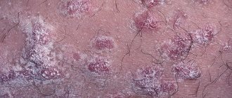
The beginning of the process is characterized by the appearance of a small pink, slightly swollen, clearly demarcated spot, gradually increasing in size and turning into a plaque. Hyperkeratosis develops on its surface, starting from the center. Gradually, almost the entire surface of the plaque becomes covered with dense scales that are difficult to remove. Only along the periphery there remains a free, slightly raised red border in the form of a roller. When the scales are removed, horny spines are often found on their lower surface, which are called “ladies' heels” and are described as a pathognomonic symptom of DLE. Removal of tightly seated scales embedded by horny spines at the mouth of the follicles is accompanied by a feeling of pain (Besnier-Meshchersky sign).
The typical localization of the rash is on the skin of the face (especially on the nose and cheeks, where the lesion may resemble a butterfly in shape), and the ears. The scalp and red border of the lips are often affected, and when the lips are affected, the process extends beyond the red border. Possible damage to the oral mucosa.
During the transition to the atrophic stage, cicatricial atrophy is formed in the center of the lesion, in the area of which there may be telangiectasia and marginal pigmentation, and on the skin of the scalp - cicatricial alopecia. The lesions are slowly enlarging.
Characterized by a long continuous course with periodic deterioration in the spring and summer. Signs of a systematic process are found extremely rarely. The prognosis for life is favorable, but the disease leads to gross cosmetic defects.
For disseminated red
lupus is characterized by the formation of several foci of skin lesions. Erythema, infiltration, follicular hyperkeratosis are much less pronounced than with discoid lupus erythematosus. In addition to the scalp, foci of disseminated lupus erythematosus are detected on the upper parts of the chest and back, on the back of the hands. The lesion usually does not lead to the formation of gross cosmetic defects, but dynamic examination often reveals signs of systemicity.
An intermediate place between the cutaneous and systemic forms of lupus erythematosus is occupied by the so-called subacute cutaneous form,
which is characterized by widespread ring-shaped lesions on the skin, which, when fused, form polycyclic, scaly areas on the edges on the chest, back and limbs with hyperpigmentation by telangiectasia in the center. In this form of the disease, moderately pronounced signs of a systemic process are revealed: arthralgia, anemia, leukocytopenia, thrombocytopenia, antinuclear factor, antibodies to DNA can be detected, however, unlike systemic lupus erythematosus, the prognosis of the disease is relatively favorable.
For deep lupus erythematosus
along with the previously described skin changes in the subcutaneous tissue there is one or more sharply demarcated dense, mobile nodular seals, the so-called lupus panniculitis.
Treatment
cutaneous lupus erythematosus should be complex.
- Glucocorticoids and cytostatics are used.
- Sanitation of identified chronic focal infection is recommended.
- Patients must constantly comply with the preventive regimen:
- avoid exposure to the sun, wind, frost;
- use photoprotective creams, and on sunny days - a wide-brimmed hat or umbrella.
Patients should be monitored by a dermatologist and rheumatologist.
Systemic lupus erythematosus
is a progressive polysyndromic autoimmune disease with the development of a hyperimmune response against components of its own cells (nuclear and cytoplasmic), especially native DNA, systemic disorganization of connective tissue and generalized vascular damage.
Mostly young and middle-aged women are affected, men are 10 times less likely.
Clinic.
SLE can be provoked by: childbirth, abortion, excessive sun exposure, hormonal disorders, infectious agents and begin with recurrent arthritis, fever, malaise, and skin rashes.
Subsequently, progressive pathological changes develop in various organs: polyarthritis with arthralgia, myositis with myalgia, polyserositis (dry or effusion pleurisy, pericarditis, peritonitis), lupus carditis, Raynaud's syndrome, lupus nephritis, pneumonitis, asthenovegetative syndrome, polyneuritis, cerebrovasculitis with mental disorders, lymphadenopathy, hemolytic anemia, leukopenia, thrombocytopenia, etc. LE cells and autoantibodies to DNA are detected.
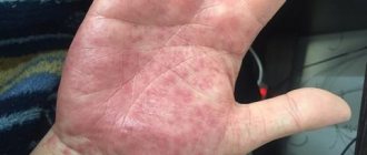
Skin lesions in the systemic form are more varied and widespread than in the cutaneous form. Sometimes (in 10-15% of patients) they are absent (lupus sine lupo), but this condition is temporary, transient. Of particular diagnostic importance is edematous erythema in the midface - the so-called lupus butterfly. Gradually, the erythema may spread to the neck and chest. On the skin of the torso and limbs there may be nonspecific polymorphic rashes: erythematous and hemorrhagic spots, urticarial elements, papules, sometimes vesicles and blisters with hemorrhagic contents. A typical manifestation is the so-called capillaritis: bluish spots on the fingertips and paronychia. The oral mucosa is often affected in the form of whitish grouped papules on a bright erythematous background. Blisters with hemorrhagic contents often appear; erosive and ulcerative rashes are less common. Dystrophic changes in the nails and diffuse alopecia are characteristic.
Treatment
systemic lupus erythematosus should be comprehensive:
- adequate anti-inflammatory and immunosuppressive therapy
- corticosteroids,
- cyclophosphamide,
- biologically active drugs
Forecast
with systemic lupus erythematosus remains generally unfavorable.
Patients should be under clinical supervision of a rheumatologist.
SCLERODERMA
Scleroderma-
an autoimmune disease that is characterized by systemic damage to connective tissue with a predominance of fibrosclerotic and vascular disorders, developing mainly in the skin and subcutaneous fat.
Scleroderma is a multifactorial disease, which is based on dysregulation in connective tissue with an autoimmune mechanism. Antibodies are detected to various nuclear components: nuclear proteins, fibroblast centromeres, as well as collagen.
Classification of scleroderma. Limited scleroderma is distinguished
(cutaneous form), which is divided into plaque, linear and small focal and
systemic scleroderma
, represented by the acrosclerotic variant (CREST syndrome) and diffuse progressive scleroderma.
Provoking factors are: hormonal disorders (menopause, childbirth, removal of the ovaries), focal infection, ultraviolet radiation, hypothermia, mechanical trauma, administration of serums, and certain medications.
Clinic
Limited scleroderma occurs more often in women aged 40-60 years. Skin rashes can be either single or multiple.
Plaque scleroderma
occurs most often. It is characterized by the first formation of one or several lilac spots of various sizes with clear boundaries without subjective sensations. Gradually, over the course of a month, the spot thickens (sclerotizes) in the center with the formation of a plaque. No compaction is observed along the periphery; the purple rim is preserved. The lesion may gradually grow, changing its color to yellowish (ivory). As long as the corolla exists, the disease is in a progressive stage. Over time (months and years), the lesion resolves and an area of atrophy forms. Thus, the disease goes through 3 stages in its course: 1) spot 2) plaque 3) atrophy. Localization of the process - any (except for the head).
Stripe (linear) scleroderma
observed more often in children. The process, as a rule, is represented by one focus, spreading linearly along the neurovascular bundle along the length of the limb or from the scalp to the forehead and back of the nose, resembling a scar from a saber strike. Quite deep atrophy of the skin and underlying tissues is typical for this form.
Fine-focal scleroderma (“white spot disease”, lichen sclerosus.) The disease is observed in women, mainly in menopausal and post-menopausal age. The process is localized on the skin of the scapular and subscapular areas of the back, on the skin of the chest, and genitals. In men, the process is more often localized on the skin of the foreskin.
A distinctive feature is the very short period of spots and plaques and the rapid onset of atrophy in the form of small areas of white color (white spots). Subjectively, it is often accompanied by itching, which is not observed in other forms of scleroderma.
Treatment
limited scleroderma pathogenetic. When the disease occurs and exacerbates, a course of treatment with benzylpenicillin is used (500 units 4 times a day, 28 million units per course. Small doses of D-penicillamine are effective (0.5-0.9 g per day, course 3-6 months. For small-focal forms, courses of unithiol are used intramuscularly and cytostatics are used.
Systemic scleroderma
Acrosclerotic variant of systemic scleroderma
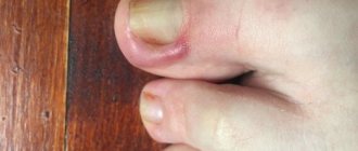
It usually begins gradually, most often with damage to the skin of the fingers. The initial manifestations resemble the symptoms of Raynaud's disease: patients complain of cold fingers, a feeling of tingling and crawling, and the skin becomes cyanotic. Sometimes the fingers turn white and become numb.
Gradually, dense swelling and sclerosis of the skin occurs, it becomes dense like wood, smooth, shiny, closely fused with the underlying tissues. The color changes to yellowish-white, the fingers are motionless, slightly bent. Gradually the process spreads proximally and reaches the shoulder. Usually after 2-3 years the facial skin is involved in the process. It thickens, acquires a waxy color, and the face becomes mask-like.
Progressing extremely slowly, the process either stops and resolves, or can take on the character of diffuse scleroderma. Possible damage to internal organs (usually the esophagus in the form of its narrowing)
Often in sclerotic skin there are deposits of calcium salts and the appearance of telangiectasia.
Considering the clinical manifestations described above, the acrosclerotic variant of scleroderma is also referred to as CREST syndrome
(which indicates characteristic clinical manifestations: C - calcification, R - Raynaud's phenomenon, E - esophagus - damage to the esophagus, S - sclerodactyly, T - telangiectasia)
For diffuse scleroderma
Characterized by an acute or subacute course of the disease with rapid progressive damage to internal organs. Such patients are seen by therapists. Skin lesions are diffuse and rapidly progressing. The development of edema of all or almost all of the skin is characteristic. The edema is very dense; a hole does not form when pressed. The skin is grayish with a bluish tint. Gradually, the skin seems to fuse with the underlying tissues. Movements are difficult, facial expressions are limited. Atrophy of subcutaneous fat and muscles develops. Raynaud's syndrome is noted. Internal organs are affected: Lungs (pneumofibrosis), gastrointestinal tract, primarily narrowing of the esophagus, kidneys, heart.
Diagnostics. Patients with suspected systemic scleroderma are subject to examination by specialists (dermatologist, therapist, neurologist, ophthalmologist), laboratory tests (characterized by increased ESR, C-reactive protein, globulin levels, detection of autoantibodies in the blood (antinuclear antibodies, rheumatoid antibodies, antibodies against cytoplasmic RNA, antibodies against collagen).etc.), X-ray (examination of the esophagus (narrowing), chest organs (diffuse pneumosclerosis), kidneys (nephrosclerosis). Echocardiography, ECG (detection of diffuse changes in the myocardium), as well as examination of vascular reflexes on brushes.
Treatment.
For the acrosclerotic variant of scleroderma, D-penicillamine is used in high doses for several years. The drug is used due to its ability to suppress excessive fibroformation and have an anti-inflammatory effect on the connective tissue of the dermis. The drug is used together with vitamin B6. The disadvantage is a large number of side effects.
Unithiol, vitamins A, E, C, D, vasoactive drugs, physiotherapeutic procedures (ultrasound, diadynamic currents, electro- and phonophoresis with lidase), massage, therapeutic exercises are also used. Cytostatics are also used.
At the compaction stage, enzyme preparations (lidase) are indicated.
DERMATOMYOSITIS
Dermatomyositis is a systemic progressive disease characterized by predominant damage to striated and smooth muscles with impaired motor function, as well as the skin. There are primary (idiopathic) and secondary (symptomatic) dermatomyositis, which develops with cancer of internal organs (paraneoplastic). The most likely immune nature of the disease.
Clinic
Idiopathic dermatomyositis
more often it develops gradually, characterized by general (low-grade fever or fever up to 38-39°C), muscle (myalgia, increasing muscle weakness) and skin manifestations.
The lesion is polymorphic in nature with a predominance of erythema and edema, mainly on exposed parts of the body. Periorbital edema and erythema with a purple tint are characteristic (symptom of “spectacles”). Skin syndrome may precede the appearance of the main symptom of dermatomyositis - muscle damage.
Damage to internal organs does not predominate in the clinical picture of the disease. Calcification of the affected tissues (Tibierge-Weissenbach syndrome) may occur.
Diagnostic criteria for dermtomyositis: typical skin manifestations; progressive weakness in the symmetrical parts of the proximal muscles of the limbs; increasing the concentration of serum muscle enzymes; myopathic changes with electromyography; picture of polymyositis on muscle biopsy; increased creatininuria; reduction of muscle weakness when treated with corticosteroids.
The differentiation of primary (idiopathic) and secondary (paraneoplastic) dermatomyositis is vital, determining treatment tactics and prognosis.
Treatment:
depending on the activity and course of the process in primary dermatomyositis:
- prednisolone for a long period with a gradual dose reduction and corrective treatment;
- cytostatics;
- aminoquinoline drugs (plaquenil, delagil, etc.);
- extracorporeal treatment methods.
For paraneoplastic dermatomyositis:
- antitumor therapy.
Types of systemic lupus erythematosus
- Acute discoid lupus erythematosus. Characterized by isolated skin lesions without other manifestations of the disease. Favorable prognosis.
- Chronic discoid lupus erythematosus. In 90% of cases, long-term isolated damage to the skin of the face and head occurs; less often, diffuse damage to the skin (trunk, arms) is possible with the formation of red plaques that are painful when pressed. This form is also not characterized by damage to internal organs, which allows for a favorable prognosis.
- Subacute cutaneous lupus erythematosus. In this condition, along with skin changes, general malaise, asthenia, pain in muscles and joints are characteristic. Damage to the lacrimal and salivary glands is also possible, resulting in discomfort in the eyes, a feeling of “sand in the eyes,” and dry mouth ( Sjögren’s syndrome ).
- Systemic lupus erythematosus. To make this diagnosis, a combination of a certain number of clinical, laboratory and instrumental signs is necessary. The prognosis depends on the number and degree of damage to internal organs.
Skin options
According to the generally accepted classification, the following cutaneous forms of lupus erythematosus are distinguished:
- Acute (localized, widespread).
- Subacute (ring-shaped, psoriasis-like, bullous, etc.).
- Chronic (discoid, deep, hypertrophic, due to frostbite, tumor, etc.)
Acute
The main clinical manifestations of acute cutaneous lupus erythematosus:
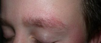
- On the cheekbones, cheeks and nose there are swollen red spots (erythema) with a cyanotic tint, reminiscent of “butterfly wings”.
- A macular nodular rash may appear.
- Rashes usually occur after exposure to ultraviolet rays (sun exposure). Allowed for several days. In their place, mild pigment spots remain.
- Some patients experience centrifugal erythema of Biette - swollen, bright red spots on the cheeks that gradually increase in size due to peripheral growth. In this case, the central part of the rash resolves in parallel.
- The mucous membrane of the oropharynx and nasal cavity may also be affected, with the formation of erosive and ulcerative lesions.
Severe course and lack of effect from outpatient therapy are an indication for hospitalization of a patient suffering from lupus erythematosus.
Subacute
The subacute cutaneous form of lupus erythematosus will be characterized by the following clinical picture:
- Papulo-squamous (nodular-scaly) or ring-shaped erythematous rashes appear.
- In the psoriasiform variant, red nodules (papules) and small plaques are observed on the shoulders and upper torso. Less commonly, the face, ears and scalp are affected.
- The surface of the spots and plaques is covered with dense scales and a slight thickening of the stratum corneum of the epidermis (hyperkeratosis).
- With the ring-shaped variant of the disease, red spotted lesions are identified, shaped like a ring, on the surface of which there is hyperkeratosis and peeling. The areas of the skin exposed to sunlight (increased insolation) are mainly affected.
- In some cases, the patient has a combined skin lesion, when signs of both forms of the disease are present - nodular-squamous and ring-shaped.
- After the rash disappears, there may be no pronounced scar changes at the site of the lesions, but long-term hypopigmentation (light areas of skin) and spider veins remain.
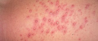
Rare forms of the disease include bullous lupus erythematosus. Mainly on the face, neck, extensor surfaces of the shoulders, and upper back, the presence of numerous vesicles of various sizes is detected, with a tense tire and serous contents. After the rash resolves, scars and dark spots on the skin (hyperpigmentation) form. If the bullous form develops, this indicates a highly active course of the disease.
Chronic
Discoid lupus erythematosus, which is a chronic cutaneous form of the disease, is manifested by the following clinical symptoms:
- The typical triad of signs is erythema, hyperkeratosis and skin atrophy.
- The lesions can be localized (facial area, ears, scalp) or widespread.
- First, intense red spots (erythema) are detected, slowly increasing in size. Then they undergo infiltration and transform into plaques that rise above the skin level.
- Hyperkeratosis (significant thickening of the stratum corneum of the epidermis) is determined on the surface of the plaques.
- If the plaques are scraped off, the patient will feel some pain.
- Active inflammation is visible near the lesions, and hyperpigmentation is present.
- After some time, rough cicatricial atrophic changes in the skin form in the center of the plaques and telangiectasia appears (persistent dilatation of small vessels).
With deep lupus erythematosus, one or several deep-lying nodes are observed, which are dense in consistency, mobile, clearly demarcated and not fused with the surrounding tissues. The skin over the nodes has a stagnant cherry tint. Erythematous and hyperkeratotic lesions may also be observed, as in the discoid form. Over time, the nodes resolve, leaving behind deep recesses due to atrophic changes in the subcutaneous tissue. Some patients experience ulceration of the nodes, which are replaced by rough, retracted scars. Calcium salts may be deposited in the nodes, and calcifications may form under the skin.
Verrucous or hypertrophic lupus erythematosus manifests itself as single plaques that protrude sharply above the skin level. The plaques show pronounced hyperkeratosis and a warty surface. The most commonly affected areas are the face, hands, forearms and shoulders. In addition, a combination of verrucous and discoid forms of lupus erythematosus is possible. It is worth noting that rashes often show resistance to local therapy.
Treatment of skin forms
The basic therapy for patients diagnosed with cutaneous or discoid lupus erythematosus is the use of antimalarial drugs and topical glucocorticosteroids. If limited damage to the skin is observed, then the administration of topical glucocorticosteroids is sufficient. To lubricate lesions on the body, hormonal creams and ointments are used. At the same time, the scalp is usually treated with lotion. If the rash is located on the face, it is recommended to use local glucocorticosteroids with weak or moderate activity.
Medicines that can be prescribed for external treatment of cutaneous lupus erythematosus:
- Flucinar. Available in the form of ointment and gel. Apply to the skin up to two times a day for 3 months.
- Triamcinolone. Use under a bandage. The lesions are lubricated twice a day. The course of treatment usually lasts about 7-10 days.
- Betamethasone. Available as cream and ointment. Use twice daily for about two months.
- Clobetasol. Refers to highly active topical glucocorticosteroid drugs. The rash should be lubricated twice a day. Duration of use is up to a month.
In some cases, it is necessary to resort to intralesional administration of glucocorticosteroids. Mainly used for localized forms that are not amenable to other therapy. Due to the risk of persistent atrophic changes in the skin, this treatment method is prescribed exclusively for indications. Triamcinolone or Betamethasone solutions can be used. It should be noted that the interval between injections must be at least 28 days.
Systemic therapy includes the use of:
- Antimalarials. Hydroxychloroquine is prescribed for oral administration for 60-90 days. During this time, significant improvement can be achieved. If necessary, treatment lasts several years. If you stop taking the maintenance dosage, the likelihood of a relapse of the disease increases by 2.5 times.
- Antioxidant drugs. An intermittent course of taking vitamin E is recommended. The medicine is taken for a week, the next week is a break. The duration of treatment is 1-2 months.
- Angioprotective drugs. You need to take Pentoxifylline and Nicotinic acid for about a month.
I would like to note that for the treatment of pregnant women, only topical glucocorticosteroids (ointments, creams, gels) are used, which belong to the first or second class in activity. Antimalarial drugs are contraindicated because they can cause the development of intrauterine defects in the fetus.
If the patient adheres to all the instructions and recommendations of the attending physician, then the likelihood of achieving remission increases significantly.
Folk remedies
The overwhelming majority of medical specialists are categorically against the use of folk remedies for the treatment of cutaneous forms of lupus erythematosus. Recent clinical studies demonstrate that without optimal medical therapy using quality medications, the disease will progress and lead to a significant deterioration in the patient's condition.
SLE symptoms, complaints and clinical manifestations
Redness (erythema) of the facial skin in the cheeks and nose
This is the most characteristic symptom of systemic lupus, which occurs or worsens when exposed to direct sunlight. Because of its characteristic shape, this erythema became known as “lupus butterfly.” Similar skin rashes may also occur in other areas. The skin of the fingers often changes color under the influence of cold from white to purple-bluish ( Raynaud's phenomenon ). of ulcers on the oral mucosa is possible alopecia ) and brittleness of the nail plates.
Pain and inflammation (swelling) of the joints
They occur frequently, but do not lead to the destruction of joint structures, as happens with rheumatoid arthritis. The small joints of the hands are most often affected, but arthritis of the large joints is also possible. Characteristic signs of inflammatory joint pain are morning stiffness (> 1 hour), increasing pain at rest and weakening with movement.
Pleurisy and pericarditis
A common symptom of lupus is inflammation of the pleural membranes lining the inside of the chest (pleurisy) and the heart sac (pericarditis), which can manifest as pain in the chest during breathing movements.
Kidney damage
When internal organs are involved in the process, kidney damage most often occurs, usually occurring unnoticed in SLE. Edema and a persistent increase in blood pressure are indirect signs of this serious complication.
Inflammatory changes in the nervous system. They can manifest themselves in a wide variety of ways (from headaches and psycho-emotional disorders to convulsive seizures).
Antiphospholipid syndrome. Also, with systemic lupus erythematosus, the formation of “immune agents” (antibodies) to the components of the blood coagulation system is possible. This condition is called antiphospholipid syndrome and is dangerous for the development of various thrombotic complications (vein thrombosis, heart attack, stroke). The presence of these manifestations in a patient at a young age requires the exclusion of SLE and antiphospholipid syndrome.
Nonspecific complaints. Many patients at the onset of the disease notice only nonspecific symptoms of the disease, such as general weakness, increased body temperature, weight loss, and lack of appetite.
Consumer Reviews
Larun about La-Cri emulsion (irecommend.ru)
“My child periodically experiences allergic dermatitis, which manifests itself as an itchy, dry crust on the skin; my daughter even woke up at night from the itching and cried. We tried several different products and none of them gave the same effect as La-Cree emulsion. This wonderful product is quickly absorbed into the skin, leaving no film effect or other unnecessary effects when it comes to a child. I'm sure moms understand me)
The emulsion effectively moisturizes and literally before your eyes, the skin becomes much more pleasant to the touch and to look at too. With daily use, my child’s skin became better and better, small wounds and irregularities quickly healed.
Plus to all this, a very affordable price, about 250 rubles. Despite the fact that the La-Cri emulsion is economically consumed and lasts for a long time.
The only thing I don't really like is the scent. But after reading other reviews, I understand that this is subjective. This is just my personal feeling. But I’m ready to forgive the manufacturer for this small drawback for the sake of the effect it produces on my baby’s skin.
And one more nuance that I personally really like, although it seems like a small thing, is the soft membrane, thanks to which a small amount of emulsion is gently squeezed out and generally creates the feeling of a more expensive product.
Definitely recommend! because If you buy more than your first package, then it’s not a sin to recommend it to others.”
Yulia Nizovtseva about La-Cri cream for sensitive skin (flap.rf)
“I’m not afraid to use it on my child’s redness, and I often use the cream myself. There is always a tube in the first aid kit just in case, and such cases happen very often. Especially in the summer, when it’s hot, there are irritations, heat rashes due to the heat, I apply the cream and everything goes away. I use it after a shower. I apply just a little onto the skin and after 5 minutes there is nothing left, the cream is easily absorbed and does not remain on clothes.
It smells nice, the texture is light, the composition is natural, it helps, what else is needed for children's skin? I’m glad that I chose Lacri cream; it cannot be compared with many children’s products.”
Sources:
- Yagodka Valentina Stepanovna, Medicinal plants in dermatology and cosmetology, Naukova Dumka publishing house, 1991.
- Molochkova Yulia Vladimirovna, Dermatology. Brief reference book, GEOTAR-Media, 2021.
- Baumann Leslie, Cosmetic Dermatology. Principles and practice, MEDpress-inform, 2021.
Diagnosis and treatment of SLE at the Yauza Clinical Hospital
Diagnosis of systemic lupus erythematosus at the initial stage of development makes it possible to achieve rapid and stable remission of the disease and avoid the development of severe complications. In the treatment of lupus, we use drug therapy (steroidal and non-steroidal drugs, biological genetically engineered drugs), methods of extracorporeal hemocorrection.
If symptoms of SLE appear, contact rheumatologists at the Yauza Clinical Hospital. Modern treatment methods, subject to preventive measures, make it possible to maintain a state of remission for years and even decades, give birth to children, and lead an ordinary normal life.
You can see prices for services
Causes
To date, the exact cause of the development of lupus erythematosus is unknown. Nevertheless, hereditary predisposition plays a decisive role in the occurrence of this autoimmune dermatosis. It has been scientifically established that genetic disorders in the functioning of the immune system are of no small importance in the occurrence of the disease. In addition, the following predisposing factors are identified that can provoke the development of pathological autoimmune reactions when the body begins to attack its cells and tissues:
- Prolonged exposure to the sun (exposure to ultraviolet rays).
- Use of certain medications (for example, antifungals, antihypertensives, antiepileptics, etc.).
- Various infections.
- Skin trauma.
- Changes in hormonal levels (in particular, estrogen levels).