Last Updated on 03.10.2016 by
Every person has moles. They have long been the most natural decoration. When a white dot appears on a mole, most patients perceive this phenomenon with great concern.
In some cases, whitening of nevus areas is not serious.
However, in most situations, this phenomenon may indicate the development of serious pathological changes in the body.
In order to understand when white spots are considered a safe sign and when not, it is necessary to study the specifics of this phenomenon.
Causes of white dots
The appearance of moles on the body is a common process that accompanies human life. Some treat such formations calmly, others consider them potentially hazardous to health, remembering that the risk of degeneration into melanoma is always present.
Nevus by its nature is a pathology that is congenital or acquired during life.
A pigment spot appears on any part of the body: face, stomach, back, limbs. In most cases, it does not pose a danger and is benign in nature. Malignancy mostly concerns atypical elements, including the appearance of white spots on the growth.
In some cases, a person feels the formation of a rod inside the nevus. It may be a manifestation of acne when a mole appears in places with a large number of sebaceous glands on:
- back;
- breasts;
- face.
A white spot appears due to a reduced number of melanocytes in the pigmented area. He talks about malfunctions in the body and requires consultation with a doctor.
Safe
White inclusions appear for a number of physiological reasons that do not pose a threat to human health.
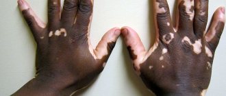
- Genetic factor. Visible lightening of the nevus may be a congenital feature of a person that manifests itself throughout life. A similar situation is often observed in vitiligo disease.
- Lightening of a birthmark may be the body’s reaction to excess ultraviolet radiation, which provokes increased melanin synthesis.
- Violation of the natural pigmentation of the skin is caused by aggressive exposure to radio waves.
- Hormonal imbalances often cause the appearance of white spots. It is mainly observed during the periods of pregnancy, lactation, puberty and the onset of menopause. Treatment with hormonal drugs for endocrine and gynecological pathologies is another reason for this situation.
- An insect bite in individual cases provokes such a reaction.
- Acne, acne. People prone to similar skin problems notice that blisters appear on the growths, containing pus inside. There may be acne. Squeezing blisters is prohibited. The necessary treatment is prescribed by a dermatologist.
White dots on a nevus, which are observed in a person throughout his life and do not change in size or shape, do not pose a health hazard.
Requiring medical attention
In individual cases, white spots that appear on a mole require medical supervision. Will help prevent the development of dangerous pathological processes.
- Lightening of pigment for no apparent reason.
- The appearance of white spots after injury to a nevus, an insect bite, or squeezing out a pimple that has formed on it.
- The appearance on the surface of a bubble filled with liquid inside, accompanied by itching.
- Lightening a mole after prolonged exposure to ultraviolet radiation or tanning in a solarium.
You should not ignore the situation that has arisen.
A timely examination by a qualified specialist and tests will help identify the type of formation. The doctor will prescribe the necessary treatment.
Possible complications
Formations of a benign nature, even with white inclusions present, may not cause inconvenience to a person throughout his life. The danger lies in the malignancy of the mole, but the degeneration is preceded by a number of factors that are impossible not to notice.
To prevent malignancy, several recommendations should be followed:
- tanning should be moderate. Prolonged exposure to ultraviolet radiation (UV rays), frequent visits to the solarium are one of the causes of degeneration;
- It is forbidden to squeeze out white formations on nevi, comb them, or tear them off;
- It is necessary to eliminate bad habits, reduce the amount of coffee consumed, and avoid stress.
Numerous moles on the body require careful attention. The appearance of unpleasant, disturbing symptoms is not always a sign of the development of melanoma, but, most likely, an indication of malfunctions in the body. Understanding the mechanisms of malignancy of nevi, preventive examinations, and a healthy lifestyle will protect you from troubles with moles, even if they have white dots.
When moles become a source of problems
A mole may suddenly begin to manifest itself as a problem:
- The greatest danger is posed by congenital large nevi (more than 1 centimeter), especially those that are inherited;
- Moles that appear suddenly, dark spots that appear after fifty;
- When a flat spot becomes slightly raised, it may soften and break off in small sections. The skin around it turns red and swells.
To prevent moles from causing health problems, you need to follow some tips:
- Try to be in the sun for a short time and at certain morning and evening hours, while solar radiation is not so aggressive. From 11:00 to 15:00 it is better not to appear in the sun at all, especially if you have sensitive skin;
- If the area of skin has already turned white around the formation, you can cover it with light-colored clothing made from natural fabrics;
- Avoid mechanical injuries to moles.
With the advent of such education, we have to be more careful and attentive to our health.
Causes of pimples on the tongue. Signs of stomatitis, candidiasis and glossitis.
Have you been fighting PAPILLOMAS for many years without success?
Head of the Institute: “You will be amazed at how easy it is to get rid of papillomas by taking it every day.
Pimples on the tongue are different from regular acne on the body, as they have a different nature. There can be different types of inflammation in the mouth. Take a close look at the language:
If the rash is not distinguished by color, but is quite painful, it is pipun (photo on the left).
Our readers successfully use Papilite to treat papillomas. Seeing how popular this product is, we decided to bring it to your attention. Read more here...
A scattering of white or red bumps that cover the entire surface of the tongue may also occur under the tongue and on the root of the tongue.
If the ulcers are small in size and do not differ in color from the general color of the oral mucosa, then the cause of their appearance could be various microtraumas of the tongue, for example, while eating seeds or slightly biting the tongue.
Such pimples are accompanied by painful sensations, interfere with eating, and sometimes even speaking.
As a rule, they go away on their own within a few days, as soon as the cause of their appearance is eliminated - the wound heals, which happens quite quickly in the oral cavity.
However, if you find white pimples on your tongue, you should consult a specialist, undergo an examination and take the necessary tests, since the cause of their formation may be:
Signs of candidiasis
With candidiasis, in addition to pimples, a white coating is noticeable, which spreads not only to the tongue, but also to the entire oral cavity.
The causes of this disease may be:
- Improper functioning of internal organs;
- Decreased protective functions of the body.
To eliminate candidiasis, antiseptics are used in combination with drugs that increase the level of immunity: multivitamin complexes and nicotinamide.
Symptoms of stomatitis
With stomatitis, white purulent plaques with a clear red border form on the tongue and cheeks. Possible bad breath.
The cause of stomatitis is commonplace - ignoring the basic rules of oral hygiene. Quite often, this disease is diagnosed in young children, who tend to put anything in their mouths.
If a child gets stomatitis, you can use the drug “Stomatofit” or any other disinfecting gel. After using it, lubricate your mouth with a weak solution of blue iodine or Cholisal gel.
Glossitis
Inflamed red pimples on the tongue indicate glossitis. There are quite a few causes of glossitis:
- alcohol consumption;
- smoking;
- hot and spicy food;
- allergic reactions of various types: to toothpaste, lipstick, mouth rinses and others;
- herpes, which can spread from the area around the mouth and lips to the oral cavity and tongue.
Rinse your mouth with a weak solution of baking soda or a decoction of medicinal herbs (celandine, sage and St. John's wort) and everything will go away. In addition, during treatment you will have to follow a diet: exclude spices, fried, spicy foods from the diet, eat warm liquid food.
If the cause of glossitis is an allergic reaction, try to determine its source and change this remedy.
How to relieve pain before the pimple goes away
Often, pimples on the tongue can cause unbearable pain, for example with stomatitis. Especially if your child has acne! There are several ways to relieve pain until the pimple heals.
- To reduce pain, lubricate your tongue with peach or rosehip oil.
- Honey applied to the affected area will help relieve pain due to stomatitis; you can also use jam from tea rose petals.
- For glossitis, soak a paper towel in vodka and apply it to the surface of your tongue for a few minutes.
Why do dots appear on a mole?
Before answering the question of why dots appear on a mole, it must be said that in the absence of other symptoms, such a nevus is still considered a benign formation. However, the reasons why such inclusions appear may be fraught with potential danger, so you need to be vigilant.
Among the factors contributing to their appearance on the body of a mole, first of all, it is necessary to include all types of damage, be it mechanical trauma, or prolonged exposure to ultraviolet radiation, as well as the influence of the external environment. And it is very important not to forget that any modified birthmark can be subject to malignancy - the process of degeneration of a neoplasm from benign to malignant. Therefore, as soon as such changes are detected, it is necessary to immediately seek medical advice.
What should I do if I have a similar but different question?
If you did not find the information you need among the answers to this question, or your problem is slightly different from the one presented, try asking an additional question to the doctor on the same page, if it is on the topic of the main question. You can also ask a new question, and after a while our doctors will answer it. It's free. You can also search for the information you need in similar questions on this page or through the site search page. We will be very grateful if you recommend us to your friends on social networks.
Medical portal 03online.com
provides medical consultations via correspondence with doctors on the website. Here you get answers from real practitioners in your field. Currently, on the site you can get advice in 48 areas: allergist, anesthesiologist-resuscitator, venereologist, gastroenterologist, hematologist, geneticist, gynecologist, homeopath, dermatologist, pediatric gynecologist, pediatric neurologist, pediatric urologist, pediatric surgeon, pediatric endocrinologist, nutritionist, immunologist a , infectious disease specialist, cardiologist, cosmetologist, speech therapist, ENT specialist, mammologist, medical lawyer, narcologist, neurologist, neurosurgeon, nephrologist, oncologist, oncourologist, orthopedist-traumatologist, ophthalmologist, pediatrician, plastic surgeon, proctologist, psychiatrist, psychologist, pulmonologist, rheumatologist, radiologist , sexologist-andrologist, dentist, urologist, pharmacist, herbalist, phlebologist, surgeon, endocrinologist.
We answer 96.29% of questions.
A person can observe not only a brown, red, blue, but also a white mole on his skin. It looks like a small oval or round spot. The boundaries of such a nevus are clear and even. Just like other moles, it is a benign formation. It does not cause discomfort to its owner, except in cases where a white mole is located on the face, spoiling the person’s appearance.
Symptoms of pathological whitening of nevus
Despite the fact that self-diagnosis is complicated, it is still possible to conduct an independent initial history of the cause of lightening of the mole.
In order to understand whether the whitening of a mole is dangerous or not, you simply need to assess its condition according to the basic parameters of malignancy.
The following symptoms may indicate the development of pathological processes that cause white spots to appear:
- violation of the clarity of the outline of the mole;
- asymmetry of skin neoplasm;
- unreasonable lightening of the central part of the mole;
- the occurrence of sharp pain during palpation, later the discomfort can become permanent;
- increase in the volume of neoplasm;
- severe itching in the mole area;
- bloody discharge from the neoplasm;
- the formation of moles with similar pathogenic signs.
What symptoms should you see a doctor for?
The reason to check the condition of a mole with a white dot in the center should be:
- change in size: sudden growth or reduction;
- the appearance of purulent, bloody, bloody discharge from the head;
- increased body temperature;
- the appearance of itching;
- pain in and around the nevus;
- skin redness;
- the appearance of microcracks on the surface.
The affected area must first be treated at home to stop the bleeding, if necessary, apply a bandage and cover with a bandage. Such measures will prevent infection. After the examination, the specialist will prescribe the necessary treatment: treatment with antiseptics and ointments. Deep injury requires a more detailed examination, dermatoscopy. If necessary, the nevus will be removed.
Dots on a mole are black
In certain cases, the presence of black dots on a mole should not cause any concern, but under one condition - the absence of other symptoms.
This violation of color integrity is explained by a violation of pigmentation. As a rule, this occurs during the period of greatest solar activity, with abuse of solarium, as well as with disruption of the hormonal system.
However, in order to exclude any negative consequences, doctors still recommend undergoing a medical examination. It’s even better to remove the problematic nevus, especially since modern medicine currently has a number of advanced ways to get rid of them. And of course, this is simply necessary to do if, in addition to darkening, other changes in the birthmark are noted.
The main causes of yellow coating on the tongue in adults
Disorders of the gastrointestinal tract
The primary reason for the appearance of a yellow coating on the tongue is a disruption in the functioning of the digestive system. With these disorders, bile is thrown into the stomach. If the stage of the disease is initial, then the mucous membrane of the tongue is covered with a uniform yellow layer. At the same time, it is of low density, translucent, and can be removed without much difficulty during cleaning. There is an unpleasant odor in the mouth, despite recent hygiene actions.
Such plaque is also a symptom of slagging in the human body , which is typical for people who suffer from constipation. In the case of a dark yellow dense or yellow-gray plaque in combination with a strong foul odor from the mouth, one should speak of the presence of serious disorders in the gastrointestinal tract. Most often these are chronic forms of diseases that last for more than one year (gastritis, enterocolitis, peptic ulcer, pancreatitis and other disorders in the functioning of the gastrointestinal tract).
In this case, a gastroenterologist will help you determine the diagnosis and prescribe treatment.
Sometimes you will need to take additional tests for the presence of intestinal infections, which may be the culprit of a yellow coating on the tongue and gastrointestinal disorders (diarrhea, vomiting against a background of elevated body temperature).
Damage to the liver, inside the hepatic ducts or gallbladder
A symptom of liver dysfunction inside the hepatic ducts are yellow deposits with a possible greenish tint. In this case, a bitterness is felt in the mouth, which temporarily disappears when eating, and then appears again.
Such plaque is also characterized by pathologies such as cirrhosis of the liver, hepatitis, inflammation of the gallbladder, parasitic liver infections, and suprahepatic jaundice. It is very important to treat the root cause of the plaque.
Inflammation of the tongue (glossitis)
In this condition, the tongue becomes inflamed. This occurs against the background of infection by fungi and bacteria. The deposits will have a white-yellow color, erosions form underneath them, which make it painful when trying to clean the tongue. In this case, a dentist will help you.
Presence of a viral infection
If you are sick with the flu, ARVI, sore throat, pharyngitis, then do not be alarmed by the appearance of strong yellow deposits on the mucous membrane. Upon recovery, the mucous membrane will return to its normal color.
In order to determine the reasons for the appearance of a yellow coating on the tongue more accurately and correctly, in any case, you need to consult a specialist. Only a doctor can make the correct diagnosis and recommend special treatment. As soon as the pathology is cured, you can forget about the unpleasant yellow coating on the tongue and bad breath.
Diagnosis and removal
A white formation on the body of an adult or child may not cause inconvenience, but may have dangerous signs that require an immediate visit to the doctor. Alarming symptoms:
- when pressing on the nevus, a compaction is felt;
- sharp blanching of the brown spot;
- the borders of the mole have lost their clarity, the edges have become blurred;
- the nevus began to grow;
- the hairs that were previously on the spot disappeared;
- the surface became rough and bumpy;
- a dark coating has appeared in the center of the mole;
- upon contact with clothing or the hand, the formation began to hurt;
- the nevus began to itch;
- a burning sensation appeared;
- without mechanical damage, the mole bleeds;
- A white halo appeared near the brown spot.
If several signs are detected, you need to urgently contact a dermatologist or oncodermatologist for examination and further examination. When visiting a doctor, the patient will be asked questions about when the nevus formed, whether it was damaged, over what period of time it increased in size, and what disorders of the internal organs the person has.
The doctor then examines the spot with a dermatoscope. This is a device that helps to examine the structure of the nevus in thirtyfold size. Dermatoscopy can be performed using a digital device that is connected to a computer. The recorded image of a suspicious nevus is transmitted to the monitor. Using a special program, indicators are automatically displayed on the monitor, based on which you can determine whether there is a process of degeneration or not.
If the mole turns white and pus or blood comes out of it, the dermatologist will refer the patient for a cytological test. It involves taking a smear. Using a microscope, the benign quality of the formation is determined. If a person has ripped off a white mole, it is necessary to bring it to the doctor. He will send it for a histological examination, as a result of which it will be determined whether the formation is benign or contains cancer cells.
If there are no malignant cells, the dermatologist will prescribe appropriate treatment. It consists of applying ointments that contain zinc, antibiotic or acetylsalicylic acid. If a malignant nevus is detected, the patient needs to have it removed. Thanks to modern methods provided by clinics, you can get rid of a tumor painlessly in 10–15 minutes. Removal methods:
- Surgical removal. The nevus is excised with a scalpel under local anesthesia. The method should not be used if the formation is on the face, as it leaves a scar.
- Laser surgery. Using a laser, the nevus and its tissue are removed. No anesthesia is required. There is no scar left after the operation.
- Cryodestruction. The cells and tissues of the nevus are frozen with liquid nitrogen, as a result of which the growth falls off. Used for small white moles.
- Electrocoagulation method. A mole is removed using current.
- Removal by radio waves. High frequency waves are used to remove moles. The method does not leave scars and is used for both large and small nevi.
Types of nevi with a white halo
There are several types of birthmarks, surrounded by a light rim.
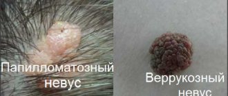
- Nevus of Sutton. It is an ordinary mole, around which there are white areas of skin that have an irregular shape or merge with each other. Such formations often appear in adolescence and can be single or multiple. They do not pose a threat to health and extremely rarely degenerate into a malignant formation.
- Nevus of Setton. This mole looks like a separate pigment spot surrounded by a large light halo. The size of the rim can reach 1 cm in diameter. In some cases, the skin around moles has a white-brown, mottled tint. Such a nevus also does not cause any inconvenience to a person.
- Vitiligo . This is a genetic disease, as a result of which the human body stops producing the pigment melanin. As a result, white spots appear around moles and in general on any areas of the skin. This disease can occur in both children and adults.
Treatment methods for a white spot on a mole
Medical assistance to a person who has discovered a clearing of a nevus without accompanying pathological causes is not needed. It is enough to observe a suspicious element and undergo examinations by a dermatologist for preventive purposes.
If pathological processes are present, treatment is prescribed immediately.
- If there is a threat of degeneration into a malignant tumor, the oncologist carries out the necessary diagnostics to determine the type of formation, prescribes complex therapy and recommends removing the mole.
- Surgical treatment of oncology is carried out. Otherwise, the risk of relapse and the development of numerous complications increases.
Nevus is removed:
- surgical excision;
- cryodestruction;
- electrocoagulation;
- radio wave method;
- laser device.
Preference is given to a method after which it is possible to conduct histology.
So the doctor will once again check the nature of the formation. Depending on the chosen method, further treatment is prescribed and recommendations for wound care are given.
Treatment
Treatment methods for skin tumors with a halo depend primarily on the diagnosis. In particular, the doctor may recommend removing ordinary nevi using a scalpel or laser, or simply not touching them, observing the dynamics for some time. If we are talking about a potentially dangerous mole, then it must be removed, including healthy skin, and carefully examined. In the future, the patient may be prescribed additional treatment designed to prevent cancer or stop malignant processes.
Reasons and features of the appearance
Red dots on the body, like moles, are often congenital; their appearance is caused by impaired functionality of the body as a result of ailments that the mother had during pregnancy. Neoplasms no larger than 1 cm in size often disappear on their own without medical intervention.
It has been proven that adults with fair hair and white skin are more susceptible to the appearance of such moles.
The reasons for the appearance have not been studied, although experts put forward several theories:
- hormonal problems;
- mechanical injuries;
- liver dysfunction;
- lipid metabolism disorder;
- abuse of exposure to UV;
- autoimmune diseases;
- red nevi are a sign of hemophilia;
- heredity.
The appearance of new spots should attract attention; this may be the cause of the disease. You need to get rid of spots if they begin to become inflamed, itchy, or take on a hanging appearance. The method of elimination will depend on what form of formation the patient has. If it is a convex vascular nevus, medication, limiting exposure to sunlight, and visiting a solarium can help. You can use traditional therapy, but there is no guarantee that the growth will disappear.
ICD code – 10
The International Classification of Diseases is usually called by the abbreviation ICD. The modern classification is already its 10th revision. Doctors all over the world use it constantly to:
- Provide a medical document to the patient when it is necessary to conceal the diagnosis;
- Fill out medical certificates, make extracts, issue patient records at the hospital;
- Prepare statistical documents.
The international classification allows not only the exchange of information, but also the preservation of medical confidentiality.
Nevi, like other diseases, have their own ICD code. It depends on the type of education:
- D22 – melanoform;
- Q82.5 – congenital non-tumor;
- 178.1 – non-tumor.
A well-known statistical fact: an adult has at least 20 moles on his body. Almost 100% of Europeans have moles.
In young children they are almost invisible; as they grow, nevi begin to appear on different parts of the body, including mucous membranes. Pregnancy can also cause them to appear.
Hello, I have had a mole on my back for a long time. Never bothered me. 2 months ago I decided to go and check. The doctor said that the nevus was absolutely normal (he did a dermatoscopy).
Just yesterday I noticed that there was red around the mole and a white dot inside. If you look at it it looks like an abscess. I ran to a dermatologist, he looked, did a digital dermatoscopy (I’m attaching a photo) and said that the mole was normal and did not need to be removed. I assumed that there was an abscess there - I tried to squeeze it out, but it didn’t work. He said that it was better not to touch it and said that he had never seen anything like this in practice, but it was definitely nothing bad and still didn’t look like a pimple.
I have a question for you, perhaps you can determine what it looks like from a photo of the mole and dermatoscopy. And is it necessary to remove such a mole? It doesn’t bother me at all, it’s been 6 years since I saw her.
Is it possible to squeeze out a mole with a white spot in the center?
The formation of a white core in a mole is not always alarming. Many people make the mistake of trying to squeeze it out. With superficial damage, internal tissues can be injured. If the process spreads to the subcutaneous fat layer, an abscess may develop. The active growth of the purulent focus begins. Necrosis often forms in its center.
Complications will negatively affect the growth. The inflammatory process will begin.
There is pain and swelling of the nevus itself and the skin around the affected area. Such changes several times increase the risk of malignancy of an initially benign growth.
When squeezing out the formation, a person can introduce an infection inside. Inflammation that develops after the incident is another risk factor for malignancy. It cannot be ruled out that after squeezing the pimple will not appear again. The area of the rash may increase significantly.
What does the appearance of a white dot on a mole mean and why is it dangerous?
A white dot on a mole is a cause for concern for the owner of an unusual growth. A person cannot understand the reason for its appearance, its nature and predict whether it poses a health risk. Such growth, which does not cause discomfort, simply needs to be controlled; if there are doubts about its benignity, consulting a dermatologist or oncologist will not hurt.
Causes of white dots
The appearance of moles on the body is a common process that accompanies human life. Some treat such formations calmly, others consider them potentially hazardous to health, remembering that the risk of degeneration into melanoma is always present.
A pigment spot appears on any part of the body: face, stomach, back, limbs. In most cases, it does not pose a danger and is benign in nature. Malignancy mostly concerns atypical elements, including the appearance of white spots on the growth.
In some cases, a person feels the formation of a rod inside the nevus. It may be a manifestation of acne when a mole appears in places with a large number of sebaceous glands on:
A white spot appears due to a reduced number of melanocytes in the pigmented area. He talks about malfunctions in the body and requires consultation with a doctor.
Requiring medical attention
In individual cases, white spots that appear on a mole require medical supervision. Will help prevent the development of dangerous pathological processes.
- Lightening of pigment for no apparent reason.
- The appearance of white spots after injury to a nevus, an insect bite, or squeezing out a pimple that has formed on it.
- The appearance on the surface of a bubble filled with liquid inside, accompanied by itching.
- Lightening a mole after prolonged exposure to ultraviolet radiation or tanning in a solarium.
You should not ignore the situation that has arisen.
Treatment methods for a white spot on a mole
Medical assistance to a person who has discovered a clearing of a nevus without accompanying pathological causes is not needed. It is enough to observe a suspicious element and undergo examinations by a dermatologist for preventive purposes.
If pathological processes are present, treatment is prescribed immediately.
- If there is a threat of degeneration into a malignant tumor, the oncologist carries out the necessary diagnostics to determine the type of formation, prescribes complex therapy and recommends removing the mole.
- Surgical treatment of oncology is carried out. Otherwise, the risk of relapse and the development of numerous complications increases.
Nevus is removed:
- surgical excision;
- cryodestruction;
- electrocoagulation;
- radio wave method;
- laser device.
Preference is given to a method after which it is possible to conduct histology.
So the doctor will once again check the nature of the formation. Depending on the chosen method, further treatment is prescribed and recommendations for wound care are given.
Is it possible to squeeze out a mole with a white spot in the center?
The formation of a white core in a mole is not always alarming. Many people make the mistake of trying to squeeze it out. With superficial damage, internal tissues can be injured. If the process spreads to the subcutaneous fat layer, an abscess may develop. The active growth of the purulent focus begins. Necrosis often forms in its center.
Complications will negatively affect the growth. The inflammatory process will begin.
There is pain and swelling of the nevus itself and the skin around the affected area. Such changes several times increase the risk of malignancy of an initially benign growth.
When squeezing out the formation, a person can introduce an infection inside. Inflammation that develops after the incident is another risk factor for malignancy. It cannot be ruled out that after squeezing the pimple will not appear again. The area of the rash may increase significantly.
What symptoms should you see a doctor for?
The reason to check the condition of a mole with a white dot in the center should be:
- change in size: sudden growth or reduction;
- the appearance of purulent, bloody, bloody discharge from the head;
- increased body temperature;
- the appearance of itching;
- pain in and around the nevus;
- skin redness;
- the appearance of microcracks on the surface.
An obligatory reason for examination by a doctor is an injury to the formation.
The affected area must first be treated at home to stop the bleeding, if necessary, apply a bandage and cover with a bandage. Such measures will prevent infection. After the examination, the specialist will prescribe the necessary treatment: treatment with antiseptics and ointments. Deep injury requires a more detailed examination, dermatoscopy. If necessary, the nevus will be removed.
Self-medication is prohibited. It can lead to suppuration of the wound and the development of an inflammatory process.
Possible complications
Formations of a benign nature, even with white inclusions present, may not cause inconvenience to a person throughout his life. The danger lies in the malignancy of the mole, but the degeneration is preceded by a number of factors that are impossible not to notice.
To prevent malignancy, several recommendations should be followed:
- tanning should be moderate. Prolonged exposure to ultraviolet radiation (UV rays), frequent visits to the solarium are one of the causes of degeneration;
- It is forbidden to squeeze out white formations on nevi, comb them, or tear them off;
- It is necessary to eliminate bad habits, reduce the amount of coffee consumed, and avoid stress.
Numerous moles on the body require careful attention. The appearance of unpleasant, disturbing symptoms is not always a sign of the development of melanoma, but, most likely, an indication of malfunctions in the body. Understanding the mechanisms of malignancy of nevi, preventive examinations, and a healthy lifestyle will protect you from troubles with moles, even if they have white dots.
Link to main publication
articles:
Loading…
Didn't find suitable advice?
or see all questions...
Source: https://ProMelanin.ru/rodinki/belaya-tochka.html
Preventing Trouble
Although signs of skin aging are everywhere, it is definitely worth supporting and listening to your body in the long run. Everyone has probably heard about the general rules and recommendations for maintaining a healthy lifestyle that require fairly low energy expenditure. This includes regular physical activity that improves blood flow through the vessels; proper nutrition, including the exclusion of harmful fats and carbohydrates, which only cause blockage of the blood passages; control the entry/evaporation of body fluids using moisturizing creams, reducing water consumption in case of excessive sweating or oily skin.
In order to avoid the unpleasant problems described above, defects and discomfort, doctors recommend, if possible, to regularly follow these “rules,” if you can call them that. They are rather small wishes, the fulfillment of which, however, can help not even to know about the inconvenience or, especially, about serious illnesses.
When should you be concerned?
The mole has crusted over, itches and hurts - what could this mean? If you notice such symptoms, you should immediately make an appointment with a dermatologist or dermatologist-oncologist.
A mole that has become crusty and itchy requires special attention. This process may indicate:
- The inflammatory process occurring in the tissues of the nevus. It is indicated by symptoms in the form of itching, redness, and an increase in size of the tumor. In some patients whose mole has darkened and crusted over, droplets of liquid or blood appear on its surface. Such wounds do not bode well. They become easily infected, which can result in the formation of an abscess on the surface of the growth.
- Degeneration (malignancy) of a nevus into a malignant skin neoplasm – melanoma. If a mole peels off and grows, then this process absolutely cannot be ignored. The degeneration of the growth is accompanied by the appearance of “spider-like threads” surrounding it. They can also “spread” in different directions, forming a kind of cobweb pattern. At the same time, the nevus loses its symmetry, becomes uneven, and acquires torn edges.
If a mole is cracked, inflamed and painful, then you should not self-medicate. The only thing that is allowed to be done at home is to treat it with Chlorhexidine or hydrogen peroxide. These are antiseptic agents that will prevent infection of the tumor.
It is also allowed to use decoctions of medicinal plants that have anti-inflammatory, soothing and disinfecting properties. These are chamomile, sage, eucalyptus, calendula.
Note. Such home procedures are allowed to be carried out for a day, maximum two. After this, you must definitely visit a doctor’s office, since traditional medicine relieves symptoms, but does not eliminate the problem as such.
Localization and possible pathologies
Light birthmarks can appear on any part of the human body. However, most often a white mole is found on the head, neck, armpits, back, and intimate areas. In addition to the area of the body, they can have different depths:
- Deep location in the dermis. They have a convex shape. The surface is smooth or rough. Coloration varies from flesh-colored to dark brown. Often such moles are covered with hairs. There is no need to be afraid of this; this is a sign of their good quality.
- Located in the upper layers of the epidermis. They have a flat shape. Color can vary from light brown to almost black. Formed in intimate areas, on the legs and palms.
- Located between the top and bottom layers of skin. Presented in the form of a circle or oval, with clearly defined boundaries. Under the influence of ultraviolet rays or hormonal imbalance, they can change their shape to a more convex one. There are no hairs on such a mole, the surface is not rough or uneven.
The popular interpretation says in which place the light nevus formed, in that place there is an organ in which inflammatory processes occur. However, the formation of colorless moles may indicate that this area of the body is subject to constant mechanical stress. For example, it is rubbed by tight clothes or shoes, injured by jewelry, or torn off during shaving. As you know, any damage can be accompanied by an inflammatory process, as a result of which a benign formation will develop into a cancerous tumor.
Clinical manifestations
Changes in pigmentation around moles are most often associated with disruptions in the functioning of cells that are responsible for the synthesis of melanin. The clinical manifestations of the spots may vary depending on the neoplasms they surround. Experts identify the following moles that tend to have a light areola:
- Nevus of Sutton. This formation does not have clear boundaries, is light in color, surrounded by lighter areas of skin that do not have pigmentation. The size of the formation can be different; when several nevi are close together, they merge together. They occur most often during puberty. It rarely develops into melanoma, but such cases have been reported.
- Nevus of Setton. It has a similar appearance to the previous variety. These neoplasms are brightly colored, have clear boundaries, are about 1 cm in diameter, and are surrounded by lighter skin. In some cases, Setton's nevi are observed, in the light brown structures of which growing hairs are visible. They can occur at any age and disappear on their own.
- Vitiligo. This disease is the sudden appearance of light areas on the skin, including around moles. They may appear and disappear. Experts associate this phenomenon with hormonal imbalances, severe stress, taking medications, and disorders of the immune system. The disappearance of stains is possible by eliminating negative factors.
Often the patient does not immediately notice a change in skin pigmentation, only after he feels itching, burning, and peeling. This is often due to friction from clothing and shoes in flexed areas - elbows, soles of feet, knees, etc.
Are black spots on a mole a cause for concern?
Black spots on moles are not that common, but they need to be kept under control. This is how papillomavirus can manifest itself. It may not bother a person, but if there is a desire and the character is benign, it can also be removed.
Such a mole may not always be melanoma. Blackheads can be the result of injury (nails, comb, etc.). If a mole is scratched, it is not necessary to go straight to an oncologist and observe for a while; in general, they quickly and without consequences take on their previous appearance.
Black and white dots on a nevus, even if they do not bother their carrier, can turn out to be much more serious than they initially seem. To avoid possible consequences and surgical interventions, it is better to consult a doctor without delay.
Changes in moles
Sometimes the typical brown nevi already present on the body may change. In particular, a white coating or an incomprehensible white dot may appear on them. Classical formations are capable of increasing in size during life (but not quickly), darkening a little and even changing shape. Sometimes ordinary pimples form on them - pustules, which at first really look like a white dot on a mole. But any changes in the structure of nevi are a reason to quickly seek advice from a dermatological office.
The appearance of a white coating on a mole may be the first indication that malingization is occurring - malignant degeneration. Other possible signs of cancer:
So, if an incomprehensible dot or plaque appears on a mole, it is better not to hesitate or wait, but to get diagnosed as soon as possible. Early detection of cancer can save lives.
Why do white moles form?
The formation of white coloration of nevi depends on the influence of both external and internal factors. White moles on the body causes:
- Heredity. The difference between white nevi is their possible appearance in a newborn baby. If parents have up to ten light moles on their body, then their child may have a similar number and location.
- Improper body care. When using cosmetics that contain synthetic and low-quality ingredients, white spots may appear. Also, if you frequently visit a beauty salon where a person undergoes cosmetic procedures, and fail to follow the cosmetologist’s recommendations, there is a risk of white nevi.
- Excessive exposure to ultraviolet rays. The appearance of lightened moles is facilitated by prolonged and frequent exposure to the open rays of the sun. Especially in the summer from 10 am to 5 pm. People with fair skin should be wary of such exposure and also limit visits to the solarium to a minimum.
- An autoimmune disease. The immune system can react aggressively to the body's own cells. As a result of protection, a mechanism is activated that changes the structure of cells.
- Thyroid diseases. A disease of the endocrine system can provoke the rapid appearance of both white and brown nevi, which vary in size and shape.
- Hormonal changes in the body. Occurs during the period of bearing a child, during menstruation, termination of pregnancy, as well as during menopause in women, during puberty in adolescents, and with frequent stress.
- Diabetes mellitus of various types.
- Having bad habits and an unhealthy lifestyle. The skin undergoes changes if a person often drinks alcohol or smokes. He also sleeps little, is stressed, and works a lot.
- Skin diseases that are in the acute stage. As a result of such diseases, not only white moles, but also dark spots and light scars may appear on the skin.
- Changes in climatic conditions. If a person moves to a place where the climate is very different from the previous place of residence, where there is more solar activity, then the immune system will react with spots on the skin.
- It is important to eat new food and water of a different quality.
Why did a light halo appear near the nevus?
Quite often, patients seek advice from a dermatologist, trying to understand what the light halo around a mole is and whether this phenomenon is dangerous. Doctors reassure that most often such changes are completely natural or provoked by non-dangerous diseases. A white halo around a nevus can be caused by:
- Hereditary predisposition. In particular, if parents have encountered such a feature on their bodies, then it may well appear in their children.
- Exposure to ultraviolet rays. Systematic visits to a solarium or beach automatically transfer a person to a certain risk group. Moles can react negatively to such irradiation if the skin is not protected with special cosmetics.
- Hormonal fluctuations. This problem can occur in adolescents, women during pregnancy, and also during menopause. Sometimes the problem can arise as a result of systematic stress and certain endocrinological diseases.
- Suffered injuries. As a result of burns, infectious processes and all kinds of injuries, the skin can partially depigment - lighten.
- Vitiligo. This is not a dangerous disease in which a person has a deficiency of melanin. As a result, discolored spots of different localization appear on the skin.
- Fungal infection. Sometimes a white circle around a nevus is a consequence of the activity of pathogenic fungi. A similar picture can be observed with pityriasis versicolor.
Of course, one should not exclude the possibility that the appearance of a white circle near a nevus is a harbinger of developing skin cancer. But, as a rule, the appearance of melanoma is accompanied by other symptoms that cannot be ignored.
In some cases, the sudden appearance of a white spot around a mole is a warning that the nevus will soon disappear on its own. Although, it is worth noting that the likelihood of such a development of events is quite small.
Atypical nevi with a white halo
Doctors distinguish a separate form of nevi, which are always surrounded by a rim of skin completely devoid of pigment. Such neoplasms are called nevus of Sutton or Setton, and they are also often called halonevus. Scientists have concluded that the appearance of such moles:
- It is often associated with a hereditary history of vitiligo.
- It is provoked by a special autoimmune reaction of the body to its own cells, nevomelanocytes.
Typically, Sutton's nevi are observed in adolescents and young adults (up to 20 years of age). Most often they are observed on the back.
Halonevuses disappear on their own several years after their appearance; a light, depigmented circle may remain on the skin for some time, but subsequently it disappears. Treatment of such special nevi is not required; they are not prone to malignancy.
Light brown and brown rings
A halo of brownish color, which is observed around moles in different parts of the body, is considered a more alarming symptom. Possible reasons for this phenomenon include:
- Seborrheic keratosis of the skin. These are benign neoplasms on the skin, which are typically characterized by thickening of the stratum corneum of the epidermis. To date, doctors do not know the exact causes of its occurrence. With this disease, spots, roughness and irregularities appear on the skin, which may resemble nevi. Most often they appear on the skin of the chest, neck, and face. Such keratomas may have a brown color (typical of moles), and there may be a brownish halo around them. Subsequently, the tumors grow, darken and become bulging. The disease does not pose a threat to health, but keratomas can become injured, become inflamed, or cause aesthetic discomfort.
- Melanoma. A cancerous tumor may well look like a mole surrounded by a brown halo. Usually, the nevus itself has an uneven color and is characterized by an uneven edge. It is worth considering that melanoma is an aggressive type of cancer and is prone to rapid growth.
- Dubreuil's melanosis. This is a precancerous condition, which is characterized by the appearance of a brown spot on the skin with an irregular shape and fairly clear boundaries. Often such a neoplasm looks like a black or dark brown blot on a lighter background. The size of the tumor can vary from 2 mm to 2 cm. Such a spot can easily develop into cancer.
- Complex nevus. This is a special mole that usually appears gradually. Initially, a brown spot appears on the skin, and after a few years a nevus forms in its center. Such a neoplasm is practically safe, but it is important to differentiate it from other conditions.
It is completely impossible to determine on your own what exactly caused the change in the condition of the nevus or the appearance of an atypical mole on a brown background. Only an experienced doctor can make an accurate diagnosis.
Reasons for a child
As a rule, the appearance of white or brown circles around a mole in children is associated with a hereditary predisposition or excessive exposure to the sun (in the absence of sunscreen). However, the risk of some more serious problems should not be ruled out. Of course, melanomas in children are not diagnosed so often, but if there are any changes in the condition of moles, parents should definitely play it safe and seek medical help from a qualified doctor.
Why does the nevus gradually lighten?
It happens that moles become lighter and even completely disappear from a person’s skin. Not a single doctor can figure out exactly why nevi lighten and how this process can be influenced. However, doctors are unequivocally confident that even such a symptom can be a very alarming sign of cancer. According to doctors, any changes in the condition of a mole are a reason for an unscheduled consultation with a dermatologist.
What does a white spot around a mole warn about?
When a white halo appears around the formation, the reason is a possible malignant degeneration. This is accompanied by itching and changes in the shape and color of the nevus.
Basically, a white dot on a mole appears during the period of hormonal changes, because it is then that the body is most weakened. It indicates possible problems associated with oncology.
If such a manifestation occurs after sunbathing, this is also a reason to consult a doctor, since most likely such a transformation indicates the appearance of melanoma. In this case, the provocateurs are the sun's rays. Consultation with a dermatologist is required to prevent serious consequences.
White halos may indicate problems in the functioning of pigment cells. Then we are talking about the disease Vitiligo. Although it often does not cause physical discomfort, psychological problems are not excluded.
Moles with a white spot around them are not a problem if:
- A nevus can have an irregularly shaped halo, be located next to each other and merge with each other. Such moles are typical for teenagers and do not pose any threat;
- The formation looks like a separate spot, with white pigment around it. The size of the halo can reach one centimeter. It may be light brown or spotted. These are also harmless manifestations.
A white spot on a mole itself is absolutely safe. But if pathological processes are present, you should consult a doctor immediately:
- Color change of nevus;
- Purulent discharge from it;
- The surface peels, cracks, swells and becomes inflamed;
- The edges are deformed.
All these changes require immediate examination.
If a mole with a white halo bothers you from an aesthetic point of view, a dermatologist will help you get rid of it. He will first reveal the status of the mole, whether it is benign or malignant. Removal methods will be similar to mole removal. The doctor will help you make the choice; in any case, after the operation, you should not neglect the observation of a specialist.
Method of treatment
The method of treating a stain primarily depends on its nature and the danger it poses. The treatment regimen is suggested by the attending physician, based on the collected medical history and test results.
Nevus removal options
The choice of option for removing the tumor is made by the doctor, who bases his decision on the size, location, type, shape and other parameters of the nevus. Various diseases are also taken into account, if any are present in the patient’s medical history. The most common options are:
- Surgical resection. The procedure is performed using a surgical scalpel. Local anesthesia or general anesthesia is first administered. This method is most effective in cases of deep formation, extensive localization, and in cases involving oncological degeneration of nevi. The disadvantage of resection is the large area of damage, the possibility of bleeding and scarring of the wound surface.
- Layer-by-layer cutting of the structure. This procedure is performed using a laser beam, which minimizes the risk of infection on the wound surface and also eliminates bleeding. Subsequent healing and scarring depends on the size of the formation and the pigmentation spot around it.
- Electrocoagulation. The thermal effect on the tumor is carried out using high-frequency currents. As a result, coagulation occurs, which prevents bleeding, and there are no traces of scarring after the procedure. However, experts cite the disadvantage of this method: the possibility of getting burns to the tissues that surround the mole.
- Cryodestruction. Removal of the tumor occurs by exposing it to low temperatures of liquid nitrogen. The procedure is marked by the complete destruction of pathological cells. However, uncontrolled cold exposure in and around the nevus tissue can have a negative impact, causing partial frostbite and structural damage, which results in a longer recovery.
- Radio wave exposure. The procedure is based on vaporization of formation and tissue using high-frequency equipment.
Timely removal of the tumor can eliminate or reduce the risk of oncological degeneration or melanin mutation into melanoma.
Conservative treatment methods
When identifying a pathology such as vitiligo, dermatologists recommend resorting to conservative treatment methods:
- Taking B vitamins.
- Treatment with photosensitizer drugs, which must be taken in courses.
- Taking immunomodulator drugs.
- In some cases, melanocyte cell transplantation is recommended.
Taking B vitamins is a conservative treatment for a white spot around a mole.
In this case, you should never use self-medication, as this may provoke the risk of malignant degeneration of the neoplasm.
Traditional methods
Alternative medicine offers its own methods for eliminating stains around springs. However, you should not use them thoughtlessly, as this can only worsen the condition. Before use, you should consult your doctor. The most common methods are:
- Lotions made from apple cider vinegar, celandine, lemon, garlic, etc.
- Red geranium baths, prepared as follows. 2 tbsp. l. herbs must be poured with 1 liter of boiling water, then left to infuse for 5-8 hours. After time, strain the infusion and add to the bath for the affected areas. It is done before bedtime. This method is also applicable in the case of vitiligo.
White dot on a birthmark
Unfortunately, medicine cannot yet say exactly why white dots appear on moles, since this issue remains incompletely studied to this day. However, some reasons that can cause such symptoms have been identified. These include:
- Genetic predisposition;
- Prolonged exposure to direct sunlight, or addiction to solariums and bactericidal lamps;
- Exposure to radiation;
- Large doses of X-ray irradiation, which is cumulative in nature;
- Unstable hormonal levels (during puberty, during pregnancy, lactation, with endocrine diseases, etc.)
- Presence of blackheads or acne;
- Insect bites.
In the absence of other symptoms, the white spots themselves are not dangerous, however, in order to eliminate any doubts, if they are detected, it is better to consult a doctor.
White coating on the tongue in pregnant women
The causes of white plaque on the tongue can be different, both physiological and indicative of some pathology.
Pregnancy is a time in every woman’s life when it is necessary to take more careful care of her health. This period is characterized by changes in hormones in the body of women, and the body’s immune system may weaken.
A white coating on the tongue may appear in a pregnant woman, but if she is in good health and nothing bothers her, the pregnancy proceeds without pathologies, then she may not worry about the plaque.
However, in the case of a denser layer of deposits with increasing bad breath, you should consult a doctor, since there are a number of reasons that cause this condition of the mucous membrane.
Causes of white plaque on the tongue of a pregnant woman:
- bad habits (smoking, drinking alcohol);
- dryness of the mucous membrane in the mouth;
- fever, in case of illness;
- fungal disease - candidiasis (thrush);
- increased loss of fluid by the body of a pregnant woman;
- treatment with certain medications;
- sexually transmitted diseases (for example, syphilis);
- damage to the mucous membrane due to lichen planus;
- insufficient oral hygiene (presence of caries).
The doctor will help identify the causes of the white plaque and prescribe special medications, if necessary, that will not affect the health of the unborn baby.
Should moles on the labia be removed?
Every person has moles on their body: for some people they are barely noticeable and do not cause much trouble, while for others they are large and require special attention.
Moles on the labia can cause a woman a lot of inconvenience.
Medicine has known about the appearance of moles in intimate places for a long time. They were described in ancient times.
For a long time, doctors believed that removing a mole (or nevus) was harmful, since not all formations may turn out to be benign.
A mole on the major or minor lip can interfere with normal sexual life. Such formations do not look aesthetically pleasing and bring discomfort to family life. A woman may become irritable and unsure of herself.
A nevus is not dangerous until it begins to grow and degenerate.
All moles must be monitored so as not to miss the process when the formation reaches the melanoma stage.
But this is already a malignant tumor and a completely different approach is needed to it.
Reasons for appearance
Various factors can trigger the appearance of new moles.
- One of them is the abuse of solariums or prolonged sunbathing.
- The formation of nevi can also be associated with the work of the body itself.
The possible causes of new moles are:
- hormonal changes;
- problems of the pancreas, liver, gastrointestinal tract;
- diseases of the cardiovascular system;
- heredity;
- skin pigmentation disorders;
- lipid metabolism problems.
Photo
What are the types of moles on the labia?
Moles on the labia minora and majora can be congenital or acquired, benign or malignant.
The size of a mole can range from a slightly noticeable point to a large spot. It depends on how deep the base of the mole is.
- Benign moles are flat or slightly raised above the epithelium.
- Acquired moles appear throughout life and the cause of their formation is the genetic characteristics of the body.
Most often, a nevus grows and appears in childhood, when pigment cells move from the deep layer of the skin to the surface.
Such moles are classified into:
- epidermal. Cells of the skin pigment melanin accumulate in the epidermis;
- intradermal. Characterized by the accumulation of melanocytes in the dermis;
- borderline. Melanocyte cells are located on the border of the upper and deep layers of the skin.
This division is conditional and serves as a guide for the specialist conducting the initial examination of the patient.
According to the classification of scientist N.N. Trapeznikov, moles are divided into two risk groups:
- melanoma-hazardous;
- melanoma-free.
Photo: degeneration into melanoma
What is the danger
The mole itself is not dangerous if it does not increase in size, does not change its color and does not cause discomfort.
The danger of a mole is that any neoplasm can develop into a malignant one at any time.
According to WHO, based on laboratory studies, it was revealed that melanoma in 50% of cases appears at the site of moles.
Causes of degeneration of moles:
- a woman’s exposure to open sunlight;
- self-medication;
- damage to a mole;
- hormonal imbalances in the body.
What to do if the histology of a mole is bad?
Is it possible to remove large moles with a laser? Find out here.
Risk factors
Various factors can trigger the process of uncontrolled cell division, the main one being injuries to moles.
Injured nevi on the labia minora or majora increase the risk of their degeneration into a malignant form.
The process of degeneration of a mole can be noticed by the following signs:
- color change;
- peeling;
- painful sensations;
- cracking;
- itching;
- the appearance of black or red spots.
Video: “How to recognize melanoma in time?”
Meaning
Alternative medicine believes that the appearance of various moles on a person’s body at a certain time indicates changes in his fate.
Moleosophy, a science that interprets moles and other changes on the body, deals with such diagnostics. Since ancient times, this has been one of the methods of fortune-telling, and now this method is used in astrological medicine.
What does a mole in an intimate place mean?
It has a sacred meaning and indicates that their owner has a stormy temperament and in everyday life can be unrestrained, overly nervous and emotional.
The location of moles can be different: on the pubis, perineum, labia.
- If a mole or mark is on the left labia, then this indicates that its owner is endowed with natural charm.
- The presence of a nevus on the right labia means enormous intelligence. Moles on the labia minora and majora foretell the birth of beautiful and smart children.
- A black mole on the labia indicates a domineering character. Such a person sets a goal for himself and always achieves his goal. People with such marks are always ready to defend their happiness and love, without renouncing their loved ones.
- Red moles on a woman’s labia indicate her stormy temperament, amorousness, and inconstancy.
Questions and answers
Very often, women ask specialists questions about the advisability of removing moles in intimate places.
Doctors clearly advise their patients to remove the mole and then undergo a histological examination of the mole, since injuries during sexual intercourse or hygiene procedures can lead to irreversible consequences.
Photo: patient examination
What to do if there are hanging formations on the labia minora?
It is necessary to contact an oncologist. Most likely, you will be prescribed laser or radio wave removal.
A hanging mole is removed quickly and painlessly.
The radio wave method is the most effective way to remove moles, in which there are no relapses.
Is it possible to squeeze out or remove a small dark mole yourself?
Nevi are a cluster of pigment cells that are benign, but carry the risk of degeneration into melanoma.
It is not recommended to remove them yourself.
Why did new hanging formations appear after pregnancy?
If you have a new mole, you should consult your doctor.
Pregnancy is characterized by hormonal disruptions, which sometimes leads to the appearance of new nevi.
What is the best way to remove large brown spots?
User reviews on the Internet say that it is better to do this with a laser.
In any case, you need to contact an oncologist and after diagnosis, the doctor will recommend the optimal method of removal.
Is it dangerous if blood comes from a mole?
In what cases is free diagnosis of moles carried out? Find out here.
What does a mole on the right thumb mean? Read on.
How are raised brown nevi on the labia majora removed?
Modern medicine offers several quick ways to painlessly treat moles - laser treatment, electrocoagulation, cryotherapy.
In this case, surgical removal of moles is more optimal.
- With this method, the nevus is excised with a scalpel within the boundaries of healthy tissue.
- Then the wound is sutured, where over time an inconspicuous scar will remain.
Have I injured a mole in an intimate place?
Injured nevi increase the risk of degeneration into a malignant form.
Be sure to see a specialist. You can decide on treatment during a face-to-face consultation with a gynecologist or dermatologist.
After a series of studies, a decision is made on the optimal method of removal.
Photo: bloodshot mole
Blood-filled formations appeared. Is it dangerous?
If there is any change in the size, color or shape of the formation, you must see a doctor, and only he will determine whether you should worry about this issue or not.
A dermatologist can provide qualified advice on moles and other formations.
If they are in an intimate place, then it is necessary to see a gynecologist, who, if necessary, will redirect the patient to another specialist.
An oncodermatologist deals with neoplasms that progress or change.
Video: “How to treat papilloma virus? Removal of condylomas, papillomas, warts"
kozha.hvatit-bolet.ru>