The area of skin around the auricle contains a huge number of sebaceous glands, as a result of which this area is especially susceptible to the formation of so-called wen. Possible ailments include papillomas, fibromas, atheromas and lipomas.
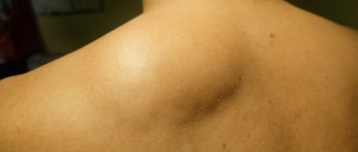
Fatty tumors that form in the ear area grow slowly and are often not cancerous. Unlike wen on the earlobes, the appearance of tumors in the area behind the ear is only 0.2% among benign tumors in the facial area. Wen behind the ear or on the lobe requires treatment, which usually involves surgical removal.
What to do if you have a wen on your ear and how to recognize the characteristic signs of this phenomenon?
What is a lipoma behind the ear?
The lipomatous node is characterized by slow growth, low cancer risk, and the cells are rarely malignant.
If you feel that the neoplasm is painless, the skin in the area of the fatty node does not change. The fat is encapsulated in a fibrous capsule, from which dense, even contours are observed during the examination. The lipoma is mobile and does not show clinical manifestations. In most cases, patients consult a doctor for aesthetic complaints.
Wen or tumor - how to determine?
Due to the lack of obvious clinical manifestations, it is impossible to distinguish fats from other tumor neoplasms.
If a lipoma is a cluster of fat cells enclosed in a capsule shell, then other tumors and neoplasms may have a completely different structure:
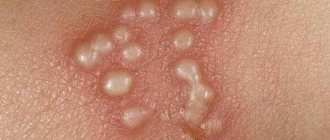
- Papillomas are a consequence of infection with the papilloma virus, forming above the surface of the skin, but differing in the stage of formation by a small cone (for the treatment of papillomas on the head, more here);
- Atheromas are cystic neoplasms of the ducts of the sebaceous glands, provoked by overloaded glands and localized along the thickness of the skin. How to identify: Lipomas or atheromas are found in this article;
- Fibroids are dense, touching little bumps formed from a small amount of fat cells and connective tissue.
Particularly noteworthy are angiolipomas with abundant vascular filling, myolipomas - tumors with a muscular structure.
Lipomas are different from swollen lymph nodes. The latter type of tumors is always painful, which indicates the development of an inflammatory process in the respiratory tract.
Causes of wen behind the ears
Thus, a cyst is formed in the glandular ducts, the cavity of which is filled with fat cells, connective tissue, cholesterol and particles of the cornea. The structure gradually becomes dominated by fat cells and a lipomatous lesion is formed.
The following factors can trigger the pathological process:
- Hereditary predisposition;
- Pustular lesions of the skin of the face, neck, ears;
- Hormonal disorders;
- Metabolic disorders;
- Obesity;
- History of subcutaneous polyps or papillomas;
- Alcohol abuse, smoking.
Of great importance in the formation of fats around the ear; Alcohol abuse, smoking:
- Lack of proper hygiene;
- Increased sweating at the hairline;
- Blockage of the hair follicle;
- Traumatic factor;
- Independent earlobe piercing for jewelry.
Symptoms
Clinical manifestations of fat in the postauricular skin and earlobes are often fatty or absent. In most cases, there is an aesthetic defect that worries women.
When tumors grow behind the ears, the blood vessels and nerve endings become compressed. Symptoms of a growing tumor include pulling pain when the tumor is localized, and patients often experience headaches.
If the lipoma is complicated by an inflammatory process, the characteristic symptoms are :
- temperature increase,
- redness in the localization of adipose tissue,
- edema,
- pain with every touch.
Diagnostics
A number of such measurements :
- X-ray;
- Ultrasound;
- MRI or computed tomography as diagnostics.
Laboratory tests are used to identify or exclude possible concomitant pathologies and inflammatory processes. In the presence of atypical symptoms indicating the development of complications, a comprehensive diagnosis is required.
Fat behind the ear belongs to the field of surgery, oncology, dermatology. If surgical intervention is necessary, a consultation with an anesthesiologist-resuscitator is possible.
How to remove a wen behind the ear?
Fat behind the ear can be treated conservatively and surgically. If the tumors are small, there are no signs of complications, possible risks are excluded, and the growth is insignificant, then they resort to wait-and-see tactics. The patient is examined by a surgeon several times a year.
Conservative treatment
Topical medications are used to treat fat :
- Vishnevsky liniment,
- Ichthyol ointment,
- Iodine network,
- Vietnamese star.
All these ointments have a pronounced anti-inflammatory and disinfecting effect. The formulation of topical medications helps to literally extract the fat content. Small new growths often open on their own after daily use of medication.
In addition, you can use popular methods. Many herbalists assign hemlock a special role in the treatment of fatty tumors. They make potions, tinctures and compresses. Products based on aloe, onion and garlic juice, celandine, and pepper are considered popular.
The disadvantage of conservative treatment is the questionable therapeutic effect :
- First of all, it is unlikely that the fat can be completely removed from the ointment. Even if an autopsy of a lipomatous tumor is performed, the capsule will remain inside.
- Secondly, long-term systematic use of local remedies is necessary to obtain results.
Surgical treatment
One of the promising areas in the treatment of fatty tumors is surgery. Oncologists believe it is necessary to remove all new growths on the body to prevent cancer.
Surgeons perform operations for special indications :
- inflammation,
- intensive growth,
- specific morphological structure according to biopsy or atypical cells,
- soreness,
- swelling,
- involvement of adjacent healthy tissues in the pathological process.
There are various surgical treatment methods :
- Scalpel dissection . A traditional method that is most often used to remove tumor tumors. Despite the trauma and likelihood of scarring, the method has a number of advantages: elimination of relapse, removal of all altered tissue, the possibility of further histological examination, accessibility and ease. After the procedure, it is important to monitor the wound surface with an antiseptic to avoid infections.
- Laser removal . A modern method based on the impact of a precision laser beam. This method allows you to painlessly remove the tumor along with the capsule shell, thereby reducing the risk of complications such as bleeding. The tissues heal quickly, which ensures high aesthetic results. The disadvantage is the high cost and limited availability of equipment.
- Electrocoagulation . A surgical method in which a lipoma burns out using an electrocoagulator - an electric current of a certain frequency. In most cases, electrocoagulation complements traditional scalpel excision.
- exposure to radio waves . A radio knife is a directed stream of radio wave radiation. Under its influence, the tumor is destroyed along with the capsule. This method is as expensive as laser removal, so not every clinic can afford the equipment for manipulation.
The indication for surgery is the patient's desire to remove a cosmetic defect.
The operation is performed under local anesthesia, but in complex cases anesthesia may be required.
Complications and prognosis
Lipomas rarely lead to complications; inflammation and injury are common.
The most dangerous complications arise when you try to open lipomas on your own.
If, for example, a lip of fat is squeezed out and a tumor develops, the situation is often the result of an infection in which healthy tissue is involved in the inflammatory process.
If the fatty tissue is a tumor after self-surgery, it is important to consult a doctor. It is important to understand that it is impossible to remove fat along with the capsule itself.
Other dangerous complications are hearing loss, inflammation and dysplasia of the lymph nodes, damage to the innervation of nerve endings in the face.
Additional information about liposuction removal by popular methods:
The prognosis for ear fat is favorable. The risk of transformation of cancer cells is minimal. A cancerous tumor occurs due to complex inheritance and the presence of other oncological centers.
- For the treatment of breast lipoma in women and men, read this article.
- You can make an appointment with a doctor directly on our resource.
- Be healthy and happy!
Fever behind the ear - what is a tumor, how is it detected and how is it treated? Link to main publication
Main symptoms
At the first stages, it is difficult to notice the wen.
How to test hearing in newborns in the maternity hospital and independently: research methods and resultsRead
The main distinguishing characteristics of lipoma from other neoplasms are:
- the lump does not cause pain;
- the surface structure of the tissues under which the wen appeared does not change;
- the fat capsule is mobile.
If the wen is injured, then pain and discomfort occurs. If the inflammatory process develops, symptoms of intoxication appear: body temperature rises and weakness appears.
Lipoma localized behind the ear: methods of treatment and removal
Primary fatty tumor (lipoma)
A fatty tumor behind the ear is a benign tumor that is painless and does not need to be removed without inflammation. In 0.2% of cases, it consists of facial sealants, never harmful, accompanied by a recurring flow.
Reasons for the appearance of wen
Oil in the ear is produced by the sebaceous glands. Causes of lipoma on the ureter:
- Carbohydrate, lipid metabolism disorders.
- Hormonal imbalance caused by endocrine pathologies - diabetes mellitus, thyrotoxicosis.
- Refusal of the sebaceous glands.
- Hypothermia, overheating.
- Violation of personal hygiene rules.
- Sebaceous gland infection from ear piercing or wearing lingerie jewelry.
- Hyperhidrosis or excessive sweating.
- Bad habits provoke individual or group behavior.
- Poor environmental conditions, bad food and water negatively affect internal organs and glands.
- Heredity factor.
You may be interested in: Laser removal of a polyp of the cervical canal: preparation for the operation and its course
Symptoms and how to distinguish them from other formations
Fat behind the ears may be asymptomatic, depending on the cause, and may be associated with signs of an underlying medical condition. Clinical picture:
- The skin remains unchanged. Redness occurs during the inflammatory process.
- Without pain. The sealant is flexible and resembles cartilage in composition. Discomfort occurs due to mechanical impact.
- If there is no infection, the disease is not accompanied by pus, swelling or hyperemia. The red color of the ball indicates inflammation.
Additional research methods can confirm the diagnosis:
- Complete blood test indicating the number of lymphocytes.
- Ultrasound, CT, and MRI determine the true size and depth of the formation on the auricle.
- A puncture is necessary to determine the content of the tumor.
- Histological examination will help exclude a malignant tumor.
Therapeutic tactics depend on the results obtained.
Treatment methods for the ears
Do you need advice from an experienced doctor? Get an online consultation. Ask your question right now.
Ask a free question
Fever in the earlobe is a painless cosmetic defect. If the formation is small, it can be treated with medications. Large tumors require surgery.
There are no drugs on the drug market that can completely eliminate the problem. They use ichthyol ointment, liniment, and Cherry ointment. The seal may dissolve, but the capsule remains.
Fats behind the ears recede quickly, so they need to be treated.
Surgery
Thanks to the possibilities provided by modern medicine, it is possible to get rid of a tumor surgically:
- The operation is performed under local anesthesia. The surgeon cuts the node with a scalpel and incubates the capsule with its contents. He stitches the wound.
- Endoscopy. During the procedure, only the contents of the capsule are removed. The capsule is left behind. Advantages: minimal bleeding. There is a risk of relapse.
- Radio wave therapy is a gentle hardware procedure. The pathological formation is cut out with a radio wave knife.
- Laser removal is a modern treatment. Suitable for treating skin lesions located on the head, behind the ears, and on the eyelids. The laser does not leave scars and does not cause bleeding. The irradiated area heals quickly.
- Liposuction. Operating principle: fat suction. Special solutions are injected into the tumor, which gives a positive effect. During liposuction, the capsule is not removed. There is a risk that the problem will recur.
Laser surgery, radio wave therapy is used for small lipomas. Large seals located behind the ear (more than 3 cm) are subject to radical removal.
Treatment at home
You can get rid of fat behind the ear at home if you use folk remedies correctly.
- Kalancho. The leaf should be cut lengthwise, and the meat should be attached to the seal, securing it with a patch. The procedure must be performed daily. The installation helps reduce the formation diameter.
- Take a small onion and bake it in the oven. Pass on a grater with laundry soap. Apply the resulting mixture to the cone and secure with plaster. Change the bandage 3-4 r/d.
- Aloe. Cut the leaf in half and apply to the cone. Can be used if there is an infection.
- Take sour cream, honey, salt in the same proportion and mix. Apply the mixture to the fat behind the ear and secure with a bandage.
- Soak cheesecloth in vodka and red pepper twitter. Apply the compress for 15 minutes.
- Take garlic with fat in a 2:1 ratio. Turn on the meat grinder. Place the resulting porridge on the hump. Secure it with plaster.
Strong pressure is accompanied by an outflow of contents. If the mass bursts behind the ear, there is a risk of infection, which connects through the blood. The risk of self-medication at home leads to growth, penetration of germs, pus, hardening in the form of a bone or cartilaginous plate. Possible complications must be taken into account.
What to do in case of inflammation of the formation
Inflammation of fat - redness in the ear area, accompanied by growth and painful sensations on palpation. The cause of the disease is a mechanical effect.
There is a risk of consequences if you try to remove the sealant yourself at home.
Alcohol lotions can be used before surgery. Folk remedies help relieve inflammation: infusion of mother and stepmother leaves, baked onions, thick porridge of sprouted wheat grains. To avoid the formation of pus, the lime must be removed.
How to avoid recurrence of wen on the earlobe
The frequent appearance of linden behind the ear can be avoided by consulting a doctor:
- The patient must lead a healthy lifestyle. Bad habits negatively affect the skin.
- Chronic endocrine disorders require corrective therapy.
- Maintaining personal hygiene reduces the risk of relapse of the pathology.
The article was reviewed by the site editors link to the main publication
Didn't find the advice you needed?
doctor or view all questions...
Item rating:
Download…
Does alternative medicine help?
Lipoma in the ear area can form in both adults and children. And since few of us like to visit doctors, the first thing that always comes to mind is to solve the problem ourselves. For this purpose, they often resort to traditional medicine recipes. In most cases, ointments and all kinds of warm compresses are powerless in the fight against wen. Moreover, independently “pulling out” their contents can be dangerous, since softening of the lipoma can lead to infection and inflammation.
Despite the fact that some folk remedies have a resolving effect, such treatment is very long-term. And prolonging the problem, especially without prior diagnosis, can be dangerous. Therefore, to prevent a small wen behind the ear from turning into a serious threat to health, it is better to consult a doctor and have it removed using radical methods in just a few minutes.
Wen behind the ear, on the lobe: what it looks like, how to get rid of lipoma
Subcutaneous benign tumors can occur anywhere in the body. One of the most common places for their localization is the area behind the ear. Such adipose tissue does not cause any negative consequences, unless it has an inflammatory process. To get rid of it, you need to know the possible causes and effective treatments.
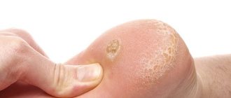
What does a wen look like behind the ear?
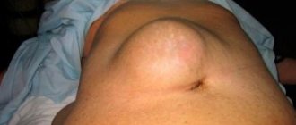
Fat or lipoma is a benign tumor in the connective tissue that, if complicated, can spread deep into the muscles and blood vessel associations. This is a mobile formation that does not cause pain. It is most often found near the cartilage or earlobe and looks like a regular pimple or a small spherical subcutaneous filling.
The borders of the lipoma are usually clear and have a round, less often oval, shape. The internal structure of adipose tissue is long: it is divided into equal parts, which are not tangible due to the layer of epidermis and the capsule shell.
The amount of fat behind the ear can vary greatly. It usually grows to the size of a pea, but can expand dramatically if left untreated and untreated. Due to a large tumor, the skin begins to sag, swelling, blood vessels may become clogged, and blood stagnation may occur. In the worst case scenario, there is a risk that the ear tissue will die and ulcers will form.
Causes of lipoma behind the ear
There can be various causes of lipoma behind the ear. Among the most common provocative factors...:
- metabolic disorders,
- organ damage,
- pathologies and anomalies of the external part of the auditory canal,
- obstruction of the sebaceous glands,
- bad habits,
- diabetes mellitus and other endocrine diseases,
- radiation therapy or radiation exposure,
- hereditary predisposition,
- bad environmental situation,
- hormonal imbalance,
- unhealthy diet (eating foods rich in carbohydrates or fatty foods),
- hypodynamic and inactive lifestyle,
- violation of personal hygiene,
- seborrhea and other skin diseases.
Worth a mention! Fever often occurs at the site of soft tissue damage after incorrect or unsuccessful ear piercings and other types of piercings.
Taking all factors into account, it is necessary to determine how susceptible your body is to lime blooms. This will allow you to diagnose fats in time and cure a disease that has not yet progressed.
Symptoms and characteristic signs of lipoma
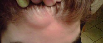
If you look at the fats near the ears, they look like thickening of the skin. Color varies from yellow to white. In the earlobe or near the ear, the lipoma usually has a diameter of no more than 5 cm. If it progresses, urgent surgical intervention is necessary.
First of all, a person may feel a slight tingling or itching at the site of the tumor. Then a growth appears that rises a few millimeters above the skin.
Other signs of fat:
- no pain,
- swelling (as a result of tightening the skin becomes shiny),
- slight redness,
- high skin temperature in the area of fat,
- tumor in capsule
- discomfort.
The patient has poor hearing due to poor blood circulation in the hearing organs.
In case of complications, the following undesirable symptoms may occur:
- spread of infection
- inflammation and abscesses,
- reddish,
- swelling and swelling
- severe pain,
- tumor growth and formation of new tumors,
- inflammation of the middle ear (in children).
The development of a purulent infection has irreparable consequences: If purulent exudate enters the blood vessels or lymph nodes, the meninges may be damaged.
You may be interested in: Polyps in the maxillary sinus: symptoms of the disease and treatment, diagnosis
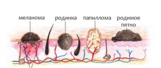
Attention! It is quite difficult to determine what is under the skin. It can also be a cyst (atheroma) or a malignant tumor. For this reason, a series of studies will be carried out first.
Fat can only be detected during a medical examination. Ultrasound, biopsy or x-ray are used as additional diagnostic methods. If the lipoma lies deep in the ear canal, an MRI (magnetic resonance imaging) is prescribed.
Differential diagnosis
If a skin tumor occurs, it is important to distinguish it from other benign tumors. Fats have symptoms similar to these diseases:
- atheroma or sebaceous cyst. This occurs as a result of blockage of the channels and the inability to secrete the sebaceous glands. Most often found in the head, lower face and back. There is pus inside the capsule that is leaking.
- Hygromatic or synovial cyst. The formation contains a serous admixture of fibrin protein or mucus. It occurs on the wrist in the tendon area. A common cause of its occurrence is chronic injury (constant monotonous movements).
- Dermoid cyst. Contains products from sebaceous and sweat glands, hair and even tooth fragments. The capsule is covered with non-pigmented skin, sometimes fistulas occur. This mainly occurs during fetal development.
- Lymphadenitis or lymphadenitis. It develops as a result of a recent illness. This disease is characterized by: soreness, enlarged lymph nodes, swelling and hyperemia. The disease can be caused by infection or inflammation.
Methods for removing wen behind the ear
If there is fat behind the ear, consult a dermatologist. Only a specialist can distinguish a lipoma from a malignant one, even if it is a small tumor.
Important! Timely treatment will bring immediate results and the consciousness will disappear within a few days.
There are several ways to get rid of fat:
- treatment with ointments and medications,
- surgical removal,
- ethnoscience.
Some bactericidal and anti-inflammatory ointments are effective only in the early stages of tumor development, when the capsule is still free. Such conservative treatment avoids purulent and inflammatory processes that destroy infection and harmful microorganisms.
Immediate lipoma removal is the most effective method. This not only eliminates the existing problem, but also prevents possible relapses in the future. The patient can choose one of several surgical options:
- Classic operation. Used to prepare large tumors.
- Invasive removal. This involves cutting no more than 30mm of fat. The operation is performed under general anesthesia, but the operation may leave scars.
- Application of laser correction. This procedure is performed under local anesthesia for a few minutes in the most sensitive and vulnerable parts of the ear. The scar heals in about 2 weeks, after which pale scars gradually remain.
- Radio wave processing. It is especially effective in preparing lipomas in the ear, since this area is difficult to reach using other methods. This method eliminates the detection of bleeding and ensures rapid healing.
- Treatment with liquid nitrogen. Performed in beauty salons. The surrounding adipose tissue is frozen and subjected to cryodestruction.
- Liposuction. It is effective where there is a lot of fatty tissue, for example on the thighs or abdomen.
The most important thing in any operation is the complete removal of adipose tissue, including the capsule. Otherwise, there is a risk of relapse. After surgery, anti-inflammatory therapy is prescribed, often with the use of antibiotics.
It's worth mentioning! A benign tumor can be removed by injecting a resorptive solution into the capsule. This is only possible if the diameter of the fat does not exceed 20 mm. However, this method is not suitable for children.
Help of traditional medicine for wen
Traditional medicine has always been popular. However, recipes using natural ingredients can only give results if the fat size is small.
The most effective recipes:
- Apply chopped aloe leaf to the tumor site. Attach to the skin with plaster. As soon as the patch dries and the juice comes out, it needs to be replaced. Thus, inflammation is relieved, the contents are absorbed and the progression of adipose tissue is prevented.
- Aloe juice can be replaced with star balm. It should be evenly distributed over a cotton swab and applied to the lipoma. Frequent redness occurs.
- Alcoholic version of golden mustache (house plant). Prepare lotions from the resulting product regularly (preferably before bed). The first results will not be visible immediately.
- Cultivate cinnamon powder in water. You can also use clay instead of cinnamon. This will help clean out the pores and reduce the size of the little limescale.
- Lubricate plantain leaves with household soap. Apply to ear at night. After some time, the swelling and redness will subside.
Ways to prevent lipomas behind the ear
To avoid getting oil in the ear area, follow these guidelines:
- Stay immune.
- Avoid tobacco and alcohol products.
- Eat a balanced diet and limit your intake of fatty foods.
- Live a healthy and active life.
- Time to cure diseases.
- Unclog your pores and use low-fat makeup.
- Clean your ears every day.
- Monitor your hormonal state.
To clean your ears, use only cotton swabs, no handy objects. It is also recommended to choose only those cosmetics that are suitable for your skin type.
General information
The growth of a tumor from adipose tissue is practically asymptomatic.
And if on the face it can be determined visually independently, then behind the ears - only by touch. As long as the wen is a few millimeters in diameter, it does not cause any inconvenience. However, in most cases, the tumor continues to increase in size and begins to compress neighboring tissues, as well as nerve endings. As a result, a person may experience discomfort and unpleasant pain. Wen can appear on absolutely any part of the face, including behind or under the ear, on the earlobe and, relatively rarely, in the auricle.
The main cause of formations on the earlobe is the use of unsterile instruments in salons during cosmetic procedures. This applies to a greater extent to women who wear earrings. Bacteria enter the punctured holes and can cause inflammation.
Wen near the ear area appears as a result of clogged skin pores. It is known that the sebaceous glands constantly produce subcutaneous fat, which is excreted from the body naturally.
However, if the pores become clogged, then it simply cannot be removed spontaneously and gradually accumulates under the skin. The result is a swelling that increases over time.
Important! An increased danger is represented by a wen that appears in the auditory opening. In addition to deteriorating sound perception, the tumor can cause the development of otitis media and more serious diseases. It should be removed as quickly as possible.
Lipoma behind the ear: causes, what to do and how to get rid of the wen, treatment methods
Disorders of lipid metabolism cause lipomas, benign tumors that can occur anywhere in the body. Fat behind the ear is not dangerous in itself, but under certain conditions (brushing your teeth, wearing a tight hat, etc.) it can lead to cancer. This happens very rarely, but there are still precedents.
Wen development
Lipo behind the ear is a subcutaneous neoplasm whose cavity is filled with fat. Due to the lack of channels, the fat content cannot be extracted, so the lipoma itself does not disappear, and over time the tumor increases in size.
This causes some discomfort when combing and wearing a headdress, which can lead to knot injuries and serious complications. If such a tumor is found behind the ear, on the ureter or in the ear canal, you should seek medical attention and appropriate treatment.
Causes
The exact causes of lipoma are not fully known. However, experts have identified several important factors:
- The formation of lipomas is often accompanied by disruption of the endocrine system, diseases of the liver and pancreas, and hormonal imbalance. After treating the underlying pathology, you can get rid of some of the resulting formations.
- Hereditary predisposition. Inheriting this predisposition increases the likelihood of developing fat at a young age.
- Malignant neoplasms, as well as smoking and alcohol abuse lead to the growth of one or more linden trees.
- Violation of fat metabolism. Research shows that lipid breakdown products are difficult to remove from the body, which provokes the formation of lipomas.
Small fat with a diameter of a few millimeters does not cause any complaints.
However, as the size increases, it begins to impair blood circulation, puts pressure on nerve endings, and can cause high blood pressure, as well as headaches and chronic fatigue.
Lipo, which forms in or near the ear canal, interferes with the perception of sounds, and its inflammation provokes the development of otitis media.
Many people believe that fat is a harmless growth that should not be ignored, but this is wrong. Although lipoma is benign, under certain conditions (for example, after injury) it can turn into a malignant tumor.
The greatest danger is trying to get rid of proliferation on your own without medical help. In this case, squeezing out fat can lead to injury and damage to the skin.
An open wound leads to infection, which in turn leads to inflammation and pus formation. In addition, if a lipoma is not removed correctly, it can not only reappear, but also become a source of infection.
The hearing organs are located in close proximity to the brain, so any inflammatory process in this area is dangerous for humans. The scale of the consequences in this case is impossible to predict.
You might be interested in: Colon cleansing diet: cleansing products and recipes
Signs of lipoma
Fever has several characteristics. The most important thing is this:
- absence of pain and inflammation;
- no change in skin color over the cone;
- the size of the round neoplasm varies between 2-4.5 cm;
- may move slightly to the side when pressing on lipomas.
Symptoms are similar in adults and children. More pronounced symptoms occur with the development of an inflammatory process or infection:
- skin redness;
- pain in the affected area;
- when pressed, a white or yellowish-green liquid flows out of the capsule (in case of purulent inflammation);
- itching in the area of inflammation is possible.
Sometimes you can find a pimple behind the ear that looks very similar to fatty tissue. A new formation of the first type differs from a lipoma in that the pimple contents easily come out of the capsule under pressure.
Possible complications
Lipoma grows very slowly and rarely turns into a malignant tumor. However, fat that forms near the hearing organ can be very unpleasant. Failure to address growth may result in the following conditions:
- Due to the size of the tumor, hearing damage may occur.
- Inconvenience and even pain may be felt when the nerve endings are compressed.
- As the tumor grows, the structure of the adipose tissue thickens, making subsequent treatment more difficult.
- The infection threatens the development of an inflammatory process that affects the middle and inner ear. In addition, the infection can spread to nearby tissues and organs, including the brain.
Diagnostic measures
A thorough examination is necessary to make an accurate diagnosis. Depending on the symptoms, the doctor may prescribe the following diagnostic measures:
- dot. The contents or tissue of the lipoma are examined for the presence of pathology.
- Advanced blood test. The number of lymphocytes was determined; an increase in their level indicates the development of a malignant process.
- CT scan. It is performed using a contrast media to obtain a more complete image. The method is mainly used to examine internal organs, but can also be used to diagnose lipomas.
Based on the examination results, the doctor makes a diagnosis and prescribes appropriate treatment. Since this type of tumor is practically not studied, the only way to get rid of it is surgery. Only this method guarantees complete removal of fatty tissue in the ear.
Treatment of tumors behind the ear
The treatment regimen for fat is determined individually in each specific case. Therapy involves the use of medications, folk remedies, and destructive methods to remove excrement.
Drug therapy
The drugs can be used to remove small fat (10-20 mm in diameter) in the early stages of its development. For example, the following:
- Vishnevskaya ointment. Used for compressors. The medicine is applied to the tumor, covered with a film, the upper side with a warm cloth. It is secured with plaster or bandage. Ichthyol ointment can be used in the same way. If after seven days the tumor has not disappeared, then there is no point in using other means.
- Iodine. Several times a day it is necessary to treat the affected area with a solution of iodine and vinegar (9%).
Folk remedies
Traditional medicine can be used to remove small tumors behind the ear. The following remedies are considered the most effective:
- Garlic. Grind a piece of garlic, place it on a piece of gauze and apply a compress to the fat, leave overnight. For severe burns, remove the compress and lubricate the affected area with tea tree oil. This helps relieve inflammation and irritation of the skin.
- Kalancho. Grind the leaf of the plant to a pulp state, prepare a compress with a bandage and apply it to the lipoma. The procedure is performed daily before bedtime until the swelling disappears. If after two weeks the tumor has not decreased, another treatment method should be used.
- Vinegar (9%) and iodine. Mix the ingredients in equal quantities - 20 ml of the resulting solution to treat growth several times a day without affecting areas of healthy skin. Treatment with this method may take several weeks.
- Ground pepper and vodka. A piece of dressing should be soaked in vodka and covered with black pepper (1 teaspoon) applied to the fat. Repeat the procedure twice a day until the skin over the tumor is depleted and the lipoma opens.
- vegetable oil and pharmaceutical alcohol. Mix the ingredients in equal proportions. Used as a compressor. The procedure is carried out a month before going to bed.
Unfortunately, both medications and folk remedies are ineffective in the fight against lipoma. In most cases, treatment must be carried out with more radical methods to get rid of fat behind the ear.
Destructive ways
Removal of lipomas is carried out only by a specialist with all the necessary means and tools. The clinic may offer one of the following methods:
- Surgical procedure. This is the simplest and most proven method for removing fat of any size; the operation is performed under local anesthesia. The surgeon makes an incision through which the entire capsule is removed. This prevents relapse. Scalpel removal has some disadvantages: a long recovery period, as well as noticeable scars and scars that remain after the operation.
- Endoscopy. The advantage of this method is that the incision is small, making the scar removed less noticeable on the skin. However, in this case, the fat is removed without skin, which increases the likelihood of recurrence.
- laser therapy. This is one of the safest and fastest methods of removing linden. Moreover, this method has a significant advantage: there are no scars after the operation, which is especially important if there is fat in exposed areas of the body. The duration of the procedure is no more than 15 minutes, wound healing occurs quite quickly and without side effects (inflammation, swelling). New growths on the scalp, face, neck, eyelids and ears are best removed with a laser.
- Radio wave therapy. The principle of influence is similar to the principle of laser action. Treatment is carried out on an outpatient basis, without hospitalization. The tumor is removed with a high-frequency knife. The operation is absolutely bloodless and painless. This is especially important for removing fat from sensitive areas such as eyelids, earlobes, etc.
- Absorption of fat (liposuction), as well as the introduction of special drugs into the lipomatic cavity, also has a positive effect from an aesthetic point of view. However, after liposuction there is still a risk of fat reappearance. The fact is that only the fat is removed, but the capsule remains under the skin, so over time it fills with fat again and the lipoma must be treated again.
Surgical treatment
There are several ways to remove it:
- A radical method - excision with a scalpel is performed for relatively large wen. The operation is performed under anesthesia (local or general), which requires hospitalization of the patient followed by a hospital stay. Through the incision, the doctor removes the capsule along with the possessed person, washes the wound with an antiseptic and sutures it. If you remove a tumor this way, you need to be prepared for the fact that a scar will remain on the skin. However, complete excision guarantees no recurrence.
- Removal using endoscopic equipment is a minimally invasive intervention. A small incision is made (less than 1 cm), and a special flexible device with a camera is inserted through it, which destroys the contents of the capsule. The doctor monitors the progress of the procedure on a monitor. After the operation, a barely noticeable mark remains.
- Laser removal is one of the innovative techniques. The laser beam seems to vaporize the lipoma, and acts locally, without affecting healthy tissue. The procedure is bloodless, the risk of infection and the percentage of relapses is minimal. There are no traces left after healing.
- The puncture-aspiration method is better known as liposuction. A needle is inserted into the fat capsule and, using a kind of pump, the pathological mass is pumped out. No traces remain after exposure. However, the percentage of relapses is quite high, since only the contents of the lipoma are removed, and the capsule remains in the same place.
- Electrocoagulation – a pathological growth is burned out using a variable frequency current. They are used for relatively small tumors. There is no tissue scarring, the healing process takes 10-12 days.
For medical reasons, lipomas that are rapidly increasing in size, inflamed, painful, or weeping tumors must be removed. In addition, it is recommended to get rid of potentially dangerous wen, such as angiolipoma. However, medical reasons are not always the reason for removal; people often make this decision on their own when the tumor is localized in open areas of the body, due to aesthetic discomfort.
Doctors' recommendation is that all tumors larger than 3 cm must be removed, this will help avoid complications in the future.
Please leave a comment:
Diagnostics
The diagnosis of ear lipoma is made based on the patient’s complaints, anamnesis (history) of the disease, and examination data. If localized in the middle or inner ear, instrumental research methods will be required. Laboratory methods may be required to clarify the nature of the tumor and carry out differential diagnosis.
Diagnosis of ear lipoma, which is located on the auricle, is carried out jointly by an otolaryngologist, a dermatologist and a dermato-oncologist.
The results of the physical examination are as follows:
- upon examination, the tumor can be visualized as a round, ball-like or hemispherical formation;
- upon palpation, the tumor is painless to the touch, often fused with the surrounding tissues, and soft-elastic.
Instrumental diagnostic methods are used if there is an ear lipoma deep in the external auditory canal or if its presence is suspected in the middle or inner ear.
Are used:
- Otoscopy – examination of the external auditory canal using an ear mirror. If the tumor has formed in the tympanic cavity, during otoscopy the bulge of the eardrum is determined;
- pharyngoscopy - examination of the pharynx if there is a suspicion of a large ear lipoma and its spread to the structures of the pharynx;
- X-ray of the skull in the area of the temporal bone - with the spread of ear lipoma, based in the middle or inner ear, destruction of the bone walls of the tympanic cavity may be detected;
- computed tomography of the skull in the area of the temporal bone - it is carried out for the same purpose as radiography, but thanks to computer slices the data will be more informative;
- audiometry – study of the patient’s hearing acuity in the presence of a lipoma in the middle or inner ear. Used when conductive type of hearing loss is suspected;
- hearing test with a tuning fork - carried out in the same case as audiometry, in order to establish the type of hearing loss;
- acoustic impendansometry – in the case of lipoma growth in the middle ear, it allows to identify impaired mobility of the auditory ossicles;
- otoacoustic emissions - the study of the ability of the ear to respond to sound signals. During the examination, a probe with a microphone is inserted into the ear canal;
- biopsy - collection of fragments of the ear wen followed by examination under a microscope.
Drug therapy
If a wen is inflamed on the back, stomach or any other area, you cannot do without the use of medications. After surgery, a specialist may prescribe drugs from the following groups:
You can also read: How to determine whether it is a wen or not
- Antibiotics. Immediately after the operation, the patient is prescribed broad-spectrum medications such as Amoxiclav, Sumamed, Cefatoxime, etc. If the inflammation does not go away, a drug is selected taking into account the sensitivity of the pathogenic microflora. Additionally, probiotics may be prescribed.
- Antiseptics. These products are used to treat the wound surface. In hospitals, hydrogen peroxide or Furacilin is most often used.
- Anti-inflammatory ointments. Levomekol, Ichthyol ointment, Salicylic ointment, Vishnevsky liniment show good results.
- Non-steroidal anti-inflammatory drugs. Drugs from this category help relieve pain and normalize the patient’s body temperature. The drugs Nurofen, Paracetamol, Panadol can be used.
Medicines should be taken strictly as prescribed by the doctor.
Any medications must be used strictly as prescribed by a doctor. Self-medication often leads to the development of serious complications.
Physiotherapeutic procedures such as UHF, ultraviolet irradiation, heat therapy, and magnetic therapy help to restore the damaged area more quickly after surgery. However, such methods are used only after acute inflammation has stopped.
Why you should see a doctor
If the wen hurts when pressed, it means that inflammation and pathological changes have begun in the body. Reasons for seeking medical help:
- Making the correct diagnosis. The specialist will prescribe the necessary tests and studies. They will allow you to exclude oncological processes in the mammary glands and subcutaneous layer. The examination list includes diagnostic imaging methods. An ultrasound, CT or MRI is required. If these examination methods are not available, an x-ray examination is indicated.
- Elimination of secondary infection when opening the wen. Surgical intervention is indicated to sanitize the wound surface, remove remnants of lipid tissue, tumor capsule, and operations to stop bleeding.
- Elimination of compression of neighboring organs, internal bleeding, disruption of other body systems.
- Development of further tactics for patient management. It is possible to take samples for histological examination.
If the lipoma begins to pulsate, becomes hot to the touch, the color of the skin over the formation on the thigh, side, abdomen or back changes, the shape changes, there is a reason to urgently contact a medical facility.
If the formation is painless when pressed, the issue of removal should be discussed with a doctor, rather than experimenting with dubious recipes.
The article has been reviewed
by the site editors
What to do if a ball appears in your earlobe
Don’t immediately start getting rid of the unpleasant lump. No one will like the sight of earlobes festering from an infection. But stop assuming the worst and making dire diagnoses for yourself.
Symptoms that distinguish lipoma from other types of tumors:
- the skin does not change color;
- the fat globule under the skin is mobile, which is easy to check by pressing with your finger;
- pain and discomfort are not felt until the wen is injured and inflamed.
If the tumor is smaller than a pea, its size is constant - there is no danger. You can simply make sure that it is really a wen at an appointment with a therapist. There are several signs that you urgently need to consult a surgeon:
- The wen grows quickly (about 1 cm in 6 months).
- The swelling became painful.
- Inflammation began and pus appeared.
- Lipoma is noticeable and affects appearance and self-esteem.
When a wen appears behind the ear, it is difficult to prevent it from being injured. Combing hair and wearing hats constantly disturbs the swollen area. In this case, it is also appropriate to visit a surgeon.
To clarify the diagnosis, a more complete examination will be required. Depending on the symptoms, these may be the following procedures:
- a detailed blood test to determine the number of lymphocytes that increase in cancer;
- puncture to check the tumor tissue or the contents of the wen for pathology;
- Computed tomography with a contrast agent gives a more complete picture of the lipoma, but it is done mainly when examining internal organs.
The doctor will draw conclusions based on the results obtained and recommend the best treatment. To date, lipoma has been little studied. The only way to cure a wen is to remove it completely.
Why does the wen hurt?
The pain of a lipoma can mean its degeneration into a malignant tumor, which in medicine is called liposarcoma and poses a serious danger to human health and life.
The degeneration of a tumor can begin due to various factors: damage to the lipoma due to external influences, weakened immunity, etc. Not every lipoma degenerates into a malignant tumor, but there is such a risk, and if these symptoms appear, you should definitely consult a doctor and undergo the appropriate tests .
Pain may occur due to the inflammatory process when an infection occurs or when a lipoma grows into the deeper layers of the skin. All of the above symptoms require qualified medical care.
Is it necessary to remove the inflamed wen?
Of course, the most common and popular method of getting rid of wen remains removal. If inflammation and pain appear, then it’s time to seek help from the clinic. The surgeon decides on excision individually for each patient; the operation is simple and lasts about half an hour.
Whether a person is prescribed local or general anesthesia, this factor directly depends on the size of the formation. The doctor makes an incision over the fatty membrane and begins to clean it out. After such an operation, a scar or scar may remain on the skin.
The laser method is used in medicine; many patients choose it because it does not leave any traces behind and effectively fights wen. There are no contraindications for this method of excision of formations. The operation lasts even less time than in the case described above.
A good method for solving the problem during inflammation of the wen is endoscopy. This technology is effective due to the fact that the incision is made not on the formation itself, but next to it. In addition, the endoscope has a small camera that displays the content on an external screen. Due to this, the surgeon will be able to control the entire operation.
The endoscope is able to maximally clean and cut out the fatty deposits that the wen creates. Due to this, the patient is protected from relapse of the disease.