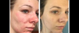
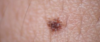
At first glance, the most harmless of all skin formations are flat moles. Without a doubt, every person has them. Most of the formations of the epidermis are brown in color. However, the color scheme can vary from dark to flesh-colored, yellow or very light shades. Pinkish or dark blue nevi are also found. The variety of shapes is also noticeable: oval, round, flat or raised.
We are so accustomed to these “flies” that we rarely pay attention to their condition and even less often compare one with another.
Nevi
Nevus - translated from Latin means mole. As mentioned earlier, they are on our body from childhood, but appear only a few months after birth. The exception in this case is birthmarks. A mole is a non-cancerous formation that may not bother you for the rest of your life. But from time to time, in some people they degenerate and cause significant inconvenience, turning into melanoma, a cancerous tumor. This is precisely their danger to human health.
Nevi actively appear after thirty years. This is why you need to carefully monitor your skin. Especially in the case when a nevus has become inflamed, which has not bothered you for many years.
Can a flat mole turn into cancer?
The chance that a typical flat mole will develop into cancer is less than one in 3,000. However, some skin growths are more prone to malignant transformation. This rule applies to:
- Atypical congenital dysplastic nevi, which differ in texture, appearance, color, size. However, the chance of developing melanoma is one in a hundred.
- Actinic keratosis is a precursor to squamous cell carcinoma. This disease precisely indicates the fact that flat moles are dangerous . They look like rough, scaly spots. This lesion usually appears in areas exposed to the sun. To prevent the development of a malignant tumor, keratosis is best treated.
- Lentigo is a malignant pigmentation that is considered as “cancer in situ.” These spots do not show invasive growth and are only diagnosed in older people. Lentigo progresses very slowly, but requires constant medical supervision.
- People with multiple skin pigmentations (more than 100) have a higher chance of developing an oncological process.
- Large birthmarks (over 8 mm) are more likely to mutate.
See also: A growing mole: 9 signs of cancer. Photo
Types of nevi
Experts conditionally divide all nevi into two types:
- Pigmented. Moles of this type are composed of melanocytes. Melanocytes are special skin cells. Pigment accumulates in these cells, which gives the mole its color.
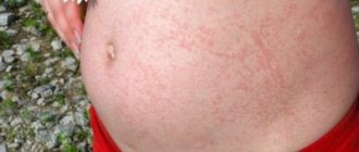
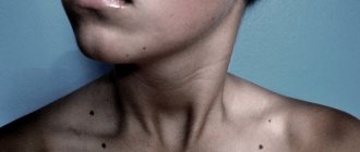
- Vascular. Moles of this type are formed when the blood vessels of the epidermis enlarge. These nevi are most common in women. Namely, the appearance on the face, back, arms and legs. Inflammation of a mole of this type should never be ignored.
Features of brown moles
The International Classification of Diseases ICD-10 assigned brown melanoform nevi the code D22 with additions based on location. In adults, there are about 50 pigmented formations. Teenagers and pregnant women become covered with nevi more often than others. The definition of “melanoform” means that the growth consists of melanocytes. There are several types:
- Convex – intradermal/epidermal.
- Flat – borderline.
- Hanging ones are papillomatous.
- Verrucous.
- Nevus of Sutton.
- Dysplastic.
Intradermal and epidermal are a light brown mole that protrudes above the surface of the skin. They are distinguished by the depth of location of the pigment cells: in intradermal cells the pigment is located deep in the dermis, in epidermal cells in the epidermis. The color of the formation may vary slightly from flesh-colored to brown. Often hair grows from them, which indicates their good quality, since the cells retain their function and do not disrupt the channels for hair growth. They may darken over time.
A border nevus is a flat, brown mole. The shade ranges from brown to black, the dimensions do not exceed 5 mm, and most often looks like a dot with rounded symmetrical edges. It is more difficult to damage such a formation. The cells are benign and rarely degenerate into melanoma.
Hanging nevi are caused by the human papillomavirus. HPV occurs in most people on Earth; the development of the disease is inhibited by the immune system. Under favorable conditions, the virus begins to multiply and warts grow on the skin.
They look like convex, loose formations of brown color of various shades. Localized on the extremities, armpits and neck. Warts may peel off. Dry skin provokes itching and self-infection of the virus in other parts of the body. Some papillomas are located on the stalk. With a weak immune response to the virus, the disease progresses and new formations appear. Treatment includes hardware removal of the growth and stimulation of the immune system.
Verrucous nevi are often confused with viral warts; outwardly they look like lobulated, rounded projections above the skin of a dark brown color. They are most often located on the face and scalp, and are also found on the body. They increase in size as the child grows, rarely exceeding 1 cm in diameter. This type is susceptible to injury and can bleed more often than others.
Sutton's nevus is a brown spot with a light, depigmented area of pink skin around it. This encircling ring is well visualized, has a distinct color, and a rounded shape. Rarely encountered, not dangerous.
Dysplastic ones have an uneven color, one part of it may rise above the other, and there is an asymmetry in size. This formation is classified as melanoma-hazardous and requires specialist supervision. The probability of melanoma if there are more than 50 moles of this type on the body and skin cancer in close relatives is 60-100%. Not every formation resembles a dysplastic nevus. Only the results of histology determine its type. An ordinary person cannot visually distinguish dangerous pigmentation from safe ones; for a diagnosis, one should contact a medical institution.
Nevus formation
Research into the formation of nevi has not yet reached a common opinion as to why exactly they degenerate. Nevertheless, several factors that provoke the formation of nevi:
- Heredity. Data about melanocytes, which subsequently transform into moles, are found in DNA. Often moles are located in the same places as those of parents and grandparents. If a mole of this particular type becomes inflamed, you should be very careful and contact a specialist.
- Hormones. The endocrine gland (pituitary gland), which is responsible for growth, development and metabolism, produces and releases a certain hormone to the body during puberty. This hormone activates the release of melanin. This leads to the production of pigment, which tends to accumulate in melanocytes. In fact, everything is ready for the formation of new nevi. A similar process occurs in the body of a pregnant woman. Pregnancy is a huge burden on a woman's health. Therefore, during its course, it is necessary to pay attention to all the little things, which include the formation of new moles. The only difference between the surge of hormones during pregnancy and puberty and the formation of nevi is that moles can form during maturation. And during pregnancy, they not only form, but also disappear.
- Ultraviolet radiation. Simply put, sunlight. Staying in the sun for a long time leads to active production of melanin. Therefore, an inflamed mole is a completely normal reaction of the body to excessive sunbathing.
- Cuts, scratches - in general, any mechanical impact on the area of skin where the mole is located. Through damaged skin, infection can enter the mole. The mole begins to hurt, itch, and sometimes peel off. These are just the main factors that trigger the process of inflammation of nevi. Some scientists are inclined to believe that both X-rays and radiation also contribute to the formation of nevi.
Factors that increase the risk of developing skin cancer
Flat brown moles can develop into an oncological form under the influence of the following circumstances:
- Constant sunlight as a source of ultraviolet radiation. In this regard, people should always adhere to the rules of exposure to the sun (use protective creams, hats, glasses, etc.).
- Even one severe burn significantly increases the chance of a malignant mole. Children and elderly people are the most vulnerable category of the population.
- Certain medications (antibiotics, antidepressants, hormones, etc.). make the human body prone to the influence of oncogenic substances, suppressing the functions of the immune system.
Important to know: Can a black mole turn into cancer? Photo
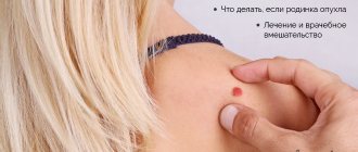
A mole is inflamed, what should I do?
A mole is just what we see on the surface of our skin. In fact, its roots go deep into the skin . It is because of this location that it causes harm to the body. Because the infection will get not only into the upper layers of the skin, but also to the entire depth of the skin. Redness around the mole indicates the initial stage of its inflammation. From the moment of discovery, you should not hesitate for a minute and consult a doctor. After all, a few days lost can turn into months of unpleasant and rather complicated treatment for you.
Nevi located on the face and other open areas of the skin are dangerous. Since in sunny weather, especially eternal and in summer, these places are exposed to ultraviolet rays . What this entails was described above. Many people do not perceive the redness of the skin near the nevus as inflammation. Namely, redness indicates degeneration from a non-malignant tumor to a malignant one. Any disease is easier to treat at the primary stage, including melanoma.
All this also applies to birthmarks. Its redness is a direct signal of the development of a cancerous tumor. Take care of your health, watch out for nevi . Inflammation of a mole can be recognized by certain signs.
Signs of inflammation
- The nevus has changed color. A mole may not only become darker, but also, on the contrary, become colorless.
- The contours become uneven and blurry. In rare cases, the reverse transformation occurs. But in any case, changing the shape of a mole is a signal for action.
- Increase in the size of the nevus.
- Redness around a mole or birthmark.
Reasons for the appearance of a crust on a mole
All birthmarks appear under the influence of changes in pigmentation and vascular system. Deformation serves as a signal to visit a doctor. The appearance of a crust on a mole is influenced by several reasons:
- mechanical injury;
- constant touching by clothes;
- getting burned;
- hormonal disorders leading to pathology.
A crust on a mole is not always a precursor to cancer. If it appears during an injury or burn, there is no health risk. Here it is necessary to eliminate the entry of pathogenic microorganisms into the wound so as not to aggravate the course of the disease.
The following symptoms indicate the possibility of developing cancer:
- hair on the formation began to fall out;
- redness of the area around the birthmark;
- secretion of lymphatic fluid;
- burning, itching, tingling;
- change in density.
If a mole has crusted over or enlarged, what does this mean, a dermatologist or oncologist can answer. Therefore, if you experience any of the above symptoms, you should immediately visit a specialist.
What to do if a mole becomes inflamed?
Having discovered an inflamed nevus, you need to adhere to the following algorithm of actions . This will help you protect your health.
- Contact an oncodermatologist. You will not be able to cure an inflamed nevus on your own.
- Do a biopsy. Find out the complexity of inflammation. Perhaps, based on the results of a biopsy analysis, if there is no risk to health, they will simply cauterize the nevus, without surgical intervention.
- If it is not possible to contact a specialist. Wipe the inflamed nevus with medical alcohol or calendula tincture until completely healed.
- Sprinkle the inflammation with streptocide.
- Treat the mole with an ointment that contains antibiotics, zinc or salicylic acid.
- Make a compress from celandine tincture. Apply the compress to the redness for ten minutes, every four hours.
- A compress of celandine tincture will also help. Celandine can be purchased at any pharmacy. Apply a cotton pad soaked in the tincture for ten minutes, three times a day.
- Lubricate the inflamed area of skin with linseed oil. Since it is this oil that promotes the speedy healing of skin diseases. By consulting with an oncologist and following these steps, you can overcome the disease and protect yourself from melanoma.
Are flat moles dangerous?
In most cases, all pigmented, non-raised spots are similar to each other and therefore do not cause alarm. But to be sure, you need to regularly conduct self-examinations, during which warning signs may be identified:
- One half of the nevus does not correspond to the other. Benign birthmarks are symmetrical, like a circle or oval.
- The edges of the mole are ragged, irregular, or cloudy.
- Healthy pigmentation is monochromatic. A combination of different colors is a bad sign.
- The large diameter of a mole, exceeding the size of an eraser from a simple pencil, should also cause concern in a person.
- If a flat mole, which has not shown any changes for a long time, has grown, begun to develop, change color, bleed, or itch. Particular attention should be paid to the appearance of elevation of one of the edges.
If any of the conditions described above are detected, it is recommended to consult a dermatologist for a more thorough examination and the necessary tests.
Removal of an inflamed nevus
If suddenly you did not have time to contact a specialist in time and the nevus began to increase in size , cause pain, change color and shape, you need to firmly understand that this process will not go away on its own. The inflamed mole will have to be removed.
Methods for removing inflamed moles
- You will have to resort to surgical removal of the nevus. Initially, you need to contact an oncodermatologist; if there is not one in your clinic, try to get an appointment with a therapist. The specialist will give you directions for removal. Then, using local anesthesia, an incision is carefully made with a scalpel and damaged tissue is removed. After the procedure is completed, the tissue that was removed from the nevus is sent for a biopsy.
- Another method for eliminating nevi is laser therapy. This is the most effective way to eliminate nevi. This happens as follows: the laser beam gradually evaporates the mole layer by layer. This process is completely painless. A huge advantage of this removal of inflamed nevi is that after the procedure there are no scars or scars left.
- Removal on the face with high-frequency current. This process also has a name - electrocoagulation. The procedure is similar in effect to laser therapy. The disadvantage of this nevus removal is the appearance of scars at the site of the mole.
Before treating an inflamed mole or birthmark, you must find out the reason that caused this deformation. The development of cancer occurs rapidly, but be patient and do not hesitate under any circumstances. Remember, you only have one health and sometimes no amount of money can improve it.
Which mole needs to be removed
Having an idea of the number of your own nevi and their appearance, it is easy to monitor their condition. Signs of the onset of pathological processes in a pigmented area of the body are as follows:
- lack of symmetry around at least one axis;
- jagged, uneven boundaries of pigmentation;
- bleeding or other discharge from the formation;
- uneven coloring of the mole, dark inclusions, the appearance of a rim or edging;
- dimensions exceeding 6 mm in diameter;
- progressive growth, the nevus may hurt and itch.
Even if one of these signs is present, it is necessary to visit a dermatologist, who, if necessary, will refer the patient to an oncologist. The doctor will recommend how to remove the formation and do its histological analysis.
Even if the mole does not show signs of the presence of cancer cells, but is located in an open area of the body, where it is constantly exposed to ultraviolet radiation, in places where clothing rubs (on the neck, arms, lower back, armpits, under the breasts, on the nipples, in the genital area), it is constantly exposed mechanical damage from razors, combs, watches, such formations must be removed.
The following formations are considered signs of benign pigmentation:
- pedunculated, width less than 2-3 mm;
- long-term existence without any changes;
- surface hair growth;
- the skin pattern is preserved on the pigmentation;
- pale color;
- the structure is softer than the skin.
- smooth edges, axis symmetry.
These signs do not guarantee the absence of pathology, but the occurrence of cancer cells in such nevi is unlikely.
Tumors should be removed in medical, not cosmetology clinics or at home, since in a specialized institution it is possible to examine the material and, if necessary, prescribe treatment, which means not to waste time in the fight against oncology.
The following hardware techniques quickly and reliably remove skin defects:
- laser;
- a liquid nitrogen;
- radio waves.
Moles can be removed surgically under local anesthesia. This mainly occurs when a malignancy is suspected. Laser removal and cryodestruction do not allow testing the material for the presence of cancer cells. These methods are used to eliminate only good moles that have been examined by a doctor, that is, the method of removing the defect is always determined by a specialist.
Pharmacies offer many drugs for skin treatment, most of them require long-term exposure, which can trigger the development of tumors. This is why you should not self-medicate.
Diagnostic and treatment methods
Therapy is prescribed only after diagnosis. Diagnostic methods are as follows:
- visual study of shape, color, size;
- taking a smear in the area of the growth;
- The discharge under the crust is taken for examination;
- computer modeling is carried out;
- fluorescence microscopy;
- biopsy of a piece of tissue for microscopic examination;
- a blood test is taken for tumor markers, which determines whether the growth is benign or malignant;
- prescribing adequate treatment.
You can carry out self-monitoring using the American Melanoma Accord system. Thanks to this system, everyone can recognize dangerous changes at an early stage. The procedure is carried out in several steps:
- “A” is the presence of asymmetry in birthmarks;
- “K” – state of the ends of the build-up;
- “K” – blurred edges, characteristic of a malignant form of oncology;
- “O” – study of color distribution, color change in relation to the original state;
- “P” – dynamic increase in size, diameter over 5 – 6 mm indicates the development of skin cancer;
- “D” - intensive growth dynamics, changes in structure, the presence of a crust and other changes are included in this parameter.
Therapy directly depends on the transformation of the mole into melanoma.
| Methodology | Treatment options |
| Radiosurgery | Radiation therapy is carried out using a radioknife. This is a non-traumatic method in which the resulting crust falls off after a week. |
| Laser removal | A safe and painless method that leaves no traces. After removing a mole with a powerful light beam, the top layer disappears after 14–20 days. |
| Surgical intervention | The method is ideal for large, hanging birthmarks that are dangerous. There may be a scar that takes a month to heal. |
| Electrocoagulation | Removal using current. A medical coagulator is used. After such an operation, it is possible to study the cut material. |
| Cryodestruction | A thin needle with a temperature sensor is inserted into the growth, then cooled to the desired temperature and abruptly removed. Freezing occurs with liquid nitrogen. With this method, a crust is formed that lasts about 30 days. |
If there is a crust on a birthmark, you should not immediately panic, tear it off or treat it yourself. Be sure to visit a doctor to make a diagnosis and prescribe treatment. Not all cases require mole removal, and timely examination can save lives.
Why do changes occur with a mole?
The main reasons why changes occur associated with moles on the body are considered:
- Heredity. Congenital nevi are sometimes passed from parent to child. The risk category includes people who have a hereditary factor for the development of malignant tumors.
- Ultraviolet. Fans of deep tanning and avid visitors to the solarium should be wary of the possible degeneration of a nevus into melanoma.
- Injury. Scratches, wounds, insect bites, and any other mechanical damage can provoke the development of pathology. Inflammation of skin tissue occurs, and active substances begin to be produced that increase cellular growth.
- Chemical exposure. If the nevus is constantly exposed to chemical-based detergents, this can lead to irritation of the formation and its modification.
- Hormonal changes. A mole changes color and size due to hormonal imbalance. This is especially true for women who are expecting a child, adolescents during puberty and people suffering from endocrinological diseases.
Whatever the reason for the appearance of the nevus, any changes in it indicate the beginning of a certain process in the skin layers. Only a doctor can say whether it is associated with a serious pathology.
Whatever the reason for the appearance of the nevus, any changes in it indicate the beginning of a certain process in the skin layers.
Diagnosis of nevus
studying a smear under a microscope is an important but dangerous part of diagnosing a nevus
A thorough examination of the patient plays an important role in the diagnosis of nevus formations. The attending physician evaluates the size, shape, localization of the nevus, the presence or absence of hair in it and a number of other features. Usually, an examination is enough to establish an accurate diagnosis and prescribe appropriate treatment. But very often there are complex clinical cases that require additional examination methods.
Smears are often taken from the surface of the tumor. Absolute indications for taking a smear: bleeding of the formation, weeping, cracks. The material taken during the smear is carefully examined under a microscope. The result becomes known the next day. But it should be said that taking a smear has a drawback: microtraumas of the nevus occur, which, under certain conditions, can further provoke malignancy.
Such studies can be carried out only in specialized clinics, where after taking a smear, the nevus will be removed. The safest diagnostic method is the use of a luminescent microscope, when the nevus is examined directly on the patient’s body, without a preliminary smear. A similar study is carried out using a dermatoscope. Before inspection, apply a small amount of oil to the formation, which will enhance the quality of the inspection. This is a very accurate and absolutely safe procedure. The only difficulty is that dermatoscopes are not available in every clinic.
Modern computer diagnostics of neoplasms is also becoming very popular, when the resulting image of the spot is stored in the computer memory, and then, using a special working programs subject it to the most thorough research. But this technique is very expensive, which somewhat complicates its introduction into widespread clinical practice.
Consequences and prevention
Flat moles aren't dangerous, but that doesn't mean you should ignore changes in size, shape, or color. Since it is not difficult to touch a mole in everyday life with nails, clothing or a razor, in the event of an injury, you should immediately consult a doctor. The consequences of removing lentigo at home often lead to negative results, one of them is melanoma. Therefore, before removing an unwanted “mark” on the body, it is better to consult a doctor.
What does a white spot around a mole warn about?
When a white halo appears around the formation, the reason is a possible malignant degeneration. This is accompanied by itching and changes in the shape and color of the nevus.
Basically, a white dot on a mole appears during the period of hormonal changes, because it is then that the body is most weakened. It indicates possible problems associated with oncology.
If such a manifestation occurs after sunbathing, this is also a reason to consult a doctor, since most likely such a transformation indicates the appearance of melanoma. In this case, the provocateurs are the sun's rays. Consultation with a dermatologist is required to prevent serious consequences.
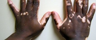
White halos may indicate problems in the functioning of pigment cells. Then we are talking about the disease Vitiligo. Although it often does not cause physical discomfort, psychological problems are not excluded.
Moles with a white spot around them are not a problem if:
- A nevus can have an irregularly shaped halo, be located next to each other and merge with each other. Such moles are typical for teenagers and do not pose any threat;
- The formation looks like a separate spot, with white pigment around it. The size of the halo can reach one centimeter. It may be light brown or spotted. These are also harmless manifestations.
A white spot on a mole itself is absolutely safe. But if pathological processes are present, you should consult a doctor immediately:
- Color change of nevus;
- Purulent discharge from it;
- The surface peels, cracks, swells and becomes inflamed;
- The edges are deformed.
All these changes require immediate examination.
If a mole with a white halo bothers you from an aesthetic point of view, a dermatologist will help you get rid of it. He will first reveal the status of the mole, whether it is benign or malignant. Removal methods will be similar to mole removal. The doctor will help you make the choice; in any case, after the operation, you should not neglect the observation of a specialist.