Causes
Chalazion can occur at any age, but according to statistics, it is more often diagnosed in adults 30-50 years old.
The main function of the eyelids is to protect the eyes from the penetration of pathogens, dust, and foreign particles. Each eyelid contains about 30-50 meibomian glands, which open through excretory ducts onto the inner surface of the eyelids. The fatty secretion produced by the glands forms the outer (lipid) layer of the protective film covering the cornea. This lipid layer ensures the maintenance of a sufficient level of moisture in the eyeball, prevents excessive evaporation of tear fluid from its surface, and ensures easy gliding of the eyelids.
The main cause of chalazion is blockage of the excretory duct of the gland. As sebaceous contents accumulate in the lumen of the gland, a benign dense nodule is formed on the eyelid - a chalazion (this is a more correct term from a medical point of view).
In turn, blockage of the lumen of the gland can occur as a result of increased viscosity of the sebaceous secretion produced, or as a result of inflammatory diseases of the eyelids caused by injuries, infectious microorganisms, wearing contact lenses, using low-quality cosmetics, or allergies.
Chalazion can occur against the background of other pathologies of the visual apparatus or diseases of internal organs, leading to weakened immunity. The most common reasons:
- barley (especially with illiterate and untimely therapy, frequent relapses);
- chronic blepharitis;
- dermatological diseases (acne, seborrhea, rosacea);
- endocrine pathologies, including diabetes mellitus;
- diseases of the digestive tract (chronic gastritis, enterocolitis, intestinal dysbiosis, bile duct dyskinesia).
Provoking factors that increase the likelihood of developing pathology include:
- failure to comply with hygiene standards, touching the face with dirty hands;
- prolonged wearing and/or non-compliance with the requirements for the use of contact lenses;
- systematic hypothermia;
- acute respiratory viral infections;
- frequent stress, chronic fatigue;
- avitaminosis.
Worth knowing! Low immunity and poor hygiene are the most common causes of chalazion in children.
The mechanism of the appearance of wen on the eyes
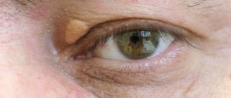
The wen near the eye on the eyelid is white or whitish in color, round in shape, and easily palpable under the skin. May form due to:
- Difficulties in metabolic processes.
- When leading an inactive lifestyle.
- Diabetes mellitus.
- Improper care of the skin of the eyelids.
- Hormonal imbalances.
- Weakening of the immune system.
Most researchers are confident that a lipoma on the face grows like a tumor, from a single cell. This is indicated by the lobed structure of the wen. Some growths are associated with problems with the outflow of sebaceous gland secretions and are located on the surface.
Symptoms
Sometimes several chalazions form on one eyelid, which are located in a chain at some distance from each other or merge to form one large seal.
A chalazion is a firm, elastic, round formation under the skin of the upper or lower eyelid. Typically, the initial stages of the disease are asymptomatic, and only in rare situations are they accompanied by physical discomfort and cause unpleasant symptoms:
- itching;
- lacrimation;
- soreness to touch;
- astigmatism (refractive error) and visual distortion resulting from the pressure of the nodular structure on the cornea.
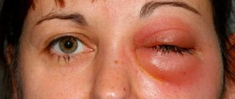
The sebaceous secretion that accumulates in the lumen of the gland is a favorable environment for the development of bacteria. Therefore, chalazion is often complicated by a bacterial infection. In this case, purulent inflammation is accompanied by:
- redness of the eyelid;
- swelling;
- throbbing pain;
- softening of the nodular structure revealed by palpation;
- increase in body temperature.
On the upper eyelid
The painless seal is located in the thickness of the cartilage tissue and is not connected to the skin. Gradually the dimensions of the compaction increase to 5-6 mm. The formation on the upper eyelid becomes noticeable (it looks like a grain of millet or a small pea) and creates a visible cosmetic defect.
On the lower eyelid
When a chalazion forms on the lower eyelid, the protrusion is directed inward, but on the outer side the eyelid may remain unchanged. Pathology of the lower eyelid is more dangerous, since the formation may be visually invisible. When it spontaneously opens, purulent contents are released onto the surface of the conjunctiva, a fistulous tract is formed, around which granulation tissue grows. The skin of the diseased eyelid becomes red and dry, covered with dried crusts.
Types of lipomas
1. Milia. These are small nodes of a yellowish-white hue. They look like whiteheads. However, they differ from them in that they do not have a duct. Milia cannot be squeezed out either in a salon or at home.
2. Xanthelasma. These are flat or convex yellow plaques. They can occur on both the upper and lower eyelids, and on the inner corners. The surface can be both smooth and slightly wrinkled. Removal of a lipoma of this nature is necessary, since it tends to grow and in some cases reaches several centimeters. They are formed mainly in women 40 years of age and older who have metabolic and liver disorders.
Reasons for the appearance of wen on the upper eyelid
Until now, medicine does not have an exact answer to the question of what factors influence the formation of lipomas on the eyelid. It is most likely that disturbances in the functioning of the body as a whole are reflected in the appearance and condition of the skin.
There are a number of reasons that indirectly affect the appearance of wen on the eyelid.
- Excessive consumption of fatty, smoked, fried foods, foods with preservatives, fast food, sweets.
- Excess weight, predominance of adipose tissue in the body.
- Diabetes.
- Hormonal changes in the body, especially in women during pregnancy and breastfeeding, as well as menopause.
- Problems of an endocrinological nature, diseases of the thyroid gland.
- Sedentary lifestyle, rare access to fresh air, insufficient physical activity, sedentary work.
- Disturbances in the functioning of the genitourinary system.
- Problems with metabolic processes.
- Genetic and hereditary predisposition to the appearance of white wen on the eyelids.
Stages of flow
The presence and severity of clinical manifestations of chalazion depend on the stage of the pathological processes:
- Formation of a chalazion capsule. It is almost always asymptomatic. The only sign is a small, painless, almost imperceptible lump on the lower or upper eyelid, which can only be detected by chance.
- Active growth of chalazion. The formation begins to increase in size and puts pressure on the eyeball, causing discomfort and deterioration of visual function.
- Inflammation. Lack of therapy at previous stages of chalazion development leads to the formation of a cyst. The mucous contents of the cyst become a favorable environment for the development of pathogenic microorganisms. As a result, an inflammatory process develops, accompanied by severe pain, lacrimation, and redness of the eye. At this stage, immediate surgical treatment is required, whereas in the absence of inflammatory processes, conservative therapy can be carried out.
Chalazion in children
Chalazion is often diagnosed in children, since most of them neglect the rules of hygiene, and many have a weakened immune system.
Features of the course of pathology in childhood:
- formation of several chalazions at once;
- frequent spontaneous opening of the formation;
- in rare cases, a chalazion may degenerate into a cyst.
Important! How to treat chalazion in children is decided solely by the doctor. Therefore, if alarming symptoms appear in a child, you should immediately contact an ophthalmologist.
Diagnostics
The affected eyelid makes blinking movements less frequently.
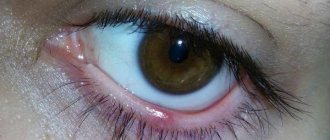
An ophthalmologist can make an accurate diagnosis based on a visual examination. With a chalazion, a compaction is found in the thickness of the eyelid, not fused to the skin. On the inner surface of the eyelid, a hyperemic (reddened) area of the conjunctiva with a central zone of a grayish tint is noticeable.
Instrumental and laboratory diagnostics for chalazion are not carried out. The only exceptions are fast-growing formations that require differential diagnosis with adenocarcinoma (malignant formation) of the meibomian gland. For this purpose, collection and histological examination of the biological material of the formation is carried out.
Chalazion is similar in appearance to styes, cysts and other eyelid diseases. Therefore, if a lump appears on the upper or lower eyelid, you need to contact a specialist. The doctor will make an accurate diagnosis and recommend the optimal treatment method for chalazion or other identified disease.
Diagnostic features
If you see any formation on the eye, the patient should go to the hospital. The doctor will diagnose:
- general and biochemical blood test;
- examination of the eyelid and eye;
- cytological studies;
- Ultrasound.
Based on the examination results, the patient is prescribed therapy. Wen is treated with pharmaceutical medications and ointments:
- Vitaon balm;
- Gistan cream;
- Videstim ointment.
They are used 2-3 times a day for a week and are effective in combating small seals on the eyelids.
Get rid of wen using folk remedies:
- compresses of chestnuts, onions and soap;
- celandine juice;
- gruel from wheat grains;
- vegetable oil;
- hydrogen peroxide;
- mixtures of sour cream, honey, salt;
- oak bark infusion.
Sometimes the patient needs to consult a surgeon, who may suggest removing the wen from the eyelid using other methods:
- Operation. It is carried out in three different ways: using a classic incision, endoscopic incision, liposuction.
- Cauterization with laser. The procedure is painless, carried out in beauty salons, does not require a hospital stay, and does not leave scars.
- Puncture-aspiration removal. Medium and large wen are removed. The procedure is carried out using a medical needle, the tissue is completely removed, but the capsule remains, which leads to relapses.
Treatment
How to treat a chalazion is decided by the doctor, taking into account the stage of the pathological process. In the early stages of the formation of a benign formation, conservative therapy is carried out, including the use of pharmacological agents and physiotherapy procedures.
If the chalazion exists for a long time, or rapidly increases in size, is complicated by infectious inflammation, and conservative methods do not have a positive therapeutic effect, an operation is performed to remove the formation.
Conservative
The duration of treatment of chalazion with medications should not exceed 14 days.
If a chalazion is detected in the earliest stages, when the sebaceous secretion has become too thick and viscous, but the capsule has not yet formed, eyelid massage becomes the main method of therapy. The procedure is carried out only by a doctor using a slit lamp and consists of squeezing the eyelid from the periphery to the edge. Thus, the sebaceous gland is freed from thick secretion.
If the capsule has already formed, but the size of the chalazion does not exceed 4 mm, treatment is carried out using pharmacological agents. Drug treatment for chalazion includes:
- eye instillation with antiseptic drops (Miramistin);
- placing mercury ointment behind the eyelid;
- treatment of the upper and lower eyelids with hormonal ointments (Hydrocortisone eye ointment);
- injections of corticosteroids or anti-inflammatory drugs directly into the cavity of the nodule (promote its resorption).
In addition to drug treatment, physiotherapeutic procedures are carried out - warming with dry heat, UHF therapy.
After each warm-up, it is recommended to massage the eyelids. The massage is carried out with light circular movements in the direction from the outer to the inner corner of the eye, from the chalazion to the base of the eyelashes. Such manipulations promote the outflow of viscous secretions and in some cases help restore the patency of the gland duct. To achieve a positive result, massage is performed for 1-2 minutes at least 5 times a day.
Attention! When the chalazion is inflamed, warming procedures are prohibited, since heat promotes the development of pathological processes. As a result, inflammation will spread to nearby structures of the eye with the development of phlegmon of the eyelid or abscess.
Surgical
The most common complication after surgical removal of a chalazion is recurrence of the disease.
Indications for mandatory surgical treatment are:
- active growth of education;
- signs of development of inflammatory processes;
- chalazion dimensions are more than 5 mm.
Surgical removal of a chalazion can be performed:
- in a classic way;
- using a laser.
Classic chalazion removal surgery is performed on an outpatient basis under local anesthesia. The operation consists of enucleating the formation along with the capsule and removing the contents of the cyst. After removal, stitches are placed on the eyelid, and the eye is closed with a tight pressure bandage. For 5-7 days after surgery, anti-inflammatory eye drops or ointment are placed in the eye.
Laser removal is an alternative method in which the formation capsule is dissected using laser beams, its contents are removed and the pathological tissue is subsequently evaporated. Laser removal is modern and less traumatic compared to classical surgery. Firstly, it does not require stitches, and secondly, it helps prevent relapses of the disease.
Attention! To prevent the cornea from being injured by the postoperative scar, the patient is recommended to wear a soft contact lens for several days after removal.
At home
Along with the use of medications, folk remedies can also be used to treat the disease. However, it makes sense to treat chalazion at home only in the initial stages of development. If the cyst has long been formed, or an inflammatory process develops, you should immediately consult a doctor and not engage in amateur activities.
How to treat chalazion at home:
- Lubricate the formation with a cut of a clove of garlic at least 4 times a day. Garlic contains many phytoncides that have a powerful antibacterial effect. Accordingly, the healing process when using the product is accelerated, and the risk of developing infectious inflammation is significantly reduced.
- Warm the affected eye twice a day. To do this, you can use a boiled egg that has not cooled down, or salt heated in a frying pan and wrapped in a linen bag. Heat should be applied for 5 minutes. After this, it is advisable to lubricate the affected eyelid with castor oil, performing massage movements.
- Wipe the sore eyelid with fresh aloe leaf juice diluted with water in a 1:1 ratio. It is better not to use undiluted juice, as it greatly irritates the mucous membranes.
- Before going to bed, apply baked onion (warm) to the affected area. Keep for 20-30 minutes. After removing the compress, do not wash your face.
- Mix 2 teaspoons each of raspberry leaves, chamomile herb, calendula flowers and dried parsley. Pour a glass of boiling water over the collection, leave until cool, and strain. Apply a cotton pad soaked in the prepared composition to the inflamed area for 5-10 minutes.
- Rinse the affected eye every 2-3 hours with a decoction of chamomile or sage (pour a tablespoon of the herb with a glass of boiling water, heat for about 5 minutes in a water bath, leave, strain).
Is it possible to be treated at home?
If a person is completely sure of the causes of the disease and that he is faced with a chalazion, he can begin self-treatment at home. This mainly applies to those patients who have already encountered the problem and whose disease has developed into a chronic form.
Expert opinion
Slonimsky Mikhail Germanovich
Ophthalmologist of the highest qualification category. Has extensive experience in diagnosing and treating eye diseases in adults and children. More than 20 years of experience.
If a person does not know the reasons for the appearance of a node on the eyelid, as well as if suppuration appears, it is necessary to immediately contact a specialist who will select a course of treatment.
Why should you see a doctor?
Despite the fact that at first glance the disease does not seem too dangerous, at the first signs you should seek help from a doctor. Sometimes a node with purulent contents can open spontaneously. In this case, there is a risk of secondary infection and worsening of the patient’s condition.
In such a situation, the doctor surgically removes the remaining tumor and prescribes antibacterial therapy. In addition, only a specialist can correctly diagnose the disease at its early stage and provide high-quality and adequate treatment without surgical intervention.
Therapy with folk remedies
It is worth remembering that treating the disease with traditional methods is only permissible at an early stage and only after consultation with an ophthalmologist.
Poultices
The most common poultice is dill poultice. To prepare it, you need to pour a few tablespoons of dried dill with boiling water so that the water just covers the plant. Afterwards, the container with dill is covered with a lid and placed on low heat. After 10 minutes, wrap the prepared dill pulp in a napkin and apply it to the affected area. It is best to do such poultices before going to bed.
Prevention
The disease often recurs, even after surgical removal of the formation. To avoid re-formation of a chalazion, it is important:
- promptly and correctly treat other eye pathologies (blepharitis, meibomitis), acute and exacerbation of chronic infectious diseases (sinusitis, tonsillitis);
- follow hygiene rules (wash regularly, do not touch your eyes with dirty hands), requirements for the use of contact lenses;
- take measures to improve immunity.
Chalazion is a rather unpleasant but treatable pathology. If the appearance of a chalazion is detected in the early stages of development, you can get rid of the problem even at home. If there is no improvement within several days, or the cyst appeared a long time ago, it is better to abandon alternative methods of therapy and consult a doctor. The specialist will select the optimal drug treatment regimen and, if necessary, remove the formation. Competent and timely therapy will help avoid various complications.
Wen removal methods
If a white or yellowish formation occurs on the eyelid, you should consult a doctor. Only a specialist will be able to accurately diagnose the phenomenon, establish the cause and recommend the best way to get rid of this problem.
The question of which doctor removes the wen is not entirely clear. If surgery is necessary, it will be performed by a surgeon. However, to determine the nature of the formation, you should consult your doctor or cosmetologist. They are the ones who can recommend the correct algorithm of action.
Medicine and cosmetology use the following methods (depending on the situation):
1. Surgical. It is necessary to get rid of the wen in this way if there are problems with closing the eyelid, it causes pain, discomfort, vision deteriorates and the cosmetic defect becomes really noticeable. The doctor makes an incision through which an endoscope is inserted into the wen. The lipoma is carefully cut off from the eyelid. The operation should be carried out as carefully as possible, the hole is made along the fold of the skin so that the scar is less noticeable. Any lipoma must be removed along with the capsule, otherwise the yellowish nodule will soon appear on the eye again.
2. Medication. A small wen on the lower eyelid can be removed like this: a medicine is injected into a small incision on the surface, with the help of which the lipoma resolves and disappears over time.
3. Laser. The most modern, advanced, safe, painless and aesthetic method. Removing a wen with a laser is a bloodless procedure; it seals the blood vessels that fed the formation, which prevents its reoccurrence. The beam acts much more accurately than a scalpel, which eliminates the appearance of scars. Tissue infection also does not occur, because the laser cauterizes the edges of the wound. The method assumes the ability to get rid of the lipoma along with the capsule without any problems. The laser beam does not leave scars, and after such an operation you can go home straight away.
4. Electrocoagulation. Excision of the affected tissue leaves a small crust, which dries and falls off in about 10 days. The pigment spot that may appear on the eyelid will soon resolve.