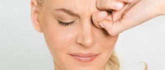
A lump on the eyelid is an unpleasant and very common problem that many people have had to deal with, even without any health problems. Such a formation near the organs of vision can act as a cosmetic defect, that is, not cause unpleasant sensations or discomfort, or be accompanied by redness, the development of a purulent process and the appearance of pain. Depending on the cause of the lump, the size of the formation can vary from a few millimeters to 2-3 centimeters in diameter.
Lump on the eyelid
Reasons for appearance
Induration in the upper eyelid can be a manifestation of an infectious disease, and a sign of a general decrease in immunity due to vitamin deficiency or mineral deficiency. In some cases, this is how pathological processes in the organs of the gastric tract manifest themselves, especially those that have taken a chronic form. The periodic appearance of various lumps, bumps or pimples on the eyelids can be caused by hyperfunction of the sebaceous glands, as well as hormonal imbalance.
The reason why education may appear on the eyelids is sometimes due to insufficient hygiene standards, for example, sleeping with unwashed makeup, using expired or poor-quality cosmetics. A seal on the eyelid can occur if the lenses are incorrectly selected, the conditions of their storage or the duration of wearing are violated, as well as if there is an intolerance to contact lenses in general. A periodically appearing pimple on the eyelid or a lump may also have neurological causes. If you experience prolonged stress, your body may react in this way.
Formations in the lower and upper eyelids can be caused by various reasons. When a white pimple appears on the eyelid, this may indicate the following ophthalmological pathologies:
Barley
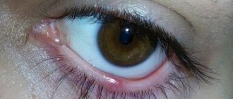
Barley is an inflammation with the formation of an abscess in the eyelash follicle or in one of the sebaceous glands located between the eyelashes. A stye begins with slight redness around the eyelash and is characterized by severe pain. After a few days, a purulent head appears in the area of inflammation, and the pain increases these days. Such formation is a sign of reduced immunity or metabolic disorders. The immediate cause of its appearance is most often the bacterium Staphylococcus aureus. The infection gets into the eye from unclean hands; sometimes all you need to do is rub your eyes or use a dirty towel.
Thus, if there is a white pimple inside the eyelid, then it is most likely a stye. It can jump up on both the upper and lower eyelids, both outside and inside the eyelid. Internal barley indicates inflammation of the sebaceous gland, and external barley indicates inflammation of the eyelash follicle. Characteristic features are:
- redness and swelling of the eyelid;
- acute pain;
- discomfort;
- feeling like something is in the eye.
The abscess opens on its own a few days after its appearance. It is strictly forbidden to open it yourself, squeeze it out or try to pierce it. It is recommended to treat stye using eye drops and ointments. It can go away on its own, but it takes a little longer. By using medications at the very beginning of the inflammatory process, you will get rid of barley in three or four days. If you stop using medications, you will experience discomfort for a week or even ten days.
For treatment, antibacterial drops are used, for example, albucid or a solution of chloramphenicol. Antibacterial ointments such as tetracycline ointment can also be used. It is recommended to wash your eyes several times a day with antiseptic solutions, for example, chlorhexidine.
How to treat
The following procedures are used to treat lumps on the eyelid:
- heat treatment (heat therapy) and massage of affected areas;
- warm compresses (2 to 4 times a day). But before this, the skin must be washed with warm water and soap. It is advisable to use baby soap, as it contains no chemicals;
- eye drops are used, but the type of drug and duration of use are determined by the attending physician;
- the cones are heated by ultraviolet light;
- in rare cases, the patient may be prescribed antibacterial drugs. As a rule, they are required if the inflammatory process has spread to the sebaceous glands. The most common drugs in this group include Tetracycline;
- corticosteroid injections are prescribed (corticosteroids are a group of anti-inflammatory drugs).
Features of treatment
First of all, when the disease has not yet spread, doctors prescribe the use of disinfectants or eye ointments. An excellent result is shown by a helium-neon laser, the effect of which is to resolve the pus accumulated in the lump. The breakthrough of the lump, as a rule, indicates a positive result of therapy.
On a note! When treating chronic diseases, doctors resort to the use of corticosteroid drugs. The drug is injected with a syringe directly into the source of inflammation, that is, into a lump on the eyelid. The duration of this type of therapy is 14 days. Throughout the entire period of treatment, the patient should be regularly examined by an ophthalmologist.
Phloxal
Surgical intervention is resorted to only in cases where drug treatment has not given the desired effect. Many people are afraid of operations, especially if they are performed on the eyes or eyelids. But the development of modern medicine involves the use of laser technologies, thanks to which the formation on the patient’s body is completely removed and with minimal trauma to the body . As practice shows, repeated surgical intervention is usually not necessary.
Video: How to remove wen on the eyelid
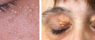
Prosyanka
Milia occurs when the sebaceous gland is clogged with dead skin particles. The signs are:
- the appearance of one or more small lumps on the upper or lower eyelid;
- absence of redness and swelling;
- painlessness.
Sometimes, with insufficient hygiene, such formations can become inflamed, which then causes pain and discomfort.
You should not try to squeeze out acne on your own, as you can cause an infection and worsen the symptoms. A qualified cosmetologist will help you remove unaesthetic pimples from your eyelid. He will select the removal method that suits you.
It is worth noting that millet appears not only on the eyelids, but also on any areas of the skin. In order to avoid occurrence, you need to monitor your diet, use high-quality cosmetics and observe personal hygiene rules. By properly and timely cleansing the skin, removing dead particles, you will avoid blockage of the sebaceous glands.
Prevention measures
Compliance with preventive measures will prevent the development of stye, xanthelasma and other pathologies in which a lump may form on the eyelid. Prevention is as follows:
- observing the rules of personal hygiene (you cannot touch your eyes with your hands, especially if your hands are unwashed);
Maintain good personal hygiene - Before each meal, be sure to wash your hands with soap. This will prevent the spread of bacteria throughout the body;
- at the slightest sign of irritation on the surface of the eyelids, it is necessary to apply a compress;
- get rid of excess weight if necessary;
- normalize your diet, stick to proper nutrition.
Disease Prevention
Experts also recommend paying attention to your immune system , that is, strengthening it in every possible way. A strong immune system will protect the body from various diseases, including preventing the appearance of bumps on the eyelids. By following these simple rules, you can protect yourself from serious health problems.
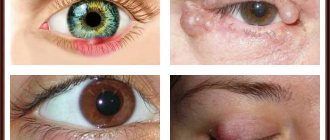
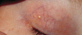
Chalazion
This white pimple on the eyelid is similar to stye, but unlike it, it does not go away on its own, appearing again and again. A chalazion is formed as a result of blockage of the sebaceous gland and the accumulation of secretions in it. The sebaceous glands produce a special secretion that is part of the tears. When this gland is blocked, the secretion continues to be produced, but cannot come out, accumulating in the ducts. This creates a compaction that gradually increases.
A chalazion can look like a pimple on the upper eyelid and also like a pimple on the lower eyelid. In appearance it resembles a small pea and is absolutely painless. When it first forms, slight redness may appear, which then goes away.
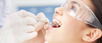
To remove this formation, you must consult an ophthalmologist. He will determine what kind of therapy is needed in your case. You absolutely cannot pierce the seal yourself!
In the initial phase of the disease, it is possible to limit oneself to local agents. For treatment, anti-inflammatory drugs for external use are prescribed: drops, ointments, antiseptic solutions. If the chalazion is large, treatment with hormonal drugs may be prescribed to prevent the growth of fibrous tissue and encapsulation of the formation.
If the disease is advanced, surgical excision of the chalazion may be required. The intervention is performed under local anesthesia and lasts no more than half an hour. In some cases, relapses may occur after removal; this depends on how successful and high-quality the removal of the seal was. Today, the most commonly used type of surgical removal of chalazion is the laser method. After surgery, antiseptics are prescribed to speed up healing and prevent relapse.
Treatment methods for lumps
When barley is diagnosed, drug treatment is indicated. The patient is prescribed ophthalmic ointment and drops. It is recommended to use an antibiotic (Tobrex, Albucid, Levomecitin) and antimicrobial ointment. The last remedy is used at night. More often, the patient is prescribed erythromycin and tetracycline ointment.
To wash the affected eye, use an antiseptic solution:
- "Chlogrexedine";
- "Miramistin".
Without treatment, barley ripens in 10 days. If therapy is prescribed, the disease is eliminated in 4 days. Most often, barley affects 1 eye. Rarely does inflammation spread to the 2nd eye. To prevent such a complication, an antibiotic is instilled into both eyes.
For chalazion, therapy is carried out only under the supervision of an ophthalmologist. You cannot pierce or squeeze out the chalazion yourself. With this diagnosis, the ophthalmologist prescribes “Tetracycline ointment”. If the disease is detected at an early stage, local therapy is indicated. To resolve the pus and eliminate inflammation, prescribe antibacterial drops:
- Ofloxacin.
- Levomecitin.
Of the ointments, erythromycin ointment is used, which is used to treat the tubercle of the cone. You can make compresses from this product. Additionally, the antiseptic Miramistin is prescribed. Another effective method in the fight against chalazion is physical therapy. If necessary, the patient is prescribed glucocorticoids. They prevent the appearance of fibrosis.
If a chalazion is detected at an advanced stage, surgical treatment is indicated. Surgical therapeutic techniques are also used when conservative treatment is ineffective. The operation is performed under local anesthetic. Its duration is 20 minutes. During the manipulation, a small incision is made in the area where the compaction is located. Then the lump with the fibrous capsule is scraped out. The possibility of reappearance of the chalazion depends on the quality of curettage of the cavity. After the operation, the patient is put on a bandage and prescribed an antiseptic.
Papilloma
Papilloma is a growth that occurs as a result of pathologically abnormal growth of epithelial cells. This disease appears not only on the eyelids; papillomas can form on any area of the skin. Characteristic features are their protrusion above the surface of the skin; sometimes they can have a stalk and seem to hang from the surface of the skin.
Papilloma is caused by a virus and is treated with special topical agents. Sometimes surgical removal of the papilloma may be required. Today, there are several methods of surgical intervention. Laser or electrical removal of growths is most often used.
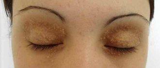
Xanthelasma
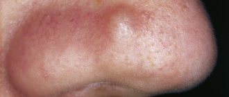
Xanthelasma is a benign formation on the eyelids that occurs as a result of metabolic disorders. Most often appears in older women. The formations look like yellowish uneven tubercles protruding above the surface of the skin. Xanthelasma is a symptom of changes in water-fat metabolism. The formation itself cannot be treated; therapy is carried out for the underlying pathology.
Furunculosis
A pimple on the eye on the lower or upper eyelid may be a manifestation of furunculosis. This disease is characterized by the formation of a painful abscess under the influence of inflammation. It is caused by bacteria. The most common cause of boil formation is Staphylococcus aureus. The inflammatory process occurs in the hair follicle of the eyelash, in the sebaceous gland.
Furunculosis is a rather complex disease; it can be accompanied by an increase in general and local temperature and general malaise. First, a slight painful redness and swelling appears, then the purulent head of the boil appears.
Maturation can take quite a long time, but under no circumstances should you crush the boil. It opens on its own when the purulent-necrotic process reaches its peak.
Antibacterial agents for external use are used to treat furunculosis. If a purulent formation appears on the upper or lower eyelid of the eye, you should consult an ophthalmologist. He will prescribe anti-inflammatory drugs to speed up the purulent process. As a rule, after opening the boil, a small scar remains on the skin.
Characteristic symptoms
Since the swelling of the lump is practically invisible at an early stage, the main symptom is itching or redness of the skin . As the lump grows, it becomes more noticeable, so the patient begins to feel the presence of a foreign body on his eyelids. Pain, as a rule, occurs only in cases when the lump begins to press on the surface of the cornea (this is especially pronounced during blinking). With the development of the inflammatory process, the area of redness gradually increases, as a result of which the inflammation spreads to the conjunctiva.
Chalazion of the upper eyelid
The patient may also suffer from a sharp deterioration in vision - the picture becomes more blurry. A purulent mass begins to accumulate in the cavity of the cone, which over time can come out through the resulting fistula on the skin and remain in the area of the corners of the eyes. The presence of purulent contents is an excellent habitat for various types of infection, which will only worsen the patient’s already difficult condition.
Malignant tumor of the lower eyelid
Video: Neoplasms on the eyelids
Complications and consequences of seals on the eyelids
If an incomprehensible lump, lump or white pimple appears on your eyelids, there is no need to try to diagnose it yourself. Incorrect treatment or lack thereof can lead to unpleasant and even dangerous consequences. Contact an ophthalmologist or ophthalmologist, he will help determine the cause of the formation of the lump, prescribe the necessary tests and determine a treatment regimen.
Independent attempts to open a formed lump or pimple may lead to additional infection and the process will worsen. In addition, it is impossible to completely remove pus by squeezing or puncturing the abscess, which means it will continue to grow. In advanced cases, the abscess can develop into an abscess. Purulent formations are very painful. It's also about aesthetics, because you can't hide your face.
Formations on the eyelids can have a mechanical effect on the body of the eyeball, disrupting blood circulation and preventing normal functioning. As a result, vision may decrease. It is also possible to develop other vision disorders, such as astigmatism. In rare cases, formations can also become malignant.
To prevent dangerous consequences for health and appearance, consult a doctor. All of these diseases can be easily cured in the early stages if adequate therapy is used.
Diagnostics
A lump on the eyelid can be of different sizes and origins, it can be a benign lump, or it can become a huge problem and degenerate into a malignant formation.
As soon as the first symptoms appear, you should immediately seek help from a special clinic for examination.
An ophthalmologist performs a step-by-step diagnosis:
- the eyelid is examined from the inside and outside, depending on the location of the formation of the seal;
- the necessary blood tests are prescribed;
- If necessary, a biopsy and x-ray are performed.
Important. Based on the examination, the specialist makes an accurate diagnosis, gives appropriate recommendations and prescribes treatment.