Where do moles come from?
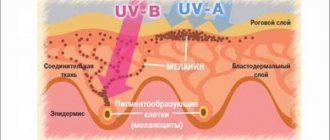
Many people believe that a person is already born with moles. In fact, this is not true. A nevus is a formation that consists of pigment cells (melanocytes). The more a person spends in the sun, the higher the likelihood of moles appearing. An interesting fact is that there are practically no such formations on the body of a baby up to 6 months old. Only some types of moles can appear on the body from birth.
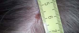
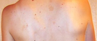
Nevi can vary in shape and size. Most often, flat formations with a diameter of no more than 2 mm can be seen on the body. Large convex nevi with a diameter of more than 1 cm deserve more attention. And if such a mole is covered with a crust, you should go to the doctor immediately, as the risk of its malignant transformation increases.
More moles can always be seen on the body of fair-skinned people. If there are really a lot of nevi, you should avoid sunbathing. The fact is that a burn increases the risk of developing pathological cells. This means that if after the beach a mole becomes covered with a dry crust, an urgent visit to the doctor is necessary.
Diagnostics
Identifying the causes of the appearance of a crust, as well as making a diagnosis, is carried out in several stages:
- Taking an anamnesis - the doctor asks the patient about the conditions under which a crust appeared on the mole, how long this process has been going on and what additional symptoms are accompanied.
- Dermatoscopy - allows you to examine the skin tumor as closely as possible, which helps the doctor determine the diagnosis and possible causes of the development of the pathology.
- Cytological examination of a smear from the surface of a mole - this analysis is most effective if any liquid is released from under the crust. Its sowing makes it possible to detect the presence of pathogenic microflora, and its quantitative and qualitative composition will indicate a possible pathology.
- Histological examination is prescribed if external signs indicate pathological processes in a mole associated with the degeneration of its cells into a malignant tumor. For histology, a section of the mole may be required, but most often it is completely removed, which helps make a more accurate diagnosis and understand whether additional examinations are needed and what therapy is required.
Dermatoscopy is a method for diagnosing a crust on a mole.
If there is a suspicion of the development of cancer, a blood test for tumor markers may be required. This analysis will reveal antibodies in a person’s blood, the appearance of which is characteristic of cancerous tumors.
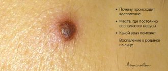
Inflammation of a mole
Any changes in the human body can lead to an increase in the number of moles, changes in the shape and size of existing formations. Thus, decreased immunity can also lead to inflammation of nevi. If a mole is covered with a crust, this does not mean at all that a terrible diagnosis will be made. However, it is recommended to consult a doctor as soon as possible.
A common seasonal cold can lead to inflammation of the mole. But convex nevi, which are located on the neck or folds, are especially common. Such formations are more susceptible to injury than others. And if such a mole becomes covered with a crust, it is possible that it was damaged.
In addition, nevi can burn in the sun. Therefore, in the summer from 12:00 to 16:00 it is better not to sunbathe at all.
Not only a crust will indicate inflammation of a mole. The area around the growth may turn red. The inflamed area usually becomes denser. And if a bacterial infection gets into the mole area, pus may appear.
How to distinguish a seborrheic mole from dangerous symptoms
A rough mole can frighten the wearer with its unusual appearance. Outwardly, it may resemble melanoma; only a doctor and diagnostic measures can accurately determine the type of tumor.
The main differences between seborrheic keratosis and an oncological tumor:
| Factor | Keratosis | Melanoma |
| Risk group | People over 30 years of age whose skin is susceptible to aging. | Can be diagnosed at any age. The patient may be a small child, an able-bodied adult, or an elderly person. |
| Form | A round or oval growth with clear boundaries. It has the ability to grow and thicken up to 1-2 cm. | Formation over 6 mm. |
| View | Flaky mole. Flat or slightly convex appearance. It may look waxy, as if painted on the body. | A spot or raised element that is in the stage of active growth. |
| Symmetry | Complied with. | Violated. |
| Color | Uniform or interspersed with white and black. | It changes and takes on several shades. |
| Borders | Smooth. | Vague with jagged edges. |
The dry growth is dense to the touch, like a dangerous nevus, but it does not hurt when touched, at rest. A painful syndrome can develop when irritated or damaged, then the skin around it itches, and secondary infection can occur.
If a person is accustomed to unusual spots on the body, he may miss cancer. Patients with keratosis are recommended to regularly visit a dermatologist and undergo diagnostics.
Melanoma is on the rise. Its increase occurs quickly: by 1-2 mm per month or more.
What to do if a mole becomes inflamed?
Self-medication in such cases is not recommended. If a mole has crusted over, only a specialist will prescribe adequate therapy. But first you will have to undergo a series of tests. The doctor must find out which infectious agent is causing the inflammation. If it is bacteria, an antibacterial ointment will be prescribed. Additionally, the damaged area will be treated with an antiseptic. Miramistin and Chlorhexidine solutions are widely used.
If the mole is located in the wrong place and is often injured, the doctor will recommend removing it. Frequent damage to nevi is one of the reasons for their malignant degeneration. Popular methods for removing moles will be described below.
How to understand that a nevus is degenerating?
If a mole is covered with a white crust, this is a dangerous sign. Melanoma is an aggressive malignant tumor of the skin that can develop rapidly. Therefore, everyone should know when to postpone a visit to a dermatologist.
There is a technique that includes a number of signs suggesting that it was a malignant tumor that we had to deal with:
- Asymmetry is the first thing you should pay attention to. A healthy mole always has the correct shape. If the round formation suddenly begins to blur, you should worry. The edges of the mole must be smooth.
- Bleeding is another dangerous sign indicating that something is wrong with the mole. If the nevus does not cause any discomfort, but blood periodically bleeds from it, you need to make an appointment with an oncologist.
- The color of the formation also matters. If a mole has darkened and crusted over, this is one of the signs of melanoma.
- You will also have to pay attention to the size. Healthy nevi can also increase in size, but if the size of the mole changes rapidly, you should sound the alarm.
When a change in nevus is dangerous
If it is expressed only by the formation of a crust, and there are no signs of inflammation, growth, pain or itching, there is nothing to fear.
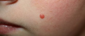
A crust on a healthy red mole is practically not dangerous from the point of view of malignancy. Red moles are called hemangiomas and extremely rarely develop into skin cancer . Damage to these nevi threatens trouble only if infection gets into the wound.
It is important! In cases where inflammation, peeling, bleeding or the appearance of bloody discharge are observed, the oncological nature of the changes can be assumed. To rule out the development of cancer, the first thing you need to do is see a dermatologist.
Mole removal methods
If a mole has crusted over, but no malignant cells were found in it, the doctor will most likely recommend removing it. Laser surgery is the most popular technique today. In this case, a powerful light flux acts as a tool.
The advantage is that after removal there is practically no scar left at the site of the mole. In addition, the likelihood of developing a secondary infection is minimized.
Moles that involve only the upper layers of the epidermis can also be removed using cryodestruction. In essence, such formations are frozen. Flat nevi are removed with a cotton swab, which is pre-moistened in liquid nitrogen. The procedure takes no more than 10 minutes. Within an hour the patient can go home.
If melanoma has been diagnosed, surgery is performed using a scalpel. In this case, the patient may be given general anesthesia. Not only the formation itself is removed, but also healthy skin with a diameter of 5 mm around it. Such actions minimize the development of relapse. Additionally, the patient may be prescribed radiation or chemotherapy.
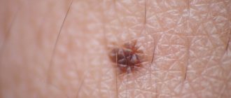
Causes of crust formation on the surface of a mole
The most common reason for the formation of a crust on the surface of a nevus is its mechanical damage. Injured cells succumb to the natural process of regeneration, where the crust acts as a characteristic barrier between the body of the mole and the environment. It acts as a kind of protection for an open wound from pathogenic microorganisms in the environment.
Mechanical damage is a possible cause of a crust on a mole
Also, crust formation occurs when a mole falls off. There are cases when the nevus spontaneously dries out and separates from the body. At the site of the formed wound, a crust appears, under which regeneration processes take place. After spontaneous removal of the scab, the mole area becomes covered with epithelial cells, leaving a small scar.
Excessive exposure to sunlight on the nevus, which provokes the formation of a burn, can also cause the formation of a crust. But the most dangerous option is the malignancy of the neoplasm into a malignant tumor, which is extremely difficult to diagnose based on appearance.
Prevention
Simple steps will help you protect yourself from dangerous consequences. Regardless of the number of moles on the body, you should make it a rule to avoid sunbathing between 12:00 and 16:00. In this way, it will be possible not only to maintain health, but also to prolong youth. If you have a large number of moles on your body, you should avoid direct sunlight even in the early morning or after 16:00.
If a mole is often injured, it is worth removing it. Under no circumstances should you seal the damaged area. The thermal effect created can harm the nevus.
It is worth regularly checking yourself and your loved ones for the appearance of new moles or changes in existing formations. Timely identified pathological formations can be removed without unpleasant consequences.