Moles on the human body can be of different shades: from light brown to deep black. Color depends on the amount of melanin (pigment substance) - the more of it, the darker the moles.
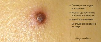
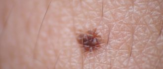
Photo 1. The color of a mole tells about its character. A black mole itself is not dangerous, but if it changes color, you should consult a doctor. Source: Flickr (Anastasia Catherine).
Causes of moles
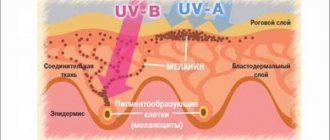
Most moles form in childhood and adolescence. Heredity and genetic predisposition play a key role. In adulthood, the appearance of new formations is facilitated by external and endogenous factors. The most important is hyperinsolation - regular and prolonged exposure of the skin to ultraviolet radiation.
Ultraviolet light is the main catalyst for the formation of melanin pigment in skin cells, and it is melanin that determines the color of the entire skin and its elements. The appearance of a black mole may be a consequence of too frequent and long exposure to the sun. Fans of artificial tanning are at risk, since solarium lamps are a source of intense ultraviolet radiation.
Changes in hormonal balance can contribute to the appearance of new nevi. For women, a risk factor is ovarian dysfunction (imbalance of sex hormones). Changes in hormonal levels can be triggered by taking oral contraceptives and hormone replacement therapy during menopause. The formation of black moles is possible during pregnancy and lactation.
When black moles become dangerous
Melanomas tend to grow rapidly. Typically, the blackened structure of a nevus, becoming a malignant neoplasm, is characterized by lumpyness, with the presence of inclusions of different shades. In addition, the nevus may turn black, red or gray. To prevent moles from degenerating into malignant melanomas, if there are multiple tumors on the body, you need to regularly examine yourself for changes in spots, and also undergo regular examinations with a dermatologist.
It is not always possible to talk about the malignant nature of a nevus. If its size is less than 4 mm, there is a smooth and even homogeneous structure, the correct shape, most likely there is no need to fear a serious illness.
The emergence of new tumors
If a black mole appears on the body, differing in color, size, structure from other neoplasms, it is recommended to consult a doctor as soon as possible. The dermatologist will write out a referral for testing, after which we can talk about the nature of the mole.
Darkening of old nevi
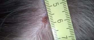
A dangerous sign may be the fact that the mole has turned black in a short period of time. Depending on what processes occur in the body, external changes in the skin may also be observed. If the old nevus has suddenly darkened, it is necessary to undergo a medical examination.
Dark spots in children
If a mole on a child’s body has darkened, the possible causes of the pathology are:
- hereditary factor. If relatives have a large number of moles on the body, it is likely that the child has inherited this feature;
- failure of the mother's hormonal system during pregnancy;
- adverse effects of environmental factors;
- the presence of infectious diseases in a mother who is breastfeeding her baby;
- disruption of the pigmentation process in the baby’s body.
Excess pigmentation can manifest itself as dark brown spots on the child’s body, as well as red, gray, and purple.
Change in color of hanging nevi
Changes in the color and surface structure of a hanging mole can occur under the influence of external and internal factors. For example, weather changes and heat can provoke excessive sweating, as a result of which the hanging nevus can change color. Mechanical damage to a mole or rubbing it with tight clothing can also cause a change in color. If these factors are absent, and the hanging nevus suddenly darkens, this can be a dangerous sign; you should consult a doctor as soon as possible.
Dangerous symptoms
Only a very few black moles pose a potential threat. The risk of malignant transformation to melanoma is low, but caution should be maintained. In any case, it is advisable to show the skin lesion to a specialist. You definitely need to see a dermatologist or oncologist if you have the following symptoms:
- The black mole increases in size. Even small growth should be alarming; a rapid increase in size is an unfavorable sign.
- The surface has changed (becomes “glossy” or loose).
- The formation peels off or bleeds.
- Unpleasant sensations appear in the form of pain, burning or itching.
- The shape has changed, the edge has become striated, uneven, asymmetrical.
- The color changes: an ordinary mole becomes black, bluish or dark brown.
Any dynamics should be alarming. There is no need to panic, but you need to make an appointment with a specialist. What you definitely cannot do is try to remove the tumor yourself using traditional medicine. Injury by mechanical or chemical influence is one of the main causes of malignant degeneration.
What is the treatment?
Subsequent therapy is based on the diagnosis revealed by the results of examinations:
- Degeneration of nevus into melanoma. In this case, a traditional operation is performed under local anesthesia to remove the formation. After this, the tumor tissues are sent for histological analysis to assess their malignancy. The disadvantage of this method is that it does not prevent recurrence of such cases. But still, today this is the surest way to get rid of malignant tumors.
- Nevus does not pose a health hazard. The patient will be offered recommendations to prevent moles from degenerating into melanomas. A harmless darkened nevus can be removed using a number of hardware methods - cryodestruction (exposure to liquid nitrogen), radioknife (removal using high-frequency electromagnetic waves), electrodestruction (removal of a nevus using electric current), laser therapy methods.
Melanoma-dangerous nevi
Listed below are specific types of black moles that are melanoma-dangerous. This means that when exposed to unfavorable factors, they can degenerate into melanoma; the risk of malignant transformation is higher than in the case of ordinary nevi.
Dysplastic nevi. This type of nevi is characterized by the presence of melanocytic dysplasia, an atypical arrangement of melanocytes. Only a dermatologist can diagnose melanocyte dysplasia. The patient needs to know that a dysplastic nevus has a smooth surface. It does not rise above the skin, or only its central part rises. The shape is irregular, with uneven edges. The coloring is uneven, with black areas located in the center.
Blue nevus. Despite the name, the formation can be not only blue, but also black. Usually has the shape of a regular hemisphere and rises above the surface of the skin. The surface is smooth, the edges are even. Typical localization is the scalp, feet and hands, and gluteal region. The risk of malignancy increases after injury, including independent attempts at removal.
Nevus Ota. This type of tumor appears only on the face. Characteristic mainly for representatives of the Mongoloid race. The color is black or bluish. The differential sign is the presence of pigmentation of the sclera, iris or conjunctiva of the eye.
Borderline pigmented nevus. The neoplasm is formed in childhood. Subsequently, birthmarks increase in size throughout life, reaching one and a half to two centimeters in diameter. The differential feature is an uneven ring-shaped coloration, with a decrease in color intensity from the center to the periphery. The color is brown, darker in the central part.
Giant pigmented nevus. Refers to congenital. Increasing in size, such birthmarks reach gigantic sizes, up to 15 centimeters or more. A characteristic feature is an uneven surface with “potholes”, nodules and cracks. Hair growth from the nevus may occur.
For melanoma-dangerous moles, the risk of malignancy is higher than for ordinary skin tumors. Such nevi and birthmarks require observation by a dermatologist or oncologist. Malignant degeneration can be caused by exposure to external factors - mechanical or chemical damage, ultraviolet irradiation.
Indications for removal of black nevus
Not all black moles should be removed. Benign formations are removed if they interfere, are located in an inconvenient place and rub with clothing. If the nevus has acquired a red, gray, black color, or dots of the corresponding shade have appeared on it, then it must be removed. This also applies to vague, rough growths that hurt and itch.
It is imperative to remove moles that:
- More than 1 cm.
- They grow quickly.
- They have a smooth and shiny outline, different from the skin pattern.
- They change shape and become asymmetrical.
- They become deformed.
- Cause pain and itching.
- They peel off and have bumps and dots on their surface.
- Bleeding.
Diagnostics
If you have a black mole on your body, it is not at all necessary to remove it, but you need to contact a specialist. The dermatologist will conduct a diagnosis, which consists of examining the neoplasm and studying it using optical or digital dermatoscopy (under magnification).
During the examination, the doctor evaluates the size of the tumor, its density, structure, consistency, surface character, symmetry, edge, color and other signs. As a rule, examination and dermatoscopy are sufficient to make a diagnosis. Biopsy and histological examination are carried out only before removal. In other situations, dermatologists prefer not to injure black moles unnecessarily.
What to do if a mole has darkened?
Let us remind you once again that the first thing you need to do is contact a medical clinic, a qualified specialist. But darkening of a mole in itself is not a sufficient reason for making a diagnosis.
The patient will be asked to undergo several diagnostic procedures. First, a visual inspection by a specialist. Then - testing (skiascopy, dermatoscopy). If the clinical picture is controversial, then an additional biopsy is prescribed.
Treatment: removal or observation?
Benign neoplasms of the skin do not require removal, but surgery can be performed for aesthetic reasons, as well as in situations where the nevus is located in an open area of the body, is exposed to ultraviolet radiation, or is injured by parts of clothing or jewelry.
Removal of benign black moles that are not melanoma-dangerous is carried out in the following ways:
- Surgical removal;
- cryodestruction;
- electrocoagulation;
- radio wave removal;
- laser removal.
Melanoma-dangerous nevi can only be removed surgically. The operation is performed in the presence of an oncologist and involves healthy skin. The removed tissues are necessarily sent for histological examination. Melanoma-dangerous nevi can also be removed in aesthetic medicine clinics if the medical institution has a dermatologist oncologist on staff. The operation is performed in a classical way; cryodestruction and other modern technologies are not used.
If malignant degeneration of the neoplasm is diagnosed, complex antitumor therapy is indicated, which includes surgical removal with wide coverage of adjacent tissues, radiation and chemotherapy.
Consequences of mole removal
Removal of a black mole does not always occur without consequences and complications. Sometimes they can be caused by the structure of the skin or improperly performed surgery.
The consequences may be as follows:
- The appearance of a tubercle at the site of the mole, which indicates the formation of melanoma.
- A compaction that may indicate pathology.
- The appearance of swelling, scars, scars, light spots.
- Relapses and formation of metastases.
- Increase in temperature and development of infection.
To avoid such complications, it is necessary to strictly follow the doctor’s recommendations, do not wet the site of the nevus after surgery and use sunscreen when in the sun. Any decorative and medicinal cosmetics can be used a week after surgery.
After the procedure
After removing a nevus, it is extremely important to protect the skin from ultraviolet radiation. You can’t sunbathe in a solarium; you should be in the sun as little as possible. It is recommended to refrain from thermal procedures. It is impossible to remove crusts at the site of the surgical wound; the skin should be provided with maximum rest. After removing a mole on the face, it is not advisable to use decorative cosmetics.
You can learn more about the diagnosis of skin tumors and methods of their treatment at a consultation with a dermatologist at the Galaktika clinic (Moscow).
Use of pharmaceuticals
Sometimes pharmaceutical preparations help get rid of birthmarks, including:
- Stefalin is an ointment that helps get rid of moles, warts and other skin imperfections. The drug contains natural ingredients that remove pigment and prevent the development and growth of moles.
- Malavit - can be produced in the form of a gel, ointment or lotion. It effectively helps get rid of nevi.
- Cryopharma - sold in an aerosol that is applied to the skin. After this, the mole freezes, dies and falls off. The therapeutic course is 10 days.
All of these drugs are sold without a prescription, but their use should be discussed with your doctor in advance.
Preparation for dermatoscopy of moles, disadvantages and advantages of the method
The analysis does not require special preparation from the patient, the study is absolutely painless.
The only relative disadvantage is that dermatoscopy subjectively evaluates the appearance of blackheads.
The result largely depends on the experience of the medical expert.
However, a modern dermatoscope with the ability to connect to a computer allows you to share the results with other doctors.
This increases the likelihood of correct diagnosis and accurate diagnosis.
Special computer programs are being developed that should facilitate the differentiation of benign and dangerous lesions of the epidermis.
Dermatoscopy is a simple, non-invasive and widely available diagnostic method.
Does not expose the patient to harmful radiation and provides valuable information for the evaluation of skin lesions of unknown origin.
However, the most informative method for diagnosing moles/nevi remains histopathology (histology).
Histopathological examination represents the gold standard for diagnosis in dermatology.
Dermatoscopy is an additional tool that allows you to assess the condition of the epidermis.
It should be taken into account that the effectiveness and information content of this method also depends on the specialist - the diagnostic accuracy of dermatoscopy correlates with the doctor’s experience.
When to see a doctor?
You should see a doctor if you notice any of the signs of malignant degeneration. Consultation with a doctor is necessary for any changes in the tumor, including changes in pigmentation, size, structure and pain, especially throbbing, “tearing” pain when pressed.
You should contact an oncologist if there is bleeding from a mole or there is purulent discharge, as well as if the skin of the back darkens without exposure to ultraviolet rays. You need to make an appointment with a doctor for any suspicious phenomena in a nevus on the back in order to diagnose the most aggressive oncology in a timely manner.
Birthmarks in history
In the Middle Ages, people were unable to explain the reasons for the appearance of moles on the body. Therefore, they did not come up with anything better than to declare harmless spots as devilish marks. The fair sex suffered the most from this. They were forced to carefully hide such marks on their bodies. It’s good that the clothes of that time contributed to this.
In the event that a mole on a beauty’s stomach or thigh was discovered by an ill-wisher, all that was necessary to physically eliminate the victim was to report the mark to the inquisitorial tribunal. Fighters against heresy and witchcraft quickly received all the necessary recognition. Although even they were not particularly needed in order to send the woman marked by the devil to the stake.
At the beginning of the 18th century, the concept changed dramatically. At this time, a legend was spreading in the highest circles of the French nobility that said that the face of the goddess Venus was decorated with a mole. It was believed that a woman with a similar spot above her lip or on her cheek was a passionate person. All the beauties, deprived of natural moles, began to use artificial ones. They were made of velvet, black leather and taffeta. Then they glued it to the face, shoulders and chest.
In 18th-century Europe, artificial moles were considered more than just decoration or makeup items. It was a way of flirting and communicating. A mole placed above the lip indicated that the woman was free. If a fly was glued to the right cheek, it was a sign that the beauty was not free.
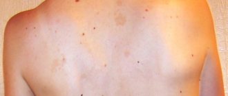
Many moles on the back
Some people who have a lot of moles on their backs immediately begin to panic, giving this phenomenon a dangerous meaning. In fact, if birthmarks appear throughout life, they do not pose any threat. But if within one to three months a person suddenly discovers an increased number of such spots, then this phenomenon should not be ignored.
You definitely need to see a doctor. A large number of moles can appear due to prolonged exposure to the sun. Sometimes the trigger can be a sudden change in climate.
In some cases, an increased number of nevi in the back area may indicate serious skin diseases, as well as malfunctions occurring with the human immune system. The phenomenon may signal malignant formations that have appeared in the tissues of internal organs.
Types of dark moles
Moles can be located on any part of the body and even on the mucous membrane. There are many varieties that are classified based on the nature of their origin. They vary in color, size and shape.
There are several types of pigment formations:
- Flat nevus. It is a collection of melanocytes in the upper layers of the epidermis. Most often, such moles are located on the stomach of women and men, hips and chest.
- Birthmark. This is a pigmented formation of light or dark color. Birthmarks are divided into three types: lentigo, coffee and Mongolian spots. Their number and size hardly change over time.
- Convex mole. Typically such formations have a rough surface. Color can be yellow, brown and black. Often a hair can grow from such a nevus. Convex moles most often form in intimate areas, on the palms and soles.
- Blue nevus. The formation has the appearance of a smooth and dense hemisphere with a light blue or rich blue color. Appears most often on the buttocks, face and limbs.
- Organoid or warty moles. The formations have a convex structure on a thin stalk. They resemble warts in appearance.