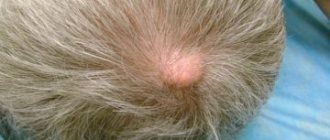
What is atheroma
Atheroma is a subcutaneous cyst, a capsule with a fat mass. It occurs at the site of the sebaceous gland due to blockage of its duct. In the medical literature, these formations are called “sebaceous gland cyst”, “trichodermal cyst”, “steatoma”, “follicular cyst”, “epidermal cyst”. They differ slightly in content, but are identical in symptoms and treatment methods.
Atheroma “breathes” through many small pores. Sometimes some of them enlarge, an opening is formed, through which, when pressed, a light mass with an unpleasant odor is released. It is impossible to squeeze out a closed atheroma. Persistent attempts will provoke inflammation.
Formation of inflammation in atheroma
Atheromas are usually single, but sometimes multiple ones occur - atheromatosis. Atheromatous formations occur in areas of the skin where hair follicles are present, so they do not occur on the head of the penis.
What it looks like:
- Single atheroma on the testicle – https://prntscr.com/wab04b.
- Multiple atheromas of the scrotal skin – https://prntscr.com/wab0ef, https://prntscr.com/wab0kl.
- Open atheroma – https://prntscr.com/wab0og.
- Atheroma under the skin of the penis - https://prntscr.com/wab0tk.
- Atheroma on the foreskin of the penis - https://prntscr.com/wab0z8.
What can be confused with. A large cyst looks like a lipoma, or wen. The latter are formed from subcutaneous fat, and not on the basis of the sebaceous gland. Lipoma does not have a capsule, only a bed. The type of tumor can be accurately determined by histological examination - studying extracted cells under a microscope. Large wen on the penis – https://prntscr.com/wab0z8.
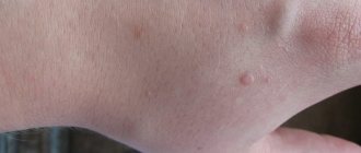
Small multiple atheromas on the penis are called Fordyce granules. This is the norm; there is no need to press or remove them. Fordyce granules - https://prntscr.com/wab1nd.
Why is it dangerous? Atheroma can fester. This happens when there is an impact, capsule rupture, infection, or rubbing with clothing. The cyst becomes liquid, hot, and painful. When localized under the skin of the scrotum, it can become a source of chronic infection, which in some cases penetrates the testicles.
Inflammation of the testicles is called orchitis - a detailed article on the treatment and prevention of orchitis.
Under no circumstances should you pierce a festering atheroma yourself. It can deepen, expand, and transform into a chronic ulcer. In some cases, the formation opens on its own and a fistula forms. If the outcome is favorable, a necrotic capsule emerges from the bed, and the wound soon heals.
- Festering atheroma - https://prntscr.com/wab2u6.
- Revealed inflamed atheroma - https://prntscr.com/wab31q.
Atheromas do not degenerate into cancer.
Postoperative period
As after any operation, there comes a postoperative period. After removing multiple atheromas on the scrotum, the doctor prescribes therapy with antibacterial and anti-inflammatory drugs. This will speed up the healing process.
If after removal a man notices a lump, then you can use local anti-inflammatory, antimicrobial and wound-healing drugs. Vishnevsky ointment helps very well. To do this, you need to apply a compress of gauze, well lubricated with ointment. In this way, you can clean the area damaged after surgery and prevent the development of an infectious process.
It is very important for men to monitor the health of their genital organs. Therefore, at the slightest suspicion of the development of atheromas, it is necessary to contact specialists for appropriate help, undergo a series of diagnostic measures and begin treatment. Remember: your health is only in your hands.
Benign formations of the male reproductive system include cavities that change the shape of the genital organs and cause superficial or internal deformation. The most common pathology of this type is a cyst in the scrotum, penis and epididymis. These formations can be combined with an infectious course. The appearance of cysts in most cases occurs in men aged 30-60 years.
Tumor and skin cysts are classified. The first include cavities that form inside the genital organs. They have an elastic structure, their walls do not extend outside the cover. A skin cyst on the penis or in another area is often accompanied by a violation of the upper skin layer, its walls are noticeably separated from the internal tissues.
Causes
Factors that provoke the formation of atheromas in the groin area:
- Damage to the structure of the hair follicle.
- Lack of personal hygiene, wearing thick underwear.
- Metabolic disorders, increased sweating and sebum production.
- Abnormal structure of the sebaceous glands and their ducts.
- Hormonal disorders.
- Exposure to cosmetics: powder, cream, deodorant.
We answer all questions:
Everything about the sebaceous glands on the penis: why they become inflamed and how to remove them.
Sometimes atheromas are hereditary. In such cases, consultation with an endocrinologist or dermatologist will be required.
Symptoms
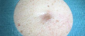
At an early stage, atheroma appears as a small ball under the skin. It gradually enlarges, becomes denser, can grow with connective tissue, and sometimes becomes calcified. The formation is painless, characterized by clear contours, mobility, and elastic structure. The diameter can be from 0.4 to 4 cm. The skin over the atheroma is of normal color. Sometimes it turns pink, and vascular veins—telangiectasia—become noticeable. Education does not cause a man any discomfort, except aesthetic.
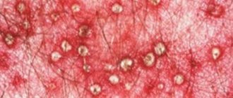
Secondary atheromas appear in individuals with increased secretion of sebaceous and sweat glands, with a tendency to form purulent acne. Such cysts are bluish in color, painful, and have a dense structure.
Dermatovenerologist Sergei Gennadievich Lenkin examines the clinical case of a young man with a dense formation on the penis
Possible complications
When a wen appears on the scrotum or genitals, you should not immediately sound the alarm and cause panic. This benign tumor is not at all dangerous and does not in any way affect the functioning of the male genitourinary system. However, you should definitely pay attention to a lipoma if the following symptoms appear:
- Pain syndrome. A benign neoplasm does not cause pain, and discomfort can only occur with strong pressure. But if the wen begins to bother you, then this may indicate the beginning of an inflammatory process developing in the epidermal tissues of the genital area.
- Intensive growth. The adipose tissue that forms the lipoma cannot immediately fill the capsule, which is the shell of the ball. If you suddenly notice that a lump on the testicles, which was previously diagnosed by a dermatologist as a lipoma, has begun to rapidly increase in size, then this should be a reason to consult a doctor as soon as possible. It is likely that the neoplasm tissue was exposed to certain negative factors, which resulted in the degeneration of the wen into a malignant tumor.
- Pain in the balls while moving. This symptom may indicate that the lipoma is located near the internal reproductive organs, constantly in contact with them while walking. Subsequently, this circumstance can provoke the development of testicular dystrophy or inflammation.
- Various dysfunctions of the reproductive system. The appearance of problems in the functioning of the organs of the genitourinary system indicates that the wen has grown to a critical size, due to which the urethra or organs responsible for potency, as well as providing blood flow in porous vessels, the spermatic cord and testicles, are pinched.
Each of these symptoms represents a developed complication caused by the presence of a lipoma in the scrotum. In this case, it is mandatory to undergo a full medical examination of the genital area and, if necessary, undergo surgery.
Diagnostics
Which doctor should I contact: a surgeon, dermatologist, dermatovenerologist. In most cases, it is useless to contact a urologist, since he will refer you to a surgeon.
The best diagnostic option is ultrasound. Signs of atheroma:
- Clear contours.
- Lack of blood flow.
- Homogeneous internal structure.
- The maximum depth of location under the skin is up to 10 mm.
Atheroma of the scrotum on ultrasound: a) – two formations of increased echogenicity, b) – a single formation with a pronounced capsule
The most accurate method for visualizing atheroma is MRI. The images allow you to examine its structure without contrast enhancement.
How to treat atheroma on the penis and scrotum
Can atheroma resolve on its own: it cannot. The contents will not escape from the capsule. As confirmation, we cite the words of a doctor from the site https://health.mail.ru/consultation/2877457/:
Atheroma is removed surgically. Indications:
- Infection.
- Multiple formations.
- Cosmetic defect.
- Large cyst size.
For large atheromas ranging in size from 10 to 40 mm, removal is most often performed by conventional enucleation. Electrocoagulation is used less frequently.
Surgical removal
Before surgery, hair is removed from the skin and the surgical area is treated with antiseptics. Then infiltration anesthesia is performed using several injections with an anesthetic. Two incisions are made along the edges of the atheroma and the skin is lifted. The most important thing is to carefully place rounded scissors under the capsule without damaging it, completely cut it and remove it from the bed. After disinfection, self-absorbing sutures are applied. If you leave a piece of the capsule, the cyst is likely to form again.
Scheme of surgical excision: 1 – skin; 2 – scissors; 3 – clamps; 4 – atheroma; 5 – excised skin flap; 6 – subcutaneous tissue
Step-by-step photos of the operation:
- Circular dissection of the skin around the atheroma - https://prntscr.com/wab691.
- Isolation of a cyst from the capsule - https://prntscr.com/wab6i7.
- Removed cyst and adjacent scrotal skin - https://prntscr.com/wab6oh.
Large atheroma on the scrotum - https://prntscr.com/wab6xr. After removal - https://prntscr.com/wab727.
The inflamed suppurating atheroma must first be opened, drained and relieved of inflammation, and only then the capsule must be peeled out. Otherwise, the risk of relapse and infectious complications increases. Inflamed atheroma on the scrotum - https://prntscr.com/wab7by. After removal - https://prntscr.com/wab7g2. It is enough to wash such a wound with hydrogen peroxide and apply a bandage. Healing will occur through natural tissue tension, no stitches are needed. An invisible scar 2-4 mm long will remain.
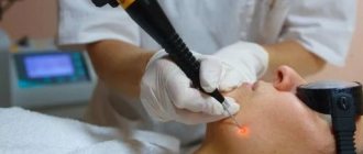
Small cysts can be removed with a laser or radio wave knife. Cryodestruction is practically not used due to increased trauma.
Removal scheme using a radio knife
The laser and radio knife bloodlessly cut through the tissue, instantly sealing the vessels and reducing the risk of infection. Such methods do not affect the testicles, since they are reliably protected by several membranes.
After removal, the formation, if necessary, is sent for histological examination, on the basis of which the final diagnosis will be established.
Rehabilitation after removal
If there is no suppuration, the patient is sent home the same day. In case of a purulent process, subsequent wound drainage and observation are necessary.
For 7-10 days after discharge, you need to treat the suture with antiseptics, take antibiotics prescribed by your doctor, and avoid physical activity. If necessary, the doctor will schedule 2-3 visits for follow-up examinations and dressing changes.
It may take 3-5 weeks for the sutures to completely dissolve, it all depends on the material used by the surgeon. During this time, a small nodule may be felt in the scrotum, and the ends of the threads are visible. They don't need to be pulled out. In case of minor inflammation, the suture should be treated with chlorhexidine 2-3 times a day and a thin layer of Levomekol should be applied. Fat-based ointments should not be used.
Levomekol has anti-inflammatory and antimicrobial effects. Price in pharmacies from 126 rubles
Traditional methods
Atheroma cannot be removed using traditional methods. Many are trying to squeeze it out. If there is a large pore, then the fat will come out of the capsule, as in the photo https://prntscr.com/wab8n6.
Such manipulation is dangerous due to suppuration. If the outcome is favorable, the atheroma will disappear, but after 2-3 months it will “grow” again, since the capsule remains in place and the fat will not exit through the glandular duct.
If the duct is recently blocked, you can try lotions with sinaflan, camphor oil, and ichthyol ointment. The lump may open or become smaller, but the expanded capsule will not go anywhere.
To delete or not to delete
As we said above, such a disease is not dangerous for the human body, but it is worth finding out the reasons for its occurrence.
The doctor may prescribe removal in the following cases:
- the neoplasm is continuously increasing in volume;
- is located in places of vascular connections and interferes with the full supply of blood to the male genital organs;
- nearby tissues are inflamed;
- sharp or aching pain when pressing on the sore area;
- unpleasant sensations, namely itching and burning;
- the neoplasm is filled with purulent discharge;
- an unaesthetic appearance that causes a number of complexes in men.
Before treating this disease, it is necessary to undergo a complete diagnosis, which includes instrumental and laboratory methods. The most common and effective type of therapy is surgery. If the cystic formation is small, then doctors may choose a wait-and-see approach. It’s just that sometimes the pathological process goes away on its own. If the doctor diagnoses a large tumor and it is growing progressively, then a surgical treatment method is necessary. There are four main types of operations.
Open surgery to remove atheroma
In this case, the patient is given local anesthesia. Doctors make an incision and remove everything that is under the skin pouch, and then remove the capsule itself. In this case, infection is possible. If doctors have the opportunity, they make a large incision and carefully remove the capsule itself. After this, the incision is sutured. The disadvantage of this procedure is the scar that may remain after the operation.
Laser removal
This technique is minimally invasive. Removal occurs due to the fact that the affected area of the skin is subject to thermal influence.
As a rule, laser removal is used when the tumor is small in size.
At the time of the operation, the skin is dissected using a laser, and the thermal effect begins to evaporate the contents of the skin pouch. As a result of such an operation, the doctor does not use any instruments other than a laser. This technique is excellent for patients who have difficulty clotting their blood. After such treatment, the patient has virtually no scars.
Removal using the Surgitron device
Removal occurs as a result of exposure to radio waves. This technique is considered the fastest and safest. Thanks to high-frequency radiation, the outer cover and internal capsule of the formations die off. During removal, the patient does not experience pain. After the procedure, there are completely no scars and the patient recovers quickly.
Prevention
Measures to prevent atheroma:
- Take proper care of your skin if it is prone to oiliness and acne. A dermatologist will give recommendations.
- Wash your intimate area with baby soap twice a day and wear comfortable underwear made from natural fabrics.
- Eat rationally, avoiding vitamin deficiency, excess sweets, and refined fats.
- Limit the consumption of alcohol, salty, spicy, canned foods to avoid disruption of metabolic processes.
Multiple atheromas on the scrotum are sometimes formed due to improper epilation or lack of proper skin care after it.
Treatment of atheromatosis
Self-medication for atheroma is unacceptable; at home it is difficult to remove residual cyst tissue after its breakthrough. Without medical intervention, there is a high probability of recurrence of the pathology and suppuration. Only surgical intervention guarantees a complete cure for atheroma. If suppuration occurs, urgent hospitalization is required.
- Classic removal. An incision is made in the capsule area with a scalpel. The cyst is enucleated, separated from nearby tissues and the wound is sutured. The stitches are removed after a while.
- Laser. Used for the removal of atheromas and spermatoceles as an effective and bloodless method. The cyst undergoes destruction, the contents evaporate. After surgery there is no scar.
- Radio waves. Used only for cutaneous cysts to burn out pathological tissue.
Treatment of cyst-atheroma is carried out under local anesthesia; the duration of the operation without complications does not exceed 30 minutes.
Treatment of testicular cysts with sclerotherapy occurs through filling the cavity with a special substance. It replaces the contents of the capsule and glues its walls. The introduction of a sclerosant is permissible in extreme cases; this type of operation can lead to infertility.