Almost every person has one or more moles on their body. As a rule, they do not cause discomfort and do not affect health in any way. But lately, more and more often, many people have begun to develop cancerous moles, which are harbingers of a terrible disease - skin cancer. Unfortunately, few people can distinguish an ordinary mole from a malignant one, which leads to the development of the disease. In this article we will look in detail at what cancerous moles look like, what their features are and how to get rid of them.
What is a malignant mole?
A malignant mole is a cancer called melanoma. It can form anywhere on the body, but most often appears in exposed areas, as they are exposed to ultraviolet radiation.
Melanoma is the most dangerous form of cancer. It is very important to monitor all moles on the body, especially if there are a lot of them. If a malignant mole is detected early, the development of melanoma can be prevented.
How quickly does malignancy occur?
The exact timing of degeneration of nevi cannot be established. Depends on the state of the immune system, the duration of influence of provoking factors of the external and internal environment. For some patients the process takes years, for others it takes several months. For some, it is enough to damage the growth once for malignancy to occur.
In medicine, there are two stages of malignancy: initiation and promotion. The first stage is triggered by certain factors: prolonged exposure to ultraviolet radiation, the sun, and regular trauma to the spot.
The structure of cells and their genetic apparatus changes. Oncogenes appear. At the second stage, atypical cellular elements begin to grow and divide. Uncontrolled proliferation ends in the formation of a mole of irregular shape and enormous size.
The development of malignant tumors occurs in four stages:
- The stage of malignancy occurs when a benign formation becomes malignant.
- The pre-invasive tumor stage is characterized by the growth of the tumor in situ.
- At the invasion stage, the tumor grows into surrounding tissues.
- The last stage is characterized by the entry of malignant cells into the bloodstream. They spread to internal organs with secondary tumor growth. Melanoma quickly metastasizes and causes the death of the patient within a few months.
Characteristic
To prevent the development of skin cancer, it is very important to know how to identify a cancerous mole. For comparison, consider the characteristics of ordinary moles and cancerous ones.
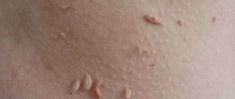
Common, harmless moles have a uniform color (brown or black) with a clear border that separates them from the rest of the body. Moles are round or oval in shape, their size is approximately 6 mm.
There can usually be from 10 to 45 moles on the human body. New ones can appear up to 40 years, and some, on the contrary, disappear with age.
Now let's talk about malignant moles. As a rule, there are a lot of them, and in appearance they are very different from ordinary ones in color, size, outline (more on this below). It happens that an ordinary mole can develop into a malignant one. In order not to miss this moment and start treatment on time, you need to undergo examination every six months or year.
Symptoms of degeneration
You can suspect that a mole is developing into cancer based on the symptoms:
- An increase in nevus is noted. Normally, the size of the spot should not exceed 2.5 cm. Birthmarks may not fall within normal boundaries. They are initially large in size. It is important to notice the increase in growth in time.
- Ulceration of the formation is observed. The appearance of an ulcer on a pigmented spot indicates the beginning of cancerous degeneration. The condition may be complicated by a bacterial infection.
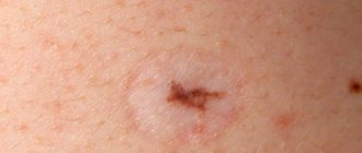
- Symmetrical, clear contours characterize a benign formation. The cancer appears asymmetrical with unclear, blurry boundaries. You can determine the characteristic by dividing the mole in half. Another option is to place the edge of a ruler in the middle of the spot.
- As the oncological process develops, the color of the growth changes. Becomes black.
- A change in the shape of the pigmented growth indicates the onset of a malignant process. A healthy mole should be oval or round.
- The smooth surface corresponds to the normal structure. Roughness and peeling are an unfavorable sign of malignant degeneration.
- Regular pathological discharge (blood, pus) indicates malignancy of the nevus.
- With cancer, bubbles with clear liquid may appear around the spot.
- The patient feels pain and itching at the location of the growth. Normally, it does not hurt or itch.
In dermatology, a simple rule is used to determine the signs of malignancy called ACCORD. Each letter of the word corresponds to a specific characteristic of a mole:
- A – asymmetry.
- K – edge (fuzzy, uneven in oncological pathology).
- K – bleeding.
- O – coloring.
- P – dimensions (more than 2.5 mm).
- D – dynamics. With malignant degeneration, the formation of brown crusts and cracks is observed.
Doctors recommend keeping an eye on large congenital birthmarks and newly appearing nevi. They occupy a leading position in malignant degeneration. Even a small growth can develop into melanoma if it is exposed to daily trauma. It is forbidden to pick out, squeeze out, or cut out pigmented skin elements. Damaged melanocytes respond with aggressive reproduction and growth. Only qualified diagnostics, correctly prescribed tests, and timely treatment will save the health and life of the patient.
Signs of malignant moles
Malignant moles (cancer cells) have some telltale signs that can help differentiate them from a typical mole. The initial stage of the disease - melanocytic dysplasia - is still treatable. Therefore, if a cancerous mole is identified and removed in time, the development of skin cancer can be avoided.
In 1985, dermatologists developed the abbreviation ABCDE, each letter of which represents one sign of a cancerous mole. Over time, this abbreviation was adapted into Russian, and it began to sound like AKORD (asymmetry, edges, color, size, dynamics). It is by these signs that a malignant growth can be identified. Let's take a closer look at each sign.
- Asymmetry. As mentioned above, ordinary moles are symmetrical. If you notice even the slightest asymmetry, you should urgently consult a doctor.
- The edges. Cancerous moles have jagged, blurry, and even jagged edges.
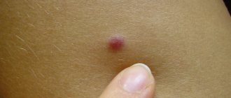
- Coloring. Common moles are usually one color (black or brown). Cancerous moles on the body can be of different shades, including red.
- Size. Ordinary moles do not exceed 6 mm in volume. If the mole is larger than 6 mm, then most likely it is malignant. In addition, cancerous moles quickly increase in size.
- Dynamics. If the mole is benign, then it does not change its color or size over the years. If you begin to notice changes, you need to consult a doctor for examination.
So we've looked at the characteristics and symptoms of a cancerous mole. If you notice at least one of these points, immediately run to the doctor to prevent the possible development of melanoma.
Risk factors
A person can live with moles all his life, and they will not bother him in any way. But there is always a risk that a standard neoplasm will begin to develop into a malignant one. Let's look at the most likely risk factors for a mole to become cancerous:
- Having severe sunburn or prolonged exposure to sunlight on common moles.
- People with white skin, light hair and eyes, and freckles are more likely than others to develop cancerous moles on their bodies.
- If there are a lot of ordinary moles on the body, then there is a very high risk that sooner or later they will begin to develop into malignant ones.
- Large sizes of standard moles. If an ordinary mole itself is large in size, then the risk of developing melanoma increases significantly.
- Hereditary factor. If your relatives have skin cancer, then you are also at risk.
To avoid the development of melanoma, it is important to take into account all these factors and, at the slightest suspicion that a mole is becoming malignant, go to the doctor.
How dangerous are moles and how not to miss their degeneration into cancer? Oncologist-surgeon tells
A mole on the face and body in different historical eras was a sign of beauty and piquancy. Modern medicine does not share the enthusiasm for them. Because pigment formations can even be of malignant origin. Which of your dozens of moles requires attention and when should you contact a specialist? Vera Grechanik, a 1st category oncologist surgeon, spoke about this
– Vera Nikolaevna, a person far from medicine, most often calls all formations on the skin moles. Please tell us what moles are and why they appear?
Vera Nikolaevna Grechanik, oncologist-surgeon. Photo from personal archive
– Moles (in medicine called nevi) are pigmented benign formations, an accumulation of pigment cells. There are congenital (they are not visible at the birth of a child, but become noticeable only under the influence of hormonal factors) and acquired (in response to any influence - solar radiation, mechanical friction, for example, clothing). As a person grows, both the moles themselves and their number grow. The appearance of new ones is often associated with changes in hormonal levels throughout life, their trauma, exposure to solar radiation on the skin, and decreased immunity.
– Which of them can develop into a malignant tumor?
– There are melanomatous moles and non-melanomatous types of moles. The following types of moles are most often predisposed to the development of melanoma: dysplastic, blue, giant congenital and borderline, i.e. there are only four main types. Remember that moles on the palms, soles and genitals are always borderline moles due to the anatomical features of the skin in these areas. They are subject to prophylactic surgical removal because these areas are most often chronically injured.
Among the serious oncological skin diseases there is cancer, and there is melanoma.
– What is melanoma?
Melanoma is a malignant skin tumor that develops from melanin-producing cells. Melanin is a pigment produced by melanocytes - skin cells that determine skin color and the possibility of tanning.
Melanoma is the queen of oncology, its behavior is unpredictable. It develops only in 30-40% of cases from moles.
Skin cancer is divided into two main subtypes: basal cell carcinoma and squamous cell carcinoma. Basalioma is treated with surgery or radiation therapy and is one of the most favorable types of tumor. It does not metastasize, but grows quite slowly (depending on the immune system). Squamous cell carcinoma in the first two stages is highly treatable; in the 3rd and 4th stages it is much more difficult to fight it, and it has a more aggressive course. It is often a disease of old age and is diagnosed in those over 70.
– Can melanoma be recognized independently?
– There is a FIGARO , it is convenient for self-diagnosis. Let's look into it. Form . Here we are talking about changing the shape of a mole. Let me give you an example: it was flat, but it became convex. And changing sizes, accelerating growth. Everything is clear and understandable. If a mole was there from birth and grew slowly along with the person, this is not accelerated growth, this is the norm. If it has never grown much, but in two weeks or three months it has doubled in size, this is the same accelerated growth. Borders . They were clear and clearly visible to the eye, but suddenly they became blurry and indistinct. And symmetry - one half of the mole became different from the other. Dimensions . Large moles are usually moles larger than 6 mm. Oh paint. Uneven - randomly located brown, gray, pink and white areas on the mole. Or discoloration of a mole. We also pay attention to this.
I also like the ABCDE , which is easy to remember.
A – asymmetry – asymmetry of formation. B – border – the appearance of an uneven edge. C – color – change in color, often darkening or cyanosis. D – diameter – change in diameter and dimensions. E - evolution - development.
Any of these changes may be a reason to consult a doctor.
– What methods of diagnosing moles exist?
– First of all, this is an examination by a specialist. If you want to know who to contact first, I would recommend consulting a dermatologist. The doctor will determine whether a consultation with an oncologist is necessary, whether dermatoscopy or mole removal is necessary.
Dermatoscopy is an examination of skin formations using a dermatoscope, a special device that allows you to “look” inside a mole without surgical intervention, evaluate its type, structure and determine whether it is benign and whether there is a need for its removal. I would like to note that the doctor’s experience is important when performing dermatoscopy.
A doctor examines a patient's mole. Photo www.illustre.ch
– What are the methods for self-control of moles?
– I usually advise patients the following way: take photographs, store them in an archive, monitor them and, if changes are noticeable, consult a doctor - a dermatologist, oncologist or therapist. Another option is to take a thin transparent sheet of paper, apply it to the mole, outline the contours and write the date and size of the mole. And so once every 3 months. The so-called map of moles will be at hand, and you can always determine how much they have changed.
– In what cases of the appearance of skin formations should a person urgently run to the doctor? Is it a warning sign if a mole gets bigger?
– It all depends on how many more moles a person has. The main thing is to make sure that they do not disturb or change dramatically. You need to run to the doctor if, for example, a mole is injured or a frequently injured mole in the sun is additionally burned. This often happens in men in the torso and arms, and in women on the legs. If a mole bothers you, hurts, itches, or causes discomfort, droplets of liquid seep out of it and a crust forms. If a mole has changed shape, color, or its boundaries have become unclear. Women are more susceptible to hormonal changes. During the period of feeding and childbirth, pregnancy, a new number of moles “sprinkles”. There is no need to panic, but it is better to visit a specialist doctor as planned.
– That is, Can injury lead to skin cancer?
– One-time – unlikely. Chronic - yes, it can. Every person in his entire life accidentally injures moles either with a razor, or with clothes, or simply scratches. Children love to “twist” moles from their parents. Long-term chronic injury over several years can lead to skin tumors.
– Is it necessary to remove moles and warts if they cause only aesthetic discomfort or is it better to avoid surgery without medical indications? People often prefer not to touch anything.
– Removal of skin lesions has two indications: medical and cosmetic. It can be removed for cosmetic purposes; even an ordinary cosmetologist can do this, but after consulting an oncologist. If the formation interferes, it is better to remove it. But if a person is comfortable with it, it doesn’t injure him in any way, outwardly everything suits him, and the doctor said that it’s not dangerous, then why remove it? You can't remove all moles.
– What methods of mole removal exist in modern medicine?
There are several of them. A surgical method in which the mole is removed with a scalpel. Laser removal method - the formation is removed with a laser like a scalpel or evaporated by a laser (if a mole - it is necessary like a scalpel, if the papilloma can be evaporated). The method of removal with liquid nitrogen is cryodestruction. Removal with an electrocoagulator - burning with electric current. Another method is radiosurgical removal: a special device acts on an area of skin with a high-frequency electromagnetic wave.
Each method has its pros and cons. There is a certain type of mole that can only be removed surgically. There is – surgical, and laser, and current, and nitrogen, and radiosurgical. The method of removal should be determined by an oncologist or dermatologist at the appointment.
I’ll tell you for sure that you can’t burn off moles at home using any homemade “folk” remedies.
– Are there proven risk factors or is the nature of skin cancer unknown?
– In general, all known risk factors for skin cancer can be divided into environmental factors and factors of the internal environment of the human body. For example, the geographic latitude of a person’s place of residence and the associated intensity of solar radiation. His phototype, heredity. Reproductive factors in women. Mole injuries. Chemical carcinogens, including hair dyes, ionizing radiation. The social status of a person and his profession, the fact that a person lives in a city or in a rural area, whether he works outdoors or whether a person spends most of his time indoors are important. It is important what medications are taken on an ongoing basis, viral infections in the past (rubella). Risk factors are the presence of diseases such as xeroderma pigmentosum, Dubreuil's melanosis, and a large number of melanoma-dangerous nevi.
Currently, exposure to the ultraviolet spectrum of solar radiation is one of the most important factors contributing to the occurrence of skin melanoma. For the occurrence of skin melanoma, what is much more important is not chronic damage to the skin by UV rays, as is typical for basal cell carcinoma and squamous cell carcinoma, but sharp and intense damage. Even a single exposure to ultraviolet light can lead to melanoma.
– Can skin cancer be cured with the advances of modern medicine?
– Basalioma can be completely cured. After 5 years, if there are no relapses, the person is removed from the register. People with melanoma and squamous cell carcinoma will never be eliminated, but a cure is possible.
– Is it true that if hair grows from a mole, then this is already a guarantee that the mole is harmless?
– No, hair growth is not a guarantee, but this is an indicator that everything is still fine with the mole. Plucking hair is strictly prohibited. It’s better to carefully cut them off, because when we pull them out, we injure the mole, it can get infected and form an abscess. And so over and over again... This is chronic traumatization.
At the same time, I advise removing a mole with hair surgically, and not with a laser, because otherwise the hair will continue to grow. It is difficult to completely excise such moles under adequate laser control.
– Many women and men do hair removal. Isn't this dangerous for those who have many moles on their arms and legs? For people with a large number of moles, which type is better to choose - hot wax hair removal, laser hair removal, or other than shaving?
– During laser hair removal, moles must be protected. This is a fairly aggressive targeted effect on a small area of the body, and there is a risk of developing skin diseases. If we are talking about waxing, sugaring, moles must be covered in advance with a band-aid with a soft base, so that when the band-aid is peeled off, it will not be injured. If we talk about laser hair removal, you need to use various cosmetics that cover moles (white cosmetic pencils, for example) and not treat moles that cannot be covered. Hair on moles cannot be removed, and professionals should be aware of this. If there are a lot of flat moles, I still recommend razors.
I often get asked questions: is it possible to do laser hair removal before going to the sea? Will laser radiation affect the skin, and will there be burns at sea? This is asked by those who don’t want to use sunscreen all the time while on vacation. If the laser allows you to individually select parameters for the phototype, hair type, and its characteristics, then the effect on the skin will be minimal. But! When on vacation, it is still necessary to use sunscreen cosmetics under any conditions. Regardless of whether there was laser hair removal the day before or not.
– What about all kinds of chemical peels on the face? Is this dangerous for moles?
– If this is done by a person who has a lot of moles on his face, then you should be careful. Using aggressive cosmetics, you need to avoid all these moles. Even superficial damage can lead to changes in its structure. If the face is clean in terms of moles and there are only small pigments, then you can do peeling. And it is best if this is done by a qualified specialist. If there are moles on the face, we should only consult a doctor.
– Aren’t peeling masks sold in regular stores dangerous?
– It all depends on the concentration of chemicals. It is not dangerous if, when used independently, large and prominent moles are not affected.
– Is it forbidden for fair people to travel to the south?
– No, it’s not prohibited. You just have to follow tanning etiquette. They are not allowed to be in the sun from 11 to 16. They are not allowed to sunbathe in the open sun. You need to lie under an umbrella, under protection, because you will still get a tan. There is no need to avoid the sea and sun. Everything just needs to be done correctly.
By the way, office workers are susceptible to this “disease”: once a year they prefer to spend two or three weeks at sea, catch as much sun as possible and come home with dark chocolates. Imagine what a blow to the skin this is!
But you need to sunbathe wisely, slowly, using sunscreens with a high protection factor.
– Can taking certain medications affect the condition of moles?
- Complex issue. Taking too many medications can cause more moles to develop. Long-term use of hormonal contraceptives by women increases their risk of developing skin tumors. The possibility of skin melanomas occurring in patients due to long-term treatment with the drug, which is used in the treatment of Parkinson's disease, has been noted. This is due to the specific metabolism of the drug in the body.
– Is a solarium more dangerous than the sun?
– If the solarium has high-quality lamps and you use the right approach, then no. But if a person comes to the procedure for the first time and just for 10 minutes, then this is bad.
But we won’t say “no” to the solarium. It is ideal to start with 1-1.30 minutes and go there not every day, but at least every other day.
There is such a myth - to prepare your skin for the sea in a solarium. If you start doing this, then a month or two or three before the rest, as I said above. If you get a quick and intense tan in a solarium in a short time, this is not beneficial, it is harmful. The tan will be superficial and will not fulfill its function of protecting the deep layers of the skin from damage by ultraviolet radiation, but rather, on the contrary, will cause additional damage to the skin. After all, tanning is a protective reaction, it is a mechanism that does not allow ultraviolet radiation to penetrate deeply and damage the skin more and more.
– How should people who have already had skin cancer protect their skin?
– They should protect their skin from the sun as much as possible, just like other people. You should also add a wide-brimmed hat to your wardrobe and use sunglasses with high-quality filters.
– Does vitiligo affect the occurrence of skin cancer?
– The absence of pigment in the area of vitiligo does not exclude the risk of developing melanoma, unfortunately. I recommend consulting a dermatologist to limit the spread of lesions as much as possible. Because, in principle, this disease itself can bring its own discomfort in a person’s life.
– Are there ways to prevent skin cancer? How to protect yourself?
– Do not injure moles. Remove dangerous ones in a timely manner. Visit specialists. Monitor yourself for newly emerging formations. Clearly know the FIGARO in order to control. Be careful with the sun. It is important to examine yourself from head to toe: scalp, feet, finger spaces, genital mucous membranes. Yes, there are moles there too. And we can’t forget about the ears!
– Vera Nikolaevna, what do you recommend to our readers?
– In medicine, the most important direction is prevention. It is very important that people take care of their health at a stage when they do not yet need medical intervention for treatment.
How is the examination carried out?
To diagnose cancerous moles, a dermatoscopy must first be performed. Using a magnifying glass and a dermatoscope, you can examine the signs of melanoma on the surface of the growth. This involves studying and evaluating the pigment of the skin and blood vessels by taking a sample of the growing mole.
The diagnosis is confirmed after a biopsy (histological analysis). Using local anesthesia, part of the mole is removed in order to carefully study its structure in the laboratory. This method is one of the most accurate.
It is possible to diagnose cancer at an early stage using a computer microdermoscopy system, but this method is not yet very widespread.
Most importantly, if you yourself notice even the slightest change in the appearance or size of your moles, you need to consult a doctor. The doctor will choose the necessary diagnostic method, and with timely examination, the risk of developing skin cancer is reduced.
Some facts you need to know about cancerous moles
If a person has more than 50 moles on his body, then he needs to monitor their condition very carefully and contact an oncologist at the slightest change.
In addition to the above signs, there are several factors that you should pay attention to:
- Darkening. A common mole may be black in color. But if it was originally brown and suddenly began to darken, then this is a cause for concern. Many people do not pay attention to the darkening of moles, since black is considered the norm.
- Inflammation. If the skin around the most common mole is inflamed or redness has formed, then you should urgently go to the doctor for examination. And under no circumstances should you treat inflamed areas of the skin with alcohol, this can only worsen the situation.
- Surface. The boundaries of the mole have already been discussed. But you should also pay attention to its surface. It should be smooth on top, without obvious roughness. If there are any, then this is a sign of the development of melanoma.
- If darkened areas of skin appear around an ordinary mole, then this is a big cause for concern. It is urgent to check with an oncologist.
As you can see, there are a lot of signs of melanoma development. It's very difficult to remember them all. Remember that any change in a standard mole may indicate that it is becoming malignant.
Treatment
Currently, the only possible treatment option for melanoma is the removal of cancerous moles. The complexity of the operation depends on the severity of the situation and the size of the formation. For small growths, half an hour will be enough.
When removing a cancerous mole, the surgeon cuts out a small area of skin (1 cm) around the mole to prevent new ones from appearing in the same place. The larger the malignant mole in volume and size, the more skin around it needs to be removed.
After the mole is excised, a sample is sent to the laboratory. There they study its prevalence level, that is, the likelihood that new such growths will appear on the body.
Treatment with folk remedies and removal
What to do if a malignant tumor is detected? Treatment of moles of this type in most cases is carried out in one way - removal. You can get rid of the growth using surgical excision or laser. The second method is used in the absence of contraindications.
Removal:
- At the initial stage of the disease (in the absence of metastases), a small area of healthy dermis, nearby lymph nodes, and part of the muscle tissue are removed along with the mole.
- In the presence of metastases, the removal of malignant skin lesions is carried out in a similar way, but after it additional treatment methods are used - chemotherapy, the use of interferons or other analogues of the immune system. Radiation irradiation is used if surgery is refused, but the result is not always positive.
The choice of treatment method for a malignant disease remains with the medical professional. He selects the most suitable option based on the patient’s condition.
Therapy with folk remedies includes the internal use of various decoctions and infusions, as well as the use of external agents.
Folk remedies:
- Dry crushed plantain leaves are poured with boiling water until a paste is obtained. Used for compresses.
- A gauze napkin is moistened with golden mustache juice and applied to the sore spot.
- Lubricate the affected areas with fresh celandine juice.
- Three times a day you need to drink thirty drops of tincture of lemongrass and eleutherococcus.
- Drink aloe juice three times a day, one teaspoon at a time.
It is worth remembering that the use of traditional medicine should be accompanied by caution. Doctors do not prohibit the use of unconventional methods for the treatment of malignant nevi. But precisely because of this, a person often misses time for therapy that will really help.
What forecasts do doctors give?
Tumor thickness is the main criterion by which oncologists make predictions. If the mole was small, then the risk of its reoccurrence is small, and the chance of life without melanoma increases.
The rehabilitation period after removal of the growth is short. A scar forms at the site of the removed mole, which heals quite quickly. The size of the scar depends on the method of removal.
Laser removal is the safest method, which leaves almost no marks or scars. But this method cannot be used in advanced cases.
It should be noted that if the operation is performed in a timely manner, then the risk of melanoma developing in the future is very small. In the future, you just need to be regularly monitored by an oncologist to avoid relapse.
When is the best time to remove a mole on the back?
It is important to know that the “fly” itself is a potential spreader of melanocytes to an area of non-pigmented skin. This may be a prerequisite for the formation of a malignant disease. But not all birthmarks on the back can be cancerous. To identify potentially “dangerous” tumors, first of all, it is necessary to consult an oncologist, who, after examination and testing, will determine whether the tumor on the back needs to be removed and will recommend an excision method.
It is better to remove a mole on the back that is constantly in contact with clothing and is injured, as well as when the first signs of cancer of a nevus are detected.
Removal can be done using the following methods:
- Radio wave method.
- Application of laser surgery (laser beam).
- Cryodestruction. Application of liquid nitrogen.
- Surgery. It is worth considering that after this procedure there will be a small scar at the site of the mole.