Purple blue spots on the skin, what are they, what medications to treat
Human skin is an external protection against the effects of environmental factors. In case of any malfunctions in the body, symptoms immediately appear on the skin indicating their presence. Usually pimples, spots, and roughness appear on the skin. Each of them indicates a particular disease in the body.
In this article we will talk about the phenomenon of purple or blue spots on the body.
Types of blue spots on the skin
Depending on whether these spots have been on the body for a long time or have just formed on the skin, they can be divided into several subgroups:
- Those that were present at birth (congenital),
- Arise immediately after birth (acquired),
- Resulting from damage to blood vessels (vascular),
- Arising from a disorder of skin pigmentation (pigmentation).
Vascular spots - causes and treatment
Their colors vary from red to purple (blue), from brown to yellow. The color of the formations directly depends on the thickening of the blood and its concentration, in what state and where this concentrated area is located: in a vessel or the blood has already entered the tissue or is still localized in the bloodstream.
- associated with one-time vascular dilatation,
- hemangiomas are formations consisting of a bunch of tiny capillaries,
- hemorrhagic - formed as a result of capillary breakage and blood splashing into the tissue, contributing to the appearance of a bluish spot.
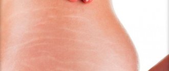
A purple or blue mesh on the epidermis indicates that these are ordinary hematomas, colloquially called “bruises”; when a person hurts himself, a purple-bluish spot forms at the site of mechanical damage.
If we talk about effective folk remedies, then in this case, bodyaga will help. The ointment should be applied directly to the site of the bruise and rubbed in with light movements two to three times a day, and the bodyagu should be used as a lotion until the bruise completely disappears. This type of stain that appears on the human body is one of the safest for health.
Which doctor should I contact if blue spots appear?
In all other cases of this kind of spots, it is necessary to urgently consult with doctors who will tell you how and how to treat them:
- Dermatologist,
- Dermato-venereologist,
- Allergist,
- Hematologist,
- infectious disease specialist
- Surgeon
- Oncologist,
who, after diagnostics and research, will make the correct diagnosis, find out the cause and origin of these formations, and also prescribe adequate therapy aimed at eliminating the root cause.
Red spots may be the result of an allergic reaction to unnatural fabrics, contaminated water, aggressive detergents, or food. In this case, the allergist may prescribe antihistamines upon visual inspection or after testing.
If the spots do not disappear, but rather grow or bleed, you should immediately consult a doctor for advice.
The etiology of spots of this color can be very different; it can be a sign of genetic diseases, as well as pathological changes in the body.
Diseases that cause purple spots
There are currently many reasons that contribute to the formation of purple spots on the body in the body. There are quite a few diseases in which one of the pronounced visible symptoms is the formation of purple spots on a person’s arms, legs or face. Some diseases are noted:
If its appearance is considered a symptom of any of the listed diseases, then treatment will be aimed specifically at them, and in parallel, you should start taking vitamin complexes containing vitamins PP, P, C.
Flaming nevus
- Firstly, such spots, if they are on the face or hands, that is, on parts of the body visible to prying eyes, cause great discomfort and inconvenience to their owner.
- It is necessary to know and remember that under no circumstances should you expose them to injury, much less try to remove them yourself, outside the hospital, since any neoplasm can degenerate into a malignant one.
Kaposi's sarcoma
Treatment of the disease is usually expressed in the elimination of clinical manifestations and alleviation of the patient’s condition.
Other reasons
The list of these diseases is far from complete, since purple (blue) spots may indicate other changes in the human body.
Therefore, for successful treatment, it is necessary to undergo diagnostics in the clinic and fulfill all the doctor’s requirements.
What diseases are accompanied by spots resembling bruises on the legs
If hematomas form on the skin with constant frequency, you should seek medical help. An experienced doctor identifies the cause of the ailment by the manifestations of other indirect signs. The table clearly shows what the indicated changes can signal.
Immune system disorders
Develops as a complication of infection, drug allergies, chronic autoimmune diseases. The immune system, coping with such pathologies on its own, produces certain antibodies. When they interact with antigens, they form complexes that circulate through the bloodstream. With vasculitis, they settle on the walls of blood vessels and provoke the development of the inflammatory process. It leads to increased penetration of the vascular wall and loss of its elasticity. As a result, hemorrhages occur in the soft tissues. If a similar process develops in the layers of the skin, spots of a characteristic color appear on the body
Bruises may appear on the abdomen, limbs, and back. With vasculitis, the pigmentation is focal: several small hematomas can be seen in one place. Over time, they merge into one large spot. There are more of them on the arms than on the legs, but they can also appear on the face and feet. Bruises are located symmetrically. With hemorrhagic vasculitis, the lower extremities swell, the patient may complain of weakness and a slight increase in body temperature
Congenital or over the years acquired disorder of the hemostasis process
Bleeding disorders
Genetic pathologies that lead to the formation of defects in blood vessels, a decrease in platelets in the blood and disruption of coagulation processes
Connective tissue pathologies, osteogenesis imperfecta, elastic pseudoxanthoma
Purpura as a complication after long-term use of steroids
Side effect of taking medications
Vitamin P deficiency
Vitamin C deficiency
Vitamin K deficiency
Complications of bacterial, viral and fungal infectious processes
Menopause in women, taking hormonal contraceptives
Estrogen levels decrease, resulting in increased blood viscosity
Large light hematomas that disappear on their own after three days. Memory deteriorates, concentration decreases, insomnia and causeless dizziness torment
Bruises can appear on the body due to hypertension and chronic tonsillitis. They can be indirect symptoms of the development of rheumatism and kidney disease. It is impossible to determine the cause of a skin defect on your own. Only a doctor can make an accurate diagnosis and eliminate the provocateur factor. The causes of blue spots are determined based on clinical signs. Differential diagnosis helps to separate bruises received during a bruise from hematomas that have arisen on the body as a result of any disease.
Treatment of neoplasms
Red vascular spots on the legs, face, and head can be quickly and easily removed using hardware treatment methods. The bottom line is that blood is allowed to bypass the diseased vessel, due to which a new network of capillaries is formed. The main removal methods include the following:
The removal procedure is painless; the patient may only feel a slight burning sensation on the area of skin with the nevus. It usually takes 4 to 5 sessions to remove an average spot. One procedure lasts on average 15 minutes. After laser coagulation, the child does not have any scars.
Red blood cells, which are located inside damaged vessels, absorb laser energy. This causes the vessels to burn and seal. The surrounding tissues remain intact.
- Sclerosis - the method involves injecting a medicinal substance into the affected area, after which gluing occurs and the affected vessel is removed from the general bloodstream system. Laser sclerotherapy takes from 5 to 30 minutes. The procedure is performed under local anesthesia. Sometimes after the procedure darkening of the blood vessels is possible, which disappears on its own after some time.
- Photocoagulation is an effective method that helps in the early stages of the development of vascular spots. The laser beam acts on the affected vessels, warms them up and glues them together.
As an alternative, cryotherapy is possible - freezing the spot with liquid nitrogen. If the vascular neoplasm has a large focal point, then doctors prescribe treatment with corticosteroid drugs.
Latest information: What are the contraindications for varicose veins on the legs?
Today, laser therapy is used in almost all cases. It allows you to quickly and painlessly remove even stains from a large lesion. No traditional methods will help in the treatment of vascular spots on the body.
Laser removal is prescribed after consultation with a pediatrician and other pediatric doctors. Doctors usually recommend removing hemangiomas that are located on open areas of the skin and cause a cosmetic defect (spots on the face) or tumors located in areas with a high degree of trauma.
General principles of treatment
Photo from the site irecommend.ru
Treatment depends on the cause of the spots on the legs. If it is a hemangioma, then it is better to remove it. This can be cryodestruction, removal by electric current, laser or excision with a scalpel.
If the cause of spots on the legs that look like bruises , then it is necessary to treat not only the skin defects, but also the disease itself. You will need local and systemic anti-inflammatory drugs, for example, Diclofenac or Ibuprofen. You also cannot do without venotonics and angioprotectors. These are Troxevasin, Troxerutin, Phlebodia.
To get rid of red spots on your legs, you need to follow a cholesterol-free diet and wear compression stockings or socks. If large blood clots form in the vessels, surgery may be required.
Minimally invasive procedures are used to eliminate spots and bruises caused by varicose veins. For example, sclerotherapy. A sclerosing substance is injected into the diseased vein, which glues it from the inside and prevents further progression of the disease.
Differential diagnosis
In most cases, with skin rashes, differential diagnosis is performed to make a final diagnosis.
Let's look at a comparison of the most common types of skin changes and their symptoms:
| Disease | Features of the rash | Symptoms (itching, peeling) |
| Allergic reactions | Several spots appear on the legs and sometimes on other parts of the body. They can rise above the surface of the skin and merge. | Patients complain of severe itching, possible runny nose, dry cough, and increased lacrimation. |
| One or more red spots appear on the leg. Gradually, bubbles with liquid contents form on them. After opening them, a weeping surface forms on the body, which turns into small red crusts. | There may be slight itching and pain when the wounds become wet. | |
| Mycosis (fungal infection) | Changes with a clear contour are most often located on the foot. | There is severe itching and peeling. |
| Hemangioma | It resembles a small red mole, but when the skin is stretched it increases in size. | Not accompanied by any subjective symptoms. |
| Diabetes | A defect on the leg most often occurs at the site of an abrasion or wound that does not heal for a long time. | There is itching and peeling. The patient complains of severe thirst and a tendency to pustular diseases. |
| Appear on the extensor surfaces of the limbs. | The rash itches and is very flaky. | |
| Photodermatosis | Several different spots form on the legs and other parts of the body, which rise above the level of healthy tissue. The rash may develop into blisters containing cloudy fluid. | Occurs in the warm season and is accompanied by slight itching. |
| Hemosiderosis | Red-brown formations appear on the skin, which gradually move from the legs to the torso. | There are no symptoms. |
| A rash with blurred borders; there may be a blister on top. | There is no itching or peeling, but there is severe pain, which is relieved by contact with cold water or an ice compress. | |
| Lichen planus | Hyperemic defects with a characteristic indentation in the center protrude above the skin level. The rash is most often localized on the legs and feet, has a purple tint and can merge into large lesions. | Itching and peeling are present. Rashes form not only on the legs, but also on the mucous membranes, abdomen, and elbows. |
| Pale red changes with blurred boundaries. They form on the legs, feet, palms and other parts of the body. About 10 new rashes appear per day, which gradually acquire a bright red color. | The patient is not worried about anything. |
Differential diagnosis allows you to minimize the risk of making an erroneous diagnosis and prescribing the wrong treatment.
Types of stains
By a spot, people mean a local change in the color and (sometimes) structure of the skin in a certain location. Doctors use the word “spot” to refer to a small area of discoloration (red, brown, black or whiter than the surrounding skin), which does not rise above the level of the rest of the skin. Elements that protrude above the surrounding skin, are flaky, have a different density from other tissues, and have a cavity inside filled with light water or pus, have completely different names. This helps doctors narrow down the “circle of suspected” diseases.
In order not to confuse the reader, in this publication we will refer to all elements of the rash as a “spot”. Let's take as a basis the classification according to which spots are vascular or pigmented.
Vascular spots
These are formations of red, purple, brown, bluish or yellow color. Their color depends on the state of the blood in a given location: in which vessel (blood or lymphatic) it is located, whether it has come out of it into the tissue or continues to be included in the bloodstream.
Vascular spots are divided into:
- those associated with temporary dilation of the vessel (for example, with allergies, infectious diseases, burns - chemical, thermal or ultraviolet);
- associated with constant expansion of the vessel: spider veins, hemangiomas;
- hemorrhagic spots - resulting from the release of blood from a capillary
Dark spots
These are spots that have developed as a result of increased or decreased levels of melanin (that brown pigment that is found in skin cells and makes our tan possible). If there is a lot of pigment, moles, chloasma, freckles, and lentigo appear. When there is not enough of it, whitish areas appear on the skin: vitiligo, leucoderma.
There is one more type of stain that occurs as a result of the artificial introduction of dye into the skin (tattoo, tattoo), but this is not a worrying problem, and we will not consider it.
Pathologies that provoke the appearance of spots on the legs
There are a lot of diseases that cause spotting of the lower extremities. These are skin and systemic diseases, infections, disruptions in the endocrine and reproductive systems. Also, a similar phenomenon is caused by disturbances in the functioning of the liver, gall bladder, and kidneys. But there are pathologies for which the appearance of spots is a mandatory symptom and one of the main manifestations.
Phlebeurysm. A very common disease. It is diagnosed in approximately half of the world's population. It occurs due to incompetent valves in the vessels and impaired blood flow. The main reason for weak vein tone is genetics. But in order for varicose veins to develop, predisposing factors are needed. These include a sedentary lifestyle, heavy physical activity, excess weight and wearing high-heeled shoes. The disease begins with swelling, heaviness in the legs, and slight pain. Then spider veins appear. After some time, the veins begin to swell and dark, hard spots appear on the legs. As the disease progresses, the pigmentation becomes black. In some places, the skin turns white and sinks, weeping, inflamed areas and trophic ulcers appear. At the initial stage, external agents are used. Severely damaged veins are treated with special injections or laser
For prevention, it is important to walk more, do gymnastics and wear high heels as little as possible. Eczema. Dermatological disease caused by allergies, metabolic disorders, malfunctions of the nervous or endocrine system
Often becomes chronic with periodic relapses. Typically presents as redness and flaking of the skin, itching and swelling. A type of disease that is localized in the lower extremities is varicose eczema. Occurs due to poor circulation and blood stagnation. The skin on the legs darkens unevenly, red spots and irritation appear, and ulcers do not heal. Manifestations are usually concentrated in the lower part of the leg. For treatment, corticosteroids, sedatives and antihistamines are prescribed. For local treatment, lotions, ointments and gels are used. If an infection occurs, antibiotics are indicated. Varicose veins require mandatory treatment of dilated veins. Purpura. Minor hemorrhages in the skin due to the release of red blood cells from the vessels. It is a symptom, not an independent disease. This phenomenon is typical for impaired platelet production, blood stagnation, coagulation problems, intoxication, and pathology of the vascular wall. The rashes appear as dark spots on the legs, like bruises. At first they are red or burgundy, then gradually darken to purple, and when they resolve, they acquire a yellow tint. At the same time, convulsions, fever, headaches and abdominal pain, and arrhythmia may occur. Therapy is reduced to reducing the production of antiplatelet bodies. Patients are prescribed hemostatic agents and aminocaproic acid. If there is a risk of bleeding into the brain, platelet infusions are given. In severe cases, excision of the spleen is indicated. Lichen. Fungal skin infection. Usually characterized by pink or red rashes, but with pityriasis versicolor. Most often they are concentrated in the upper torso, but sometimes they spread to the legs. Pigmentation changes shades to yellow and pink, the affected areas merge with each other, forming extensive lesions. At the same time, the skin peels and itches. Often chronic with relapses in spring and summer. For treatment, local antifungal agents are used. In severe cases, antimycotic drugs are prescribed for oral administration.
If a dark spot appears on your leg, you cannot ignore it. Periodically occurring defects indicate malfunctions in the body. Unusual pigmentation can signal a problem in any organ. Therefore, when it appears, you should visit a dermatologist. He will identify skin diseases or refer you to other specialists. If it turns out that there are no serious pathologies, then you can cope with the problem with the help of bleaching agents and cosmetic procedures.
Any types of spots that appear on the skin require special attention. If necessary, timely elimination of the cause that caused them should be started.
Age factor: children and elderly
Large purple spots on the skin of older people quickly form even with small impacts, since the walls of blood vessels become irreversibly thin after 50 years. Another defect is formed due to liver pathology, although the color of the spots is often brown rather than purple.
Thrombocytopenia also affects pensioners - this condition is called senile purpura, in which minor hemorrhages are diagnosed even without receiving a blow or the influence of other mechanical influences.
Anxiety grows when a rash is detected in a child, but pediatricians attribute most disorders to temporary defects. Rashes often appear due to a previous illness (cold), and the formations themselves always have a pale tint.
When a bright purple rash occurs, there is a risk of developing purpura. The most dangerous condition is meningococcemia, which develops due to sepsis. Children infected with the infection often die.
A bruise-like rash is also found with nevus flammosa (its congenital form). Damage appears in the womb, so they are immediately detected by obstetricians after birth.
The damaged structure of the baby's epithelium is not restored as it grows up, and sometimes it deteriorates even more - it turns darker. Conditions for deterioration: injury, excitement, infection.
Harmless blue spots on the baby's body appear after birth. They are located in the sacral area, sometimes on the butt, since excess melanin accumulates in a small body. The medical name is Mongolian spot, which is also diagnosed in representatives of other races. The skin defect disappears without a trace by the age of 2 years.
Spider veins are a normal variant in the elderly. The older the body, the less tone the circulatory system has.
Conclusion
Wine rashes often indicate serious health problems. Immediate contact with doctors is indicated when purple spots suddenly appear on the legs: there is an increased risk of blood clot formation.
Doctors warn! Shocking statistics - it has been established that more than 74% of skin diseases are a sign of parasite infection (Accarida, Giardia, Toxocara). Worms cause enormous harm to the body, and the first to suffer is our immune system, which should protect the body from various diseases. The head of the Institute of Parasitology shared the secret of how to quickly get rid of them and cleanse your skin, it turns out that’s enough. Read more .
If you take preventive measures in time (smear your feet with Troxevasin, avoid a sedentary lifestyle, avoid injuries), then dangerous consequences can be effectively prevented.
«>
Vascular spots causes and treatment
Their colors vary from red to purple (blue), from brown to yellow. The color of the formations directly depends on the thickening of the blood and its concentration, in what state and where this concentrated area is located: in a vessel or the blood has already entered the tissue or is still localized in the bloodstream.
Latest information: How to cure varicocele in adolescents and methods of treating the disease
Vascular spots are divided into:
- associated with one-time vascular dilation;
- hemangiomas are formations consisting of a bundle of tiny capillaries;
- hemorrhagic - formed as a result of capillary breakage and blood splashing into the tissue, contributing to the appearance of a bluish spot.
A purple or blue mesh on the epidermis indicates that these are ordinary hematomas, colloquially called “bruises”; when a person hurts himself, a purple-bluish spot forms at the site of mechanical damage.
Most often, they go away after some time without any special treatment, leaving not a trace on the body. If the bruise is painful or swollen, you can use any ointment that has absorbable properties, for example: Troxevasin.
If we talk about effective folk remedies, then in this case, bodyaga will help. The ointment should be applied directly to the site of the bruise and rubbed in with light movements two to three times a day, and the bodyagu should be used as a lotion until the bruise completely disappears. This type of stain that appears on the human body is one of the safest for health.
Features of treatment
Any hematoma can be treated at home. If the bruise has just formed, you need to apply cold to it. It will stimulate vasoconstriction, reduce the severity of blood flow, and relieve swelling. Such treatment will be effective only if no more than an hour and a half has passed since the formation of the blue spot.
Important! When using ice, avoid direct contact with the skin and wrap it in a light cloth or handkerchief.
When the swelling of the tissues around the bruise subsides, dry heat should be applied to the stain (a bag of heated salt, a boiled egg, a piece of gauze folded in several layers, heated with a hot iron). Warm compresses made from a decoction of plantain or chamomile help to resolve hematomas. You need to warm up the bruises at least four times a day. The duration of one session is 15 minutes.
Red spots on the body
Red spots may appear on the body for several reasons:
Common allergies, stress, incorrect dietary habits, serious diseases of internal organs, hormonal disorders.
A disease such as scleroderma is very often missed. It can be limited and systemic. When systemic, not only the skin is affected, but also internal organs. Psoriasis is very unpleasant and almost incurable. Red spots with white scales are very itchy and require scratching, leaving scars. Such spots appear when the immune system malfunctions, and a war begins between leukocytes and skin cells. >No one yet knows the causes of psoriasis. The beginning of the process can be caused by severe stress, allergic substances, frequent changes of place of residence or sudden climate change, and careless self-medication. Psoriasis may not itch, but eczema (an allergic disease) is always accompanied by a terribly itchy rash or blisters. This disease can appear after childbirth in women or in cases of gastrointestinal dysfunction.
The causes of allergic spots are: cosmetics, household dust, polluted air, indoor and plant allergens, food products, medications, etc. Not only does itching appear here, but also difficulty breathing, which can lead to death. You can't hesitate in this case. Immediate hospitalization and timely assistance will help save a person’s life. Slightly flaky, oval, pinkish spots that appear on the shoulders, back and thighs indicate pityriasis rosea. They don't always itch or itch. May be the result of a cold or weakened immune system. This is a seasonal disease and most often occurs in spring or autumn. Infectious diseases such as measles, chicken pox, rubella, and scarlet fever are also characterized by the appearance of red spots. If signs of one of these diseases appear, the victim must be isolated, and quarantine is established in the team where the patient is identified. To avoid complications, you need to adhere to the regimen prescribed by the doctor and the quarantine time.
Red rash in children
Spots on a child’s legs most often appear due to:
- Allergies: to new laundry detergent, to diapers or diaper products, new bath products, after an insect bite, in response to cold or sudden alternation of heat and cold. In this case, the spots rise above the skin and almost always itch, which disrupts sleep and nutrition. Following a hypoallergenic diet, taking sorbents (Atoxil, Smecta, Activated Carbon) and antihistamines (Fenistil, Suprastin, Erius) helps.
- Enterovirus infection. In this case, the rash is most often located not only on the legs, but also on the arms, mainly on the fingers. It looks like blisters surrounded by a red border, almost always very itchy. In this case, there may be an increase in temperature, similar blistering rashes on the oral mucosa, there may be nausea, vomiting, cough, loose stools (the set of symptoms depends on the type of virus acquired through food, water, or airborne droplets).
- Rubella. In this case, red spots are located all over the body, not just on the legs. At the same time, the temperature increased, nasal congestion appeared, and the eyes turned red. The rash does not itch or flake off.
- Corey. Here, red spots on the legs appear last: first, the rash appears on the face, and then goes down, merging with each other. In addition, the temperature rises, snot begins to flow from the nose, conjunctivitis and swelling of the eyelids are noted.
- Scarlet fever. This is a disease that is caused by the same bacterium that causes tonsillitis. Red spots, when pressed on, they appear better, appear on the 1st-3rd day of the disease, are located on the cheeks, on the sides of the body, and not on the legs, but in the groin.
- Meningococcal infection. This is a life-threatening condition that can begin with the fact that after 1-2 days of a runny nose (or even without it), spots appear on the legs and buttocks, the temperature rises, and the spots spread to other parts of the body. At first they may be red, without pustules or blisters. Then they darken, acquire a star-shaped shape, and merge. Dark spots on the legs that do not disappear when a clear glass is pressed on the skin may appear immediately, without a stage of red rashes. The sooner an ambulance is called, the greater the child’s chances of survival.
- Pityriasis rosea. Its manifestations are described above.
- Pityriasis versicolor. There are several similar spots, they are brown-red, cover different parts of the body, do not merge, and the skin above them peels off. The shape of the spots is different, they tend to merge, do not itch or hurt. When the rash disappears, whitish areas remain in these areas.
Children may develop spots due to ringworm and other mycoses, but these causes are more rare than those described above.
What are dangerous conditions?
Excessive rashes on the back, as well as other parts of the body, are a sign of a complex disorder in the body. During this period, they refuse to visit the bathhouse and sunbathe.
Diseases with a risk of acquiring disability:
- Thrombocytopenic purpura. The disease is dangerous due to the risk of internal hemorrhages, including in the brain. Laboratory confirmation of purpura is an extremely low number of platelets in the blood. Vascular dysfunction is often inherited, but sometimes occurs spontaneously.
- Capillary toxicosis. A sharply manifested symptom is the result of the entry into the body of an allergen (usually a food product) to which the person is intolerant. It often develops in children as a hypertrophied reaction to sore throat.
- Venous dilatation of veins (from stage 3). Although the defect appears in many patients in old age, the greatest risk is in those who have a complication. Advanced varicose veins lead to the appearance of dark areas of the dermis, which sometimes turn black. In this case, immediately consult a doctor: a change in shade is a symptom of necrotization. Appears in the arm area in 5% of cases.
- Kaposi's sarcoma. A malignant process that spreads chaotically throughout a person’s integument. The main danger is the rapid degeneration of small “marks” into 5-centimeter (in diameter) nodes.
A small red rash, especially in the legs, is sometimes the initial stage of thrombocytopenia, but more often the phenomenon occurs due to excessive exercise.
Intensive scratching of the skin with nails can also cause pinpoint but harmless changes. If vascular tone is disturbed, then formations - red moles - can remain on the body for a long time.
Facial erythema causes redness of the epithelium, and its complicated form causes a purple tint to the damaged dermis. Sharp but temporary changes are also possible with the development of drug photosensitivity in the chronic stage.
Hemorrhagic spots
Pigment spots - due to the fact that they are brown - are similar to hemorrhages that occur with vascular diseases, with pathology of the coagulation system:
- Thrombocytopenic purpura. Here the rash is not only located on the legs, it occurs spontaneously, after sleep, it is asymmetrical, both dark spots and purple and yellowish-green spots are visible (like the phases of a bruise).
- Hemorrhagic vasculitis. Here, brown spots appear immediately on the legs, symmetrically on both. The shape of the rashes is different, they tend to merge. The disease also affects the joints, the stomach may hurt, and the functioning of the kidneys and brain may be impaired. This pathology requires urgent medical correction. Read more about the symptoms and treatment of vasculitis.
Dark spots
Dark pigmentation is disorders such as melasma or melanosis, blue-gray dispigmentation.
Melanosis is promoted by any long-term and severe chronic disease. This causes the deposition of melanin in the skin. Common pathologies are:
- Endocrine melanosis, which occurs with insufficiency of the adrenal cortex, dysfunction of the gonads, diabetes mellitus, thyrotoxicosis, etc.
- Hepatic melanosis, developing against the background of impaired liver function.
- Cachectic melanosis in tuberculosis.
- Uremic - occurs in chronic renal failure.
Becker's melanosis
Or it is also called Becker's nevus. Often occurs in boys aged 10 to 15 years. Men and women are rarely affected by them.
The reasons for the appearance of nevus are still not clear. There are suggestions that this may be due to a hereditary predisposition to this type of pigmentation or the body’s reaction to ultraviolet radiation.
Dubreuil's melanosis
It appears as a flat, dark-colored spot, possibly slightly raised above the skin. The size on average reaches 5 cm, but after a few years it grows to 10 cm.
The color varies from light brown to dark and sometimes black. This melanosis is considered a precancerous condition. Often accompanied by papillomas and nodular elements.
The damaged areas are dense, with peeling and erosion. The skin around such a formation reacts with the appearance of redness, freckles, and foci of keratosis, which are indicators of the degeneration of melanosis into melanoma (a malignant tumor).
The causes of Dubreuil's melanosis are:
- age;
- abuse of ultraviolet radiation;
- skin sensitivity to light;
- skin injury;
- overdrying of the skin.
Latest information: Apple cider vinegar for swelling
Urticaria pigmentosa
A common disease in children. Hives look like red-pink spots that develop into blisters with liquid. The spots are very itchy.
And after opening the blisters, brown-brown marks remain on the skin. Urticaria occurs more often in children. As a rule, the spots disappear during puberty.
If an adult falls ill with urticaria, the situation is complicated by the appearance of systemic mastocytosis, which often leads to disability or death.
The causes of urticaria pigmentosa are still being studied. Presumably, provoking factors are:
- immune system response;
- stress;
- climate change;
- inflammation and infections;
- genetic predisposition.
Coffee-colored spots - Nevus Spilus
The spots are never hairy and have dark or black dots on the surface. Presumably the reason for the formation is heredity.
Freckles
These are small dark spots on the face or body. Pigmentation becomes more noticeable in the warm season with solar activity. Freckles may disappear as you age.
It is more common in people with light hair, eyes, and skin. Scientists have proven the dependence of the appearance of freckles on hereditary predisposition.
Recklinghausen's disease
The spots look like clusters of freckles, appear in unusual places and take on a café-au-lait hue.
Such formations can appear from birth or in childhood. The color varies, but brown shades predominate.
Rarely does the spot acquire a gray-blue color. Formations appear on the surface of the arms, legs or torso in an amount of at least 5 pieces. The patient is affected by nerofibroma, which subsequently spreads to other organs - nervous tissue, adrenal glands, etc.
Such spots degenerate into cancer from 3 to 15% of cases. The nervous system and musculoskeletal system suffer. Epilepsy, depression, fatigue occurs, vertebrae are destroyed, cysts appear, etc.
The mutating gene of chromosome 17, which is inherited, is to blame for the appearance of this disease.
Nevus Ota and Ito
Ota manifests itself as a single spot of black-bluish or dark blue color in the eye area, upper jaw or cheek. Sometimes you can find merging spots. Even less commonly, pigmentation can be bilateral.
Scientists have not yet determined the cause of nevus of Ota. Often this disease affects people of the Mongoloid race and very rarely Europeans and the Negroid race.
Nevus of Ito is similar to nevus of Ota. The only difference is in its location - neck, chest and shoulder blades.
Dark spots
As mentioned above, this is a rash associated with the brown pigment melanin, which is normally found in our skin, but is distributed evenly throughout it. The word “pigmentation” refers to spots due to excess melanin. This:
- Moles are brown or black spots located anywhere on the body, from which in some cases hair grows;
- Melasma: large dark spots that are more common in pregnant women on the linea alba and on the face, but can accompany some internal diseases and are located on the legs;
- Freckles: multiple small brown spots on the legs of various shades of brown, usually located on areas accessible to light on people with fair skin;
- Lentigo - dark brown and black spots on the legs, appearing either before 10 years of age, or in old people on open areas of the body. If such a rash element itches or peels, this indicates a malignant process;
- Secondary hyperpigmentation. They occur at the site of epilation and depilation, after removal of various formations with laser or electrocoagulation - if after the procedure, the legs felt ultraviolet rays without sunscreen in the first month. They may occur due to a deficiency of certain vitamins, such as ascorbic acid, retinol, nicotinic acid.
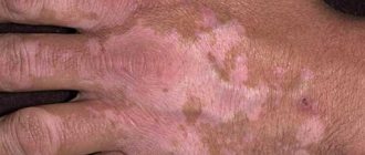
Pigmented areas also include depigmented, that is, light areas. This:
- Vitiligo: white spots of various shapes and sizes that arise as a response to frequent stress and chronic diseases of the abdominal organs (especially the liver). The following effect is observed on them: the white spots seem to be outlined in a color darker than the surrounding skin, which gradually merges with the normal skin.
- Leukoderma: This is the appearance of light patches of skin as a result of a systemic disease. If white spots without clear boundaries appear on the torso and legs, this may indicate syphilis.
Origin of purple spots on the skin
Most often, a bruise on the knee or other part of the leg occurs after a blow. Sometimes one awkward movement is enough for a dark blue spot to appear on the skin the next day. Applying cold immediately after an impact reduces the magnitude of the possible consequences.
A bruise is internal bleeding. Essentially, small vessels rupture and require time to recover. The size of the bruise depends on a number of factors: the force of the blow, the strength of the blood vessels, the elasticity of the upper layers of the dermis, etc. Particularly delicate individuals can develop a spot on the skin even after a slight pinch.
Where do hematomas come from without a reason:
1. Genetic diseases of the circulatory system. There are a number of diseases that lead to a lack of platelets: hemophilia, thrombocytopenia, von Willebrand disease. Problems with platelets affect the rate of healing of wounds and injuries. A small blow is enough to leave a large bruise on your leg for a long time. It is also impossible to exclude the onset of varicose veins, which in most cases is transmitted through the female line from mothers to daughters.
2. Hypovitaminosis. Vitamin C strengthens vascular walls, vitamin K monitors blood clotting, vitamin P regulates normal blood flow, preventing the formation of vascular plaques. A lack of any of these vitamins can lead to internal bleeding, including bruising. Even if you have persistent dark circles under your eyes, you should start taking multivitamins.
3. Lack of important microelements (calcium, cobalt, selenium). Like vitamins, some microelements are responsible for the elasticity and strength of blood vessels.
4. Malfunction of the liver. What is this vital organ responsible for? Cleansing the body? Not only! The liver regulates blood clotting, so any disturbances in its functioning lead to internal and subcutaneous hemorrhages and hematomas. Those at risk include those who suffer from hepatitis or cirrhosis of the liver.
Peripheral arterial disease
Peripheral artery disease (PAD) is a narrowing of arteries outside the core of the body. It restricts blood flow to some external parts of the body, including the limbs.
PAD often affects the legs, and people may experience symptoms such as pain, cramping, tingling and weakness. Decreased blood flow can also cause the legs and feet to gradually turn blue or appear purple. However, some people with PAD may have no symptoms.
Anyone with symptoms of PAD should see a doctor. Without treatment, this disease can progress and lead to serious complications such as severe ischemia and gangrene.
In very severe cases, the doctor may need to amputate part of the leg or foot. PAD also increases the risk of heart disease, heart attack and stroke.
Lifestyle changes, medications, and surgery can slow or stop the progression of the disease and reduce the risk of complications. Lifestyle benefits include regular exercise, stopping tobacco smoking and eating a healthy diet.
Prevention
To prevent red spots on the calves, under the knees, and in the shin area from appearing again, it is necessary to strengthen the walls of the blood vessels. It is their increased fragility and permeability that leads to bruising. To strengthen it, you need to take venotonics (Troxevasin, Detralex), follow a diet, and maintain normal physical activity.
To reduce the likelihood of dark spots appearing on your legs, you must follow these recommendations:
- refuse to take a hot bath, but a contrast shower, on the contrary, is useful;
- reduce time spent in the sun;
- do not visit baths and steam rooms;
- avoid injury;
- do not lift heavy objects;
- give up sports involving static loads on the legs, for example, weightlifting, tennis;
- wear compression stockings, do not wear things that squeeze your legs, hips, or waist;
- lead an active lifestyle, swimming is especially beneficial;
- choose comfortable shoes without heels;
- monitor your weight; excess body weight is an additional burden on the venous system.
After a hard day of work, if you feel tired in your legs, you need to lie down for 30 minutes, but keep your limbs elevated. This promotes blood flow and reduces the likelihood of congestion.
Spots on the legs most often indicate problems with blood vessels. You cannot get rid of stains on your own, since such therapy is ineffective. The patient will eliminate the consequences, but not the cause - the underlying disease that caused the pigmentation. At this time, the disease will progress, and even more severe complications are possible. Therefore, if spots appear, you should immediately consult a doctor.
Author: Oksana Belokur, doctor, especially for xVarikoz.ru