Epidemiology
The wide variety of clinical manifestations of epidermophytosis, the duration of the course, the frequency of relapses, the presence of erased forms, latent infection and carriage are the most significant epidemiological factors. The abundance of fungi in the falling scales, their high viability and germination on a variety of objects contaminated with organic substances contributes to a wider spread of epidermophytosis.
The source of infection is a sick person and a carrier of epidermophytons. Transmission occurs through direct contact, as well as through various infected things: underwear, shoes, sports items. Baths, swimming pools, swimming schools and showers in large cities are often places where athlete's foot is infected. Floors, utensils, benches and especially wastewater from baths, without sufficiently systematic and effective disinfection, are most significant in the spread of this disease.
FAQ
The described nail symptoms are very similar to onochomycosis. It is the same?
In some cases, athlete's foot nails can be caused by candida. And, as you correctly noted, nail candidiasis occurs - onychomycosis.
I can’t figure out how to properly decoction or infusion and what to make for hand baths. Are you itching and cracking a lot?
Be sure to consult a dermatologist and find out the diagnosis! To prevent infection and suppuration, and at the same time get rid of itching, buy dried chamomile flowers and a string at the pharmacy. Pour 2 tablespoons of sushi into a liter jar. Pour a liter of boiling water over everything and leave it covered, wrapped in a towel for half an hour. Strain the infusion, cool to a comfortable temperature. Dip your hands two to three times a day until you recover. After the procedure, do not wash your hands; dry them with a paper towel or napkin.
Causative agent of athlete's foot
Pathogens are divided into two main groups, differing from each other in the uniqueness of their cultural and microscopic characteristics, viability, parasitic activity and epidemiological characteristics.
A long-known and frequent causative agent of epidermophyton in the groin area, axillary folds and torso is Epidermophyton inguinale, which differs significantly from other epidermophytons and trichophytons.
The second group of epidermophytons consists of fungi with velvety-mealy crops; According to mycological characteristics, they are very close to the trichophytons of the gypseum group. The most common representative of this group of fungi is Epidermophyton Kaufmann-Wolf; its variants are much less common - Epidermophyton gypseum, Epidermophyton niveum. They cause a variety of lesions on smooth skin, primarily athlete's foot, interdigital spaces, smooth skin of the torso, nails, and also very often sensitization of the body and various allergic rashes, especially on the arms and torso.
• Epidermophyton inguinale
The colonies are round, mealy, at first dome-shaped, raised, smooth, later folded; in the center there is sometimes a small crater-shaped depression, at the bottom of which a small navel rises. Large folds are radial in nature; they are intersected by smaller ones; a mature culture takes on a cerebriform appearance due to the random arrangement of folds. The color of young colonies is yellowish-lemon, mature cultures become gray, almost always powdery, and small cracks are sometimes observed at the tops of some folds.
Along with these crops, there are velvety-mealy colonies, lumpy in the center, flat, yellowish or grayish along the periphery, as well as tall, lumpy, line-like colonies with a flat or finely folded, mealy periphery.
Pleomorphic changes are quite frequent; fluff often appears in several places at once; it is loose, tall, and can be easily and completely removed from the surface of the crop.
Under a microscope, young cultures have smooth, clearly septate mycelium; In mature cultures, large intercalary chlamydospores and sometimes chains of them are found.
In old, especially line-shaped parts of the colony, chlamydospores are exclusively polymorphic and large, up to 25-30 µm in diameter, granular, similar to those of Achorion Schonleini. Pear-shaped aleuria are not always found, located on the sides of the mycelium. At the ends of the mycelial branches there are very characteristic 4-5-cell spindles, measuring 20-35 x 6-8 µm, with larger cells located at the free, slightly rounded end.
The spindles are located on the mycelium singly or in groups of 4-7 elements, reminiscent of banana fruits in their arrangement. In old cultures, the shape of the spindles is dramatically changed, they break up into separate segments.
• Epidermophyton Kaufmann-Wolf
The colonies are rapidly growing, velvety-mealy, smooth, surrounded on the periphery by a narrow rim of gray young shoots. Mature crops are densely powdery, have a dome-shaped center, folded or lumpy, sometimes slightly recessed; often the colony is divided into large sectors by 4-6 radial grooves.
The mushroom is extremely polymorphic, has a number of variants - fluffy, gypsum-mealy, cerebriform - of various shades.
The color of the colonies is white, but sometimes yellowish, pinkish or brown crops are found. The edges of some crops are uneven and consist of tongue-shaped, velvety or mealy outgrowths that do not penetrate into the nutrient substrate.
Microscopically, a long, branched, septate mycelium is observed; Thin curls and spirals are quite common at the ends of the mycelium; plexuses in the form of nodular organs are observed along the course; Polymorphic niterkalar chlamydospores are also found; there are more of them in the older parts of the mushroom culture. Round aleuria, 2-3 microns in size, are visible in large numbers; they are located on the sides or at the ends of the mycelial filaments, often directly attached to its walls or on thin stalks. Aleuria is quite unstable, easily falls off, and is freely located in crops. The spindles of Epidermophyton Kaufmann-Wolf are quite distinctive; their length is 20-30 microns, width – 5-7 microns; They are divided into 5-6 cells by transverse partitions, the middle ones being the widest. In old cultures, spindles are rare, but chlamydospores are much more common.
Therapeutic measures
Treatment of athlete's foot is difficult even for specialists, because dermatophyte fungi are extremely tenacious. It is necessary to take into account all the nuances and approach the issue in a comprehensive manner. Moreover, it will take a long time (up to 1 year) and a lot of patience. Since the rapid onset develops into mycotic eczema, therapy is aimed at relieving inflammation and itching and tissue regeneration. Of particular importance is the treatment of athlete's foot, as the main source of infection.
Modern medicine offers effective methods of treatment, both with medications and physical therapy.
The following will help you achieve a quick recovery:
- Antifungal ointments and creams. Zalain cream has proven itself to be excellent. Its active ingredient, sertaconazole nitrate, has a wide spectrum of action. A 2% cream can deal with both mycosis and bacteria in a short time. It is enough to lubricate the affected areas twice a day with an even thin layer for 2-4 weeks, retreating 1 cm from the edge of the lesion. The medication has no contraindications and is suitable for nursing mothers, pregnant women and children. Interacts well with other medications. Affordable and available without a prescription.
Externally
- Zalain analogues: sertaconazole, terbinafine, fluconazole, miconazole, sebozole, fucis, bifosin, micoflucan, nizoral, ketoconazole, imidil, etc.
- Antiseptics (externally): miramistin, weak solution of potassium permanganate, furacillin.
- Regenerating: salicylic-zinc and sulfur-salicylic ointments.
- Hormonal: dexamethasone, prednisolone.
- Traditional medicine: infusions and decoctions of medicinal plants (baths, lotions, rubs).
Inside
- Antipruritics: loratadine, tavegil, diazolin, suprastin, pipolfen.
- Antibiotics: grisefulvin, cycloserine, natamycin.
- General strengthening: vitamin complexes + minerals, vitamins B, A, E., herbal teas.
Physiotherapy
- ultraviolet irradiation;
- electrophoresis;
- laser therapy.
Symptoms and clinical picture
Symptoms of epidermophytosis are very diverse, which is associated with the characteristics of the pathogen, the localization of lesions, the condition of the diseased organism, and its specific reactivity.
Athlete's foot is divided into the following three groups:
1) epidermophytosis of large folds with the predominant pathogen Epidermophyton inguinale;
2) athlete's foot and hands with the most common pathogen Epidermophyton Kaufmann-Wolf;
3) epidermophytis - allergic manifestations associated with antigenic irritation by various epidermophytons.
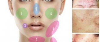
In case of epidermophytosis of large folds, the lesions are localized in the inguinal and femoro-genital folds, in the axillary areas and under the mammary glands of women, less often - on the trunk and on the scrotum. The disease begins with the appearance of red, slightly swollen, often itchy spots, covered along the periphery with small blisters and crusts. The lesions are sharply outlined, often merging into extensive garland-shaped rashes with a pinkish flaky center, with reddish edges, sometimes covered with crusts. In places of closest contact of the folds, the lesion is erythematous or intertriginous in nature. Complications such as eczematization and pyoderma with regional pyogenic lymphangitis are sometimes observed.
Athlete's feet and hands are divided into several forms:
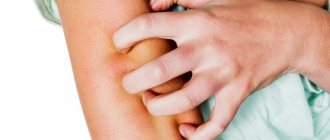
• Interdigital intertriginous athlete's foot is characterized by redness, maceration, erosion and cracks deep in the interdigital folds. Intertriginous epidermophytosis of the hands is rare, its course is basically similar to the previous one.
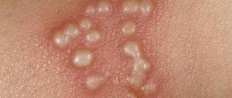
• Dyshydrosiform athlete's foot is characterized by the appearance of vesicles of various sizes from millet grain to pea, cherry and larger with clear or cloudy serous-purulent contents. The number of blisters varies - on the hands they are often multiple and small, on the legs they are larger with more pronounced inflammation. Bubbles are localized on the soles, skin of the fingers, along the edge of the foot, and less often on the heels. When the bubbles dry out, pinkish-reddish spots form in their place, surrounded by a “collar” of exfoliated epidermis, which are subsequently covered with large or fine lamellar peeling.
• Squamous and hyperkeratotic athlete's foot occurs most often on the soles and along the edge of the foot. It is characterized by pinkish spots at the sites of drying blisters, covered with layered peeling of varying thickness. Along with this, hyperkeratotic lesions occur in the form of extensive grayish-brown thickenings or diffuse calluses with cracks on the surface.
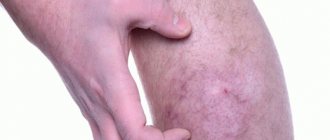
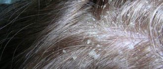
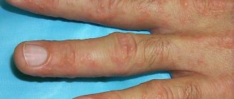
• Athlete's nails occur mainly on the feet - on the 1st and 5th toes, which are most often injured by shoes. Onychomycosis accompanies various forms of athlete's foot and is manifested by the deposition of horny masses of a yellowish tint under the edge of the nail, due to which the surface of the nail becomes spotted, striated, yellowish in color; the nail does not fit tightly to the bed; its loose masses are easily separated and sometimes crumble.
• The acute form of epidermophytosis is a rapid exacerbation of dishydrosiform or intertriginous epidermophytosis. It is accompanied by fever, headache, malaise, vesicular-bullous rashes on the soles, feet and fingers with hyperemia and swelling of the skin of the feet and legs, and an almost constant rash of epidermophytids.
The polymorphism of epidermophytosis is enhanced by the layering of some clinical forms onto others, infectious complications, and the presence of atypical and erased variants of it.
Atypical, erased manifestations of epidermophytosis occur in various clinical forms of the disease. They are manifested by barely noticeable peeling, slight maceration and slight rejection of the epidermis; inflammatory phenomena are almost absent.
Active forms of epidermophytosis are complicated by pyoderma, perifolliculitis, boils, ecthyma, lymphangitis, lymphadenitis, phlegmon appear, accompanied by fever, general weakness, malaise, loss of performance, etc.
A complication of athlete's foot eczematization occurs with weeping, redness, swelling of the skin and itching. Diffuse, often eczematous complications last a long time and remain in some people even after treatment of epidermophytosis.
• Epidermophytids are characterized by a variety of clinical manifestations; localization can be regional or generalized, often symmetrical, sometimes scattered over other parts of the body. The rash is accompanied by fever up to 38°C, headache, malaise, pain in the limbs, loss of appetite, and leukocytosis.
Their clinical manifestations are different: either in the form of erythemosquamous or erythemovesicular itchy lesions, or in the form of dishydrosiform vesicles and blisters or reddish, sharply defined spots and ring-shaped lesions merging into garlands.
Epidermophytids sometimes resemble exudative erythema multiforme, pityriasis rosea, erythema nodosum, and scarlet-like rashes in their rashes. Fungal elements are usually not found in epidermophytids.
The course of epidermophytosis is very long, relapses are repeated, especially in the spring and summer months of the year; clinical manifestations of relapses are varied, depending on the severity and intensity of the process.
Immunity to repeated infections with athlete's foot is not observed. Immunological changes are revealed by the presence of antibodies, agglutinins, complement-fixing antibodies and precipitins. They are more pronounced in acute forms, in epidermophytids and common forms of damage. Allergic reactions to epidermophytosis are most striking during exacerbations and epidermophytosis.
Laboratory diagnostics
Laboratory diagnosis of epidermophytosis, easy in typical cases, especially with lesions of large folds, is difficult in erased forms and complications of pyogenic infection.
For research in epidermophytosis, it is more convenient to take scales of peripheral lesions. Blister covers are also suitable for research, while fungi are found inconsistently and rarely in liquids.
Large-plate scales from calloused lesions are less suitable for research; moreover, they require long-term processing by boiling. Pathological material from patients with epidermophytosis sometimes contains quite a lot of fungal elements, sometimes, on the contrary, they are found alone. Therefore, long-term (2-3 hours) treatment of scales with 20% alkali in a test tube is recommended; sometimes the material is left in alkali overnight, followed by washing the centrifuged sediment with distilled water. For epidermophytosis, it is permissible to boil the drug in alkali on glass, provided that liquid is constantly added.
Despite the diversity of clinical elements, the picture of epidermophytons in pathological material is quite uniform. In typical cases, the fungus has the appearance of branching, distinctly septate mycelium threads; besides them, sometimes in the form of their continuation there are chains of round and rectangular cells, as well as individual polygonal double-circuit fungal spores. The arrangement of mycelium and spores is bizarre; in thick preparations one can see several floors of fungal elements penetrating the epithelial layers and intertwining in different directions.
In epidermophytosis of large folds (inguinal, axillary, etc.), smooth mycelium is rare; chains of round, multifaceted, polymorphic spores of different sizes (4-7 μm in diameter) predominate. They are arranged in the form of branching chains, reminiscent of a necklace or rosary; Findings of even branching mycelium are far from logical.
In scales from foci of flaccid lesions, erased forms and callous dishydrosiform lesions, along with typical mycelium and spores, and sometimes without them, the so-called “mosaic” fungus is found. It represents a plexus of branching chains and segments of transparent homogeneous particles of round, rectangular, often multifaceted, shapes of different sizes. Sometimes this kind of plexus seems to be connected with a typical mycelium and serves as its continuation.
The mosaic fungus is found quite often together with real fungal elements, which, in contrast, appear double-circuited, granular, vacuolated, and not homogeneous and transparent. Findings of mosaic fungus in scales serve as a prerequisite for more thorough and repeated studies, leading to the identification of true fungal elements.
When examining a nail affected by epidermophyton, the fungus appears in the form of chains and piles of round and multifaceted spores; branching mycelium is rare.
In the initial forms of epidermophytosis, the vesicle cover is dominated by smooth, branching, distinctly septate, densely intertwined mycelium; disputes, on the contrary, are rare. It is not possible to determine the type specificity of the pathogen by the type of fungus in the pathological material. Epidermophytons with different cultural characteristics in the pathological material are quite similar. Sometimes in the pathological material one can see elements characteristic of fungal cultures: spindle-shaped thickenings at the ends of the mycelium, spiral ends of the filaments.
To obtain pure cultures of epidermophytons, pathological material should be pre-treated with antibiotics, taken for sowing, scales from the most recent lesions, inoculated on solid media - beer wort agar, Sabouraud medium, on meat-peptone agar or peptone water even without carbohydrates, on yeast and legume media, the latter for pigment detection.
Epidermophytons are distinguished by good growth, their colonies appear on the 3-5th day, subsequently they grow luxuriantly over the entire surface of the medium. Epidermophyton inguinale cultures are distinguished by less wide and powerful colonies with an upward growth tendency.
The pathohistological picture varies in detail depending on the shape of the lesions. With inguinal athlete's foot, parakeratosis is observed; neither acanthosis nor serous effusion in the epithelium with the formation of vesicles is observed. The mycelium of the fungus is located in the stratum corneum, the filaments of the fungus are thick, septate, and branched. With athlete's foot and hands, the histological picture is also very diverse; it is determined by the depth, localization and intensity of the process.
Causes
To fall for the bait of epidermophytosis, a healthy person needs to try very hard. But to people who have certain risk factors, athlete's foot literally sticks to itself. These conditions include:
- low immune defense (presence of chronic diseases and severe infections, frequent colds, poor nutrition, overwork, stress, self-medication, etc.);
- high body weight;
- work associated with dangerous or harmful working conditions;
- errors in personal and public hygiene;
- disorders of the skin structure that are inherited (excessive sweating, improper functioning of the sebaceous glands);
- climatic features (hot weather, high humidity).
Treatment of athlete's foot
In acute stages, anti-inflammatory drugs and treatment methods are used: warm baths, lotions made from a 3% solution of boric acid, potassium permanganate (1:1000), etc.
Desensitizing therapy and elimination of local skin irritations in the area of epidermophytosis lesions are useful. Fungicides and antibiotics (griseofulvin) are used.
The information presented in this article is intended for informational purposes only and cannot replace professional advice and qualified medical care. If you have the slightest suspicion that you have this disease, be sure to consult your doctor!
Prevention
The main preventive measure is to maintain the acid-base balance of the skin. To do this, just lubricate your feet with a nourishing cream at night.
- other preventative measures:
- wash your feet every day with bactericidal soap;
- do not wear someone else's shoes;
- do not walk barefoot in showers or swimming pools, wash your feet and shoes with disinfectants after the pool;
- do not wear shoes with poor air exchange;
- include more fruits and vegetables in your diet;
- do not abuse beer.
If a herbal bath is used as a preventive measure, use one plant. Alternate the decoctions, take a break for 3-4 days. Herbal infusions destroy fats. Make sure that the broth is not saturated. For sensitive skin - very light.