First signs of danger
It is not always possible to immediately recognize which moles are truly dangerous to health, even from a high-resolution photo. However, a change in the shape or color of the nevus, the appearance of new formations, or loss of hairs from the nevus may indicate danger.
Reasons for the degeneration of a mole into melanoma
Scientists cannot determine the exact reasons that result in the degeneration of a mole into melanoma. It is believed that malignancy occurs under the influence of the following provoking factors:
- Intense ultraviolet radiation;
- Frequent sunburn;
- Regular mechanical impact on the mole area;
- Chronic diseases of internal organs.
The degeneration of a mole into melanoma most often occurs in people with fair skin and red hair, who have a large number of moles on the body and in the presence of a complicated family history.
Which moles are potentially dangerous?
There are certain categories of birthmarks that are prone to transform into a malignant form. All of them refer to abnormal skin lumps.
1. Nodular pigmented nevi: usually brown or black moles, round and flat.
2. Skin pigmented nevi: they have a raised appearance, pale color, and sometimes a hairy surface.
3. Connecting nevi combine elements of different formations.
4. Halo nevus is a pigmented area of skin surrounded by a discolored white ring.
5. Dysplastic nevus (otherwise known as Clark) is a specific neoplasm.
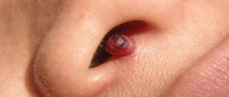
6. Spitz nevus: looks like a tumor-like growth on the skin. This spot is pink (but a combination of different colors is possible), dome-shaped, prone to bleeding. May have a hole through which liquid leaks. 7. Blue nevus has one of the shades of blue, shows well-defined borders, any size (but most often does not exceed 1 cm), looks like a lump under the skin.
Summary or briefly about the main thing:
A very small number of unevenly colored moles turn out to be melanoma. The likelihood of a malignant mole tends to zero if there are no other signs of melanoma. If doubts persist, be sure to see an oncologist and have a dermatoscopy done. If you are in doubt whether to go or not to go, it is better to go. Communication with a living doctor who is responsible for his every word, as a rule, dispels all fears.
If you still have questions, the following will help you:
- In-person appointment with an oncologist
(St. Petersburg) - Mole removal
with histology (St. Petersburg) - My online consultation (from anywhere in the world)
Other articles:
- Is sunbathing dangerous to life? Truth and fiction
- How to remove a mole without a scar? 12 tips
- Recurrence of nevus. What to do if a mole appears again?
- Dermatoscopy - what is it?
Useful article? Repost on your social network!
The number of large moles on the body is increasing: what to do
Question: what to do if large moles constantly appear? The first thing to do if many birthmarks appear on the body is to examine the skin and evaluate the symmetry, edge, color and size of the new growths.
If you identify at least one danger criterion, you should consult a doctor. The doctor will conduct an examination, and only after that a decision is made about the need to remove the nevus.
If moles appear in large numbers, a medical examination is necessary in any case.
At home
The main thing to do at home is to regularly inspect new formations. In most cases, this is enough to prevent complications. There is no need to try to get rid of birthmarks with the help of medications or folk remedies, this can have a bad effect on your health.
Some important skin care tips:
- Avoid direct sunlight on your skin (it is not recommended to sunbathe between 11 a.m. and 4 p.m.).
- Use sunscreen - it is better to choose a cream with a protection factor of at least 15 (SPF 15).
- Cover large birthmarks from the sun with a bandage.
- Do not scratch or injure the formations.
- Do not exfoliate areas of the skin where there are nevi.
What does a melanoma mole look like?
The process of degeneration of a mole into melanoma does not occur asymptomatically. Signs of malignancy appear in the very first stages, so if you pay close attention to your health, it is extremely difficult to miss them. Please note the following changes:
- The appearance of uneven color, change in shade;
- Change in skin pattern - the surface of the mole becomes shiny and completely smooth;
- Redness appears around the mole;
- Contours become blurred, symmetry is disrupted;
- The structure of the mole changes - it may become denser or softer;
- A burning sensation of itching in the area of the tumor;
- The appearance of cracks, pitting of the surface.
If you have these symptoms, immediately call the contact center of the Yusupov Hospital and make an appointment with an oncologist-dermatologist. With timely and adequate treatment, the chances of recovery increase many times over.
Mole with jagged edges
One of the signs of a non-hazardous formation turning into a dangerous one is a change in contours. It often has blurred edges and scalloped borders.
There are non-dangerous types of nevi - dysplastic. Only a specialist can make a correct diagnosis.
A mole with uneven edges can be dangerous if there are additional signs of melanoma:
- accelerated changes in size;
- the presence of clearly defined asymmetry;
- the appearance of highly indented boundaries.
The most basic reasons for the appearance
Most researchers answer the question of where moles come from in the following way. During the embryonic period, a special type of cell is formed in the neural crest of the embryo - melanoblasts. Once formed, they migrate to the basal layer of the epidermis. If the migration process is disrupted, a form of albinism develops. Sometimes the cells do not reach the epidermis, but remain in the submucosa.
Melanoblasts serve as the precursors of pigment cells, or melanocytes. They can also differentiate into neurons, glial and smooth muscle cells. Scientists do not yet know what differentiation depends on.
Thus, during the period of intrauterine development, the basis for the formation of nevi is laid. After birth, under the influence of provoking factors, they begin to stand out against the background of clear skin.
Sometimes a child is born with visually noticeable nevi, but this happens rarely (about 1% of cases). Such moles are called congenital.
But most nevi appear as they grow older. The following are the reasons for the appearance of new moles:
- Ultraviolet radiation. Stimulates the production of melanin - and, as a result, the appearance of pigment clusters.
- Hereditary factor. There is a correlation between the number and shape of nevi in close relatives.
- Radiation and X-rays. They promote increased melanin production, as does ultraviolet light.
- Skin trauma. The healing process of a wound or inflammation leads to active regeneration of cells, including pigment cells.
- Changes in hormonal levels. Neoplasms appear during puberty, pregnancy, and menopause.
- Age factor. The intensity of the formation of new nevi or the disappearance of old ones is associated with a person’s age.
A number of researchers also identify other factors - viral infections, nervous disorders. But there is no clear correlation between their effects and the formation of nevi. Most likely, they are associated with changes in hormonal levels, which leads to the appearance of new moles.
Melanocytes are the cause of moles
If moles begin to appear all over your body, this is a cause for concern and a visit to the doctor. But most often the active formation of nevi is explained by natural causes:
- frequent visits to the solarium;
- active sunbathing;
- use of ultraviolet radiation for medical purposes.
Melanocytes, which make up pigmented nevi, are formed as a protective reaction to an irritant. Therefore, doctors do not recommend excessive sunbathing - the more moles, the higher the risk of their degeneration. According to official statistics, 86% of melanoma cases are associated with exposure to ultraviolet radiation.
Ultraviolet sources can be not only natural, but also artificial. It's all about the wavelength: if it is less than 380 nm, it can cause tanning. This property formed the basis for the creation of equipment for solariums.
Can moles be inherited?
There are many cases where the shape, size, number and location of nevi in close relatives are the same. But why this happens is not known for certain.
Apparently, moles and birthmarks on the body are caused by reasons related to genetics - DNA contains information about future nevi. But this is just a theory.
How to figure it out?
If after reading this material you still have doubts, the oncologist should say the last word on the mole during a face-to-face consultation. Not a dermatologist, not a surgeon, and certainly not a cosmetologist. It is optimal if the doctor knows the technique of dermatoscopy and has this device at hand.
Very often I hear the following objection regarding a visit to the doctor about a mole: “I don’t want to go to the oncologist, in case he says that I have cancer!!!” Unfortunately, this position borders on absurdity. After all, if there is cancer, it needs to be diagnosed and treated, and not “hidden in the sand.” This is especially true for melanoma, which at stage 1 is completely curable in almost 100% of cases.
External differences between dangerous and safe nevi
It is almost impossible to determine on your own what dangerous and safe moles look like. To do this, they need to be examined using special equipment - a dermatoscope, which magnifies the image tenfold. It is quite difficult to assess risks with the naked eye.
Nevertheless, dangerous moles have a number of characteristic signs:
- Asymmetry. Draw an imaginary line through the center of the nevus: the resulting halves should be symmetrical. If one side of the mole develops more actively, this is an alarming sign.
- Ragged, uneven, fuzzy edge. Safe moles have a clearly defined border with smooth edges, melanoma does not.
- Uniformity of color. The shade of a normal mole ranges from light brown and gray to black, but it should be the same. If bluish, brown, or red spots are noticeable on the surface, you should see a doctor.
- Variability. Normally, education remains stable throughout life. Any changes in size, shape, or color increase the risk of degeneration.
- Presence of a leg. Dangerous nevi grow randomly and are unable to form a clear base.
Experts also use the ugly duckling rule. If all nevi on a person’s body are similar, only a few differ in color, shape, structure, they need to be checked first.
The size also helps to identify dangerous moles: a nevus with a diameter of up to 6 mm is considered safe. Larger lesions have an increased risk of malignancy. Although it is impossible to say unequivocally whether a 2 mm mole is dangerous: it is important to take into account not only the size, but also other factors.
How to understand when it's time to see a doctor
Every person, regardless of gender, age and health status, should conduct a complete self-examination of the body once a month. At the same time, it is important to know which moles are worth paying attention to and which are completely safe. Dermato-oncologists advise monitoring the following changes:
- growth of the tumor in area or height;
- compaction;
- increase or decrease in pigmentation;
- bleeding;
- hair loss;
- ulceration;
- the appearance of subjective sensations - itching, burning, tingling in the mole;
- the formation of satellites - small dots around the central nevus;
- hair loss;
- hyperemia - local increase in body temperature;
- enlargement of regional (nearby) lymph nodes.
Suspicious and atypical moles should be shown to a doctor. It is better if it is a narrow specialist - a dermato-oncologist. But if the clinic does not have the doctor you need, you can visit a dermatologist, surgeon or oncologist.
Every adult should be examined by a dermato-oncologist at least once a year. If a mole bothers him, a visit to the doctor should be an emergency.
Cancerous moles
Directly cancerous, i.e. malignant moles are melanoma, basal cell and squamous cell carcinoma.
Melanoma
Another name is melanoblastoma. It is divided into several types.
Superficially spreading (up to 70%)
- size up to 2 cm;
- the color is brown and dark brown uneven;
- localization on the skin of the chest, back and limbs;
- vertical and/or horizontal growth;
- at the level with intact skin;
- the edges are not smooth, wavy;
- various shapes (round, hexagonal);
- the surface is lumpy.
It remains in a dormant state for a long time and often occurs against the background of pigmented nevi. At the initial stage of the disease, it is difficult to understand the specific nature of the pathological focus, which makes timely diagnosis difficult.
Melanoma may look no different from a regular mole, so you need to pay attention to any new birthmarks
Nodular (nodular)
It is considered the final stage of development of the surface form.
- localization – head and neck;
- polyp-type node;
- the surface is smooth, shiny, uneven;
- tendency to ulceration;
- color scheme similar to the surface form.
Lentigo melanoma (melanotic freckles)
It always occurs de novo, that is, on unchanged skin. It can extremely rarely occur against the background of nevi or other precancerous diseases. It appears more often in men of advanced age (over 70 years), in young people it is registered in less than 1% of cases.
- localization – face and neck;
- multiple nodules;
- dark blue, dark brown and light brown;
- diameter 1.5-4 mm;
- the edges are not smooth, wavy.
Acral-lentiginous
- localization – palms, soles, nails;
- uneven edges;
- color gray and black.
It occurs more often in people of the Negroid race. It grows extremely slowly. Has a relatively favorable prognosis.
Basal cell skin cancer
It is also divided into several forms.
Nodular
The most common type of pathology. Characteristics:
- dense multiple nodules prone to fusion;
- diameter 2–5 mm;
- localization – facial skin;
- color ranges from pale pink to yellow-brown;
- has a tendency to ulcerate.
Surface
Peculiarities:
- rounded superficial lesion;
- diameter 1 cm or more;
- on the surface there are papillomatous growths and ulcerations;
- occurs on the skin of the trunk and limbs;
- yellowish color.
Basal cell skin cancer in the initial stages can take the form of a mole
Scleroderma-like
Relatively favorable form.
Peculiarities:
- fast growth;
- does not protrude above normal skin at the beginning, and then, due to endophytic growth, is pressed inward like a scar.
Fibroepithelial
Has a relatively favorable outcome.
Characteristics:
- node shape (color varies widely);
- diameter 1-3 cm;
- tightly elastic;
- localized on the body.
Squamous cell carcinoma
Has three forms.
Superficial
Difficult to identify and most common form.
Characteristics:
- single spot or nodule;
- color is variable, often whitish;
- there are multiple crusts on the lesion;
- protrudes above the surface of the skin;
- has a tendency to erosion.
If you suspect possible skin cancer, you should consult a dermatologist
Infiltrative (endophytic)
- multiple dense nodes;
- crusty;
- tendency to form deep ulcers.
Papillary form (exophytic)
- looks like cauliflower in appearance;
- rises above the skin;
- tendency to bleeding and ulceration.
Melanoma and all forms of squamous cell carcinoma most often develop regional and distant metastases in a short period of time (2-4 months), so they are the most dangerous. Malignant moles do not make themselves felt for a long time, and for this reason the patient is often hospitalized in the hospital against the background of severe paraneoplastic syndrome.