Surgical removal of lipoma
It would be more correct to call this procedure “traditional”, because any removal of tumors is a surgical procedure. Before the procedure, you will need to undergo an examination, the results of which will be needed by the surgeon. Most often, it will be enough to take a general blood and urine test and do fluorography. Depending on the general health of the patient, additional examinations may be required: blood sugar test, electrocardiogram, etc. - as prescribed by a specialist.
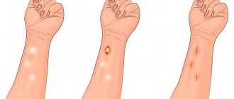
The operation itself on a small tumor is performed under local anesthesia. The surgical field is injected with anesthetic, disinfected, an incision is made through which the tumor is removed along with the capsule, and sutures are placed. The whole procedure takes no more than 15 minutes. The contents of the capsule are sent for histological examination, which is necessary to exclude a malignant process.[box#2]
If the operation is performed on open areas of the body - face, neck, arms - the surgeon may apply a cosmetic suture with an absorbable thread. In this case, there is no need to remove the stitches. Regular sutures are removed after 10-12 days, by which time the histology result is usually ready. If you are not prone to keloid scars, the scar will appear as a small light stripe that will become invisible over time.
Treatment of wen and fibroma
Methods for removing lipoma
- Removing the wen surgically. An incision is made above the tumor using a scalpel, after which the capsule with the wen is removed, and then a cosmetic suture is applied.
- Removal of lipoma with laser. Suitable for all types of wen, this method has virtually no contraindications, and it does not cause cosmetic complications. Removal occurs under local anesthesia in minutes, and recovery and healing takes a week.
- Fat deposits are removed using electrocoagulation - either electrically or surgically. The lipoma is removed along with the capsule under local anesthesia.
Progress of the operation: how the lipoma is removed
Many of our patients laugh: they say, well, what kind of operation is this, just to remove the wen... However, despite all the “uncomplicatedness”, lipoma removal is a surgical intervention, and therefore you need to take it seriously and follow the surgeon’s recommendations.
Anesthesia is not always used for such an operation, especially when it comes to using a laser. The surgeon makes a small incision (with a scalpel or laser), and through this incision the “culprit of the operation” is carefully removed. As mentioned above, the lipoma must be removed completely - the capsule itself and all its fatty contents.
In addition, even with such a simple intervention, it is necessary to ensure sterility (a laser helps a lot here). There is a separate question about the invisibility of a postoperative scar: you must agree that a small but noticeable scar will spoil the face or neck. The laser makes it possible to make the incision very thin and the scar invisible. Therefore, our surgeons very widely practice the use of a laser knife - almost always when there are no contraindications.
After removing the lipoma, a sterile bandage is applied to the intervention area. As a rule, the surgeon’s control over the healing process of the incision is not required, with the exception of complicated clinical cases when a lipoma with inflammation or suppuration was removed.
The cost of removing a lipoma, like other tumors, will pleasantly surprise you. You can get rid of an unsightly wen as soon as possible by calling us right now.
Radio wave method for removing wen
In fact, removing a tumor using the radio wave method is the same surgical operation, only the tissue is cut not with a metal scalpel, but with a “radio knife”, which, while cutting the skin, simultaneously seals (coagulates) the capillaries. Thus, the cut is bloodless. Despite all the advantages of radio wave surgery, there are contraindications to its use:
- the diameter of the neoplasm is over 6 cm;
- acute infectious diseases;
- elevated temperature;
- diabetes mellitus, epilepsy;
- the presence of metal implants or a pacemaker.
Atheromas on the scalp are most susceptible to trauma followed by inflammation. In this case, the radioknife is most effective. The operation is performed under local anesthesia in the same way as with a regular scalpel. If the size of the neoplasm does not exceed one centimeter, stitches do not need to be applied; in other cases, the wound is sutured. The advantage of this method is painless postoperative recovery, which is achieved through nerve coagulation.
Treatment methods
Fibrolip therapy is carried out by surgical extraction. Indications for surgical measures:
- rapid growth;
- large diameter;
- compression of tissues and nearby organs;
- cosmetic defects;
- soreness.
Small and medium-sized fibrolipomas are excised on an outpatient basis using local anesthesia. Bulk tumors or those formed in hard-to-reach areas of soft tissue require serious surgical removal with subsequent hospital stay for the patient.
Modern techniques:
- endoscopic removal. The surgeon makes a neat small incision and removes the soft tissue fibrolipoma capsule. Cosmetic aesthetics are preserved;
- laser. The rays melt the tissue, cut out and extract the formation. The technique prevents bleeding and infection of the wound in the postoperative period;
- liposuction. A special instrument is used to puncture the bladder and suck out its contents. Due to the preservation of the capsule inside the body, the risk of relapse of the disease is high;
- excision with a scalpel. Traditional surgery is performed if the malignant nature of the formation is suspected, or if the fibrolipoma has a large diameter. Scars remain;
- radiosurgery. Using high-frequency rays, an incision is made and the seal is removed from the subcutaneous fat layer.
The choice of technique is made by the doctor after examining and assessing the condition of the tumor.
Folk remedies are not able to reduce or resolve lipofibroma.
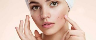
Wen on the face - causes and treatment
The most common types of wen:
- milia are small in size, filled with a white composition;
- lipomas are a mobile type of neoplasm, constantly increasing in size.
The reasons for their occurrence and the method of removal depend on the type of tumor. Wen can appear on any part of the body, and the face is no exception.
The reasons for their appearance include:
- genetic factor
- nutritional imbalance, lack of minerals, vitamins and other nutrients,
- disease of the urinary and endocrine system.
You can get rid of “white wen” at home by squeezing them out, since they have no way out. First you need to make a puncture with a clean, sterile needle and squeeze it out. The extrusion site is wiped with an antiseptic swab both before and after the procedure.
READ ALSO: Rash on the stomach of a child (29 photos): causes with explanations, small and red, after a high fever
If there are a large number of “white wen”, then it is advisable to get rid of them in a cosmetology salon using: laser therapy or chemical peeling.
You can get rid of lipomas by surgical excision. Removing them at home can lead to either overgrowth or tissue damage. During the removal procedure, a laser is used to cauterize the tumor. Tissue healing occurs within 2 weeks.
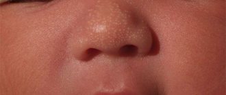
White wen - causes
White wen on the face is called milia. They can appear on the skin either singly or in several dozen at once, thereby forming a rash. They are not harmful to health and are considered to be a cosmetic defect. Lipomas of this type are absolutely painless, but can cover large areas of facial skin.
Their favorite place is the chin and cheeks; they rarely appear on the eyelids. The appearance of “white wen” on the face is accompanied by improper skin care. Most often appear on combination or oily skin types. Excess fat and lipid metabolism disorders are the main reason for their appearance.
Subcutaneous
Subcutaneous wen should be called lipomas. They are a capsule of fat that, when pressed, becomes mobile. Subcutaneous neoplasms are most often painful, since during the growth process they can affect nerve tissue.
Their origin is similar to the reasons for the appearance of milia. However, in some cases they occur due to diseases such as diabetes mellitus, dysfunction of the endocrine system, or genetic disposition.
Wen on the face of a newborn
The causes of a neoplasm in a child may be heredity or blockage of the sebaceous glands. They can appear on the cheeks, forehead, eyelids, lips, and on the body itself.
There is no point in getting rid of them at home. It is necessary to consult an experienced doctor and surgeon. Particular attention should be paid if the wen is inflamed. Because this may be a neoplasm of a different nature.
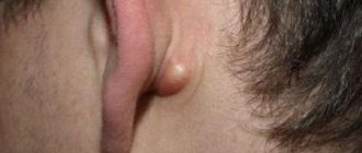
Wen on the nose, lips, and cheeks are not particularly dangerous. They arise in most cases due to underdevelopment of the sebaceous glands, and, as a rule, disappear over time on their own.
If the tumor is located in the neck area, especially with suppuration, you should consult a doctor as soon as possible. Since as the lipoma grows, the vessels may be compressed, which will lead to poor circulation.
Benign tumors: wen (lipoma, atheroma)
A benign tumor in its structure resembles the tissue from which it originated (adipose and connective tissue, epithelium, etc.) and can develop from any tissue, with partial preservation of the specific function of the tissue. They appear as slowly growing neoplasms, gradually squeezing adjacent structures and tissues, but without penetrating into them.
In the early stages of the development of a benign tumor, X-ray studies are carried out to help determine the nature of the tumor, its size and stage. Ultrasonic echolocation is performed to find a tumor of an internal organ. A biopsy is also used.
Treatment of tumors depends on its type, location and general medical history. The most common method is surgical removal, due to a proven technique and a low rate of relapse of the disease - for example, removal of wen. Surgical methods also include cryocoagulation (destruction of the tumor by low temperature exposure). In the case of inoperable tumors (for example, hormonally active), drug therapy is used, which consists of suppressing hormone synthesis.
Signs of a benign tumor:
- slow growth of the tumor, different localization is possible;
- no metastases occur, do not grow into neighboring tissues;
- After removal of the tumor (usually surgically), a complete cure follows; relapses are rare.
The reasons for the appearance of a benign tumor can be different:
- genetic changes, cell damage, reproduction and death;
- external factors that can cause cell DNA changes;
- smoking, alcohol abuse;
- drugs, chemicals;
- radiation exposure can cause cell deformation;
- injuries;
- viral infections;
- disturbances in the functioning of the immune system;
- hormonal changes that can cause, for example, breast tumors;
- unbalanced diet, eating too fatty, sweet and spicy foods.
Stages of benign tumor:
- Initiation. Two genes are minimally mutated; it is difficult to find a tumor at this stage.
- Promotion. Growth of mutated cells.
- Progression. The emergence of a tumor from mutated cells.
Types of tumor
Epithelioma
. They are formed mainly on the mucous membranes of the skin. Tumors of a yellow, white hue. Treated with surgery.
Adenoma
. Arises from glandular epithelium. If an adenoma forms in the area of the sella turcica, then the signs are as follows: endocrine disorders, signs of ophthalmic neurology.
Thyroid tumor
. Occurs in one of the lobes of the gland. Symptoms: weakness and fatigue, shortness of breath, weight loss, knots when palpated.
Sometimes a benign kidney tumor occurs, which can develop into a malignant one.
Fibroma
. Formed in tendons, uterus, mucous membranes, on the skin, in the mammary gland.
A non-cancerous tumor is characterized by the formation of a breast lump.
A tumor of the uterus is accompanied by the following symptoms:
- bleeding of the uterus;
- menstruation lasts for a long period;
- pain during sexual intercourse and in the lower back.
Leiomyoma
. Develops in the esophagus, uterus, bladder.
Osteoma
. Develops from bone tissue.
Chondroma
. Formed from cartilage tissue.
Papilloma
. Quite common; forms on the skin in the nose, intestines, respiratory tract, and has the appearance of warts.
Lipoma
.
It arises and is formed from fat cells in the subcutaneous tissue, but sometimes it can also occur in the internal organs; The common name is “ wen
”. Let's look at it in more detail.
As a rule, a lipoma occurs when the sebaceous gland changes and fills it with difficult-to-break-down high-density fats. It can grow to reach significant sizes. May pose a health hazard by compressing surrounding tissue, including nerves and blood vessels. Occasionally, with uncontrolled growth, a lipoma can transform into liposarcoma (a malignant tumor), so treatment should not be delayed. Ultrasound is used to detect internal lipomas.
Lipoma can form on the head and neck, on the chest and back, on the arms and legs, and in several places at the same time. As a rule, it is painless and can be felt as a soft lump. It grows locally and does not penetrate other tissues, but it can grow both outward, forming a tubercle, and inward, pushing the tissue apart and causing pain. The true size of a lipoma cannot be determined by external signs; this requires an ultrasound. Lipoma enlargement occurs regardless of overall weight change. And if it usually does not cause inconvenience to men, such a cosmetic defect causes problems for women.
The true cause of a wen can only be diagnosed by a specialist, so self-medication is not recommended. Occasionally there are cases of self-healing, but you should not hope for this, and when the first symptoms appear, you should consult a doctor.
Treatment of lipoma is carried out mainly surgically. The usual method involves a scalpel incision in the skin over the tumor, which is then mechanically removed (with a needle using an endoscope) along with the surrounding capsule, after which a cosmetic suture is applied to the incision. A more progressive and effective method is laser removal of lipomas; This surgical intervention is performed for all types of wen, has almost no contraindications and leaves virtually no postoperative scars. The operation lasts a few minutes using local anesthesia, the rehabilitation period and complete healing lasts about a week. The argon plasma electrocoagulation method is also used; the differences lie in the method of influence. Just in case, after surgery it is advisable to examine the extracted tumor for its malignancy.
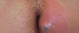
In addition to lipoma, wen also includes atheroma
, with which it is sometimes confused, is a type of epidermal cyst located under the skin and consisting of a liquid secretion. Usually occurs in places with increased sweating when the duct of the sebaceous gland is blocked, which then stretches. Most often it occurs on the face, which causes some inconvenience, especially for women. In its normal state, atheroma does not pose a danger, but when an infection occurs, inflammation occurs with suppuration, which can spread to nearby tissues.
Treatment is carried out using the same methods as for lipoma. Laser removal of atheromas is preferable, with a minimum of postoperative consequences - no scars, fairly quick recovery after surgery (about a week).
Methods for preventing atheroma include personal hygiene, abstaining from eating too fatty, sweet and spicy foods, regular cosmetic facial cleansing and the use of cosmetics that reduce oily skin and sweating.
Each method of removing wen has its own characteristics, advantages and contraindications; and which one to use should be decided by the doctor. The result in any case will be the same - a complete cure.
Published 10/25/2014 · Link | Category: Diseases and injuries
« Previous article
Next article »
Laser lipoma removal
Unfortunately, laser removal of tumors has its own contraindications. This method is not used during pregnancy and breastfeeding, diabetes, the presence of infectious skin disorders in the area of intended exposure, or immunodeficiency states. Deep-lying, large or branched neoplasms, as well as those whose structure includes vessels and nerves, are not removed using a laser.
In general, the operation is similar to the previous methods. The surgical field is anesthetized, the skin over the tumor is cut with a laser beam, and small vessels are cauterized at the same time. Then the capsule with its contents is removed using a clamp and cut off with a laser. At the end, the wound is sutured and a sterile bandage is applied. The entire procedure takes 15-40 minutes, and during the rehabilitation period you only need to follow the rules of asepsis to avoid inflammation of the wound.
Review of methods for eliminating the disease
Conservative treatment of fibrolipoma has no effect; this method of treatment is not used. Some specialists prescribe expensive drugs that provide short-term relief. The main method of removing fibrolipoma is surgical.
- The use of a toxic drug that destroys the structure of lipoma tissue. Indications for the procedure: no pain, diameter up to 0.03 meters. Several procedures are necessary to completely remove the seal.
- High frequency radio waves. Prolonged use of radio waves on tissue formation leads to complete destruction.
- Puncture-aspiration procedure. The procedure is carried out using a needle or syringe. The process is aimed at sucking out the contents of the tumor. Many experts are ardent opponents of this method of treatment. Efficiency and quality leave much to be desired. Frequent relapses.
- Laser surgery. Removing compaction with laser beams. The indication is a defect up to 0.05 meters in diameter. The positive thing about this operation is the absence of scars. After the intervention, the patient must be observed by the attending physician. Lipofibroma may appear in neighboring areas; this process must be monitored.
- Surgical intervention. It is carried out last, at the last stage. There are indications for the procedure: pain on palpation; stretch marks on the skin; large sizes, contributing to compression of the underlying tissues and organs; intensive tumor growth; disorders of the condition, disorders in the clinical blood test.
The surgery to remove fibrolipoma is performed under general anesthesia. The tissue is dissected, an inspection is carried out, followed by removal of the formation along with the capsule. Applying a suture and processing the surgical field is the final result.
In the postoperative period, it is necessary to take antibiotics and anti-inflammatory drugs, and further observation by the surgeon.
Fibrolipoma of the armpit is an unpleasant but not dangerous disease. Correctly selected treatment tactics make it possible to avoid complications. If the size of the wen does not exceed 3 cm, you can get rid of it using traditional methods. Large lipomas are removed surgically or using a laser. The armpit is a delicate area and can become damaged due to constant changes of clothing. In this case, you must immediately go to the hospital to avoid consequences. There are no medications that help the tumor resolve. There are injectable medications that act on the capsule and stop the growth of the formation. Warming compresses will help eliminate inflammation and wen. The recipe is quite simple. It is made on the basis of Dimexide, diluted in equal quantities with boiled water. Apply 2 times a day. Contraindications: suppuration and skin defects. Treatment at home is possible after consultation with a doctor. The doctor gives instructions and recommendations that are safe and suitable for the individual patient.
Fibroadenoma is a tumor located in the mammary gland, found among the female population. Fibrolipoma must be treated exclusively surgically. If cancer is suspected, breast resection is performed.
Laser destruction
One of the most modern hardware methods to get rid of a tumor is laser removal of lipoma, after which all traces disappear within two weeks. This is an innovative technology for eliminating benign tumors, which eliminates the patient’s exposure to radiation.
The operation itself lasts no more than half an hour. Instead of a scalpel, a laser is used, which cuts the surface and burns its contents without affecting healthy tissue.
Removing a wen with a laser does not require the use of general anesthesia and has undeniable advantages:
- the operation itself is carried out quickly, bloodlessly and painlessly, there is no need to suture the wound;
- a targeted effect allows you to completely destroy pathological cells;
- since there is no direct skin contact with the instrument during the procedure, the possibility of infection is eliminated;
- traces from the laser beam gradually disappear;
- The rehabilitation period lasts no more than two weeks.
Unfortunately, this technology has disadvantages:
- with large sizes and deep location in the tissues, the laser is not able to completely destroy the lipoma;
- the use of a laser does not make it possible to obtain samples of materials for biopsy;
- there is a certain percentage of risk that the wen will grow back;
- the cost of the procedure is high, so you should clarify in advance how much it costs to remove lipomas.
Like any surgical intervention, laser fat removal has a number of contraindications. These include the presence of the following diseases:
- diabetes;
- HIV infection;
- oncology;
- inflammatory processes on the skin;
- herpes in the acute stage;
Also, lipomas are not removed during menstrual periods and during pregnancy and lactation.
Diagnostic methods
Diagnosing fibrolipoma is not difficult, except for a tumor located deep in the tissues. In such cases, they resort to special additional methods.
- X-ray. The diagnostic method is based on the use of radioisotope rays. When studying the image, you can notice light formations with smooth, clear contours. Tumors in the chest and abdominal area cannot be seen without contrast. A special gas is used that gives a control X-ray picture.
- CT scan. A modern technique that allows you to assess the extent of the lesion, determine the presence of fibrolipomas, and differentiate it from other diseases.
- Ultrasound. A diagnostic method that allows one to identify the structure of a formation due to hypoechogenicity.
- Biopsy. Necessary to clarify the nature of the tumor - is it benign or malignant. The procedure requires a device with a thin needle that takes a small amount of tumor tissue. The resulting material is sent for cytological examination.
Symptoms
Wen is characterized as a fairly soft volumetric formation under the skin. Often there are several of them throughout the body. The size of each wen can range from tiny to huge (as big as a child’s head). But the most common are lipomas with a diameter of 1.5 to 5 centimeters.
A particularly severe case is when a seal forms in which liquid accumulates. The result is a huge swelling with a pedicle that stops blood circulation; as a result, the tumor leads to tissue death and causes disruption of the function of one or another organ.
Therefore, it is very important to remove lipomas on the head, as well as lipomas on the stomach or other places, even if they do not yet cause significant discomfort.
How to determine diagnostically?
When localized subcutaneously, the diagnosis of lipoma is rarely difficult; it is determined by palpating the tumor. However, conventional palpation is not enough; ultrasound is indicated.
For lipomas with internal localization, a large-scale instrumental study is carried out. The “gold standard” is magnetic resonance imaging, which allows you to study organ tissue layer by layer.
Laboratory methods are rarely informative; they allow us to exclude an inflammatory process, impaired liver and kidney function.
Thanks to careful diagnosis, a probable lipoma is differentiated from malignant tumors, vascular aneurysms, helminthic infestations, and other neoplasms that pose a threat to the health and life of the patient.
The morphological structure of the tumor is important, but if oncological risks are suspected. If a fragment of the neoplasm is preserved, a histological examination is possible.
Radio wave and puncture-aspiration methods
Relatively small lipomas can be removed using radio waves under local anesthesia. This procedure is carried out using special equipment that allows one to influence a tumor of this kind through intense radio wave exposure.
READ ALSO: Pimples with white heads
This method has many advantages:
- absence of blood during conduction;
- painlessness;
- complete absence of complications after the procedure (no infection, swelling, pus or infiltrates);
- minimum period of postoperative rehabilitation (healing takes place within a week or two);
- good cosmetic effect (no scars on the wound);
- no need for stitches;
- no rehabilitation in a clinical setting is required (after the operation the patient can go home);
- After the operation, the patient can work calmly.
The radioknife removes all manifestations of the tumor and, almost always, eliminates relapse in the patient. After the tumor is removed, it is automatically sent for histological analysis.
Another, but no less effective, is the puncture-aspiration method of lipoma removal. This procedure involves inserting a needle into the tumor itself, after which a special pump removes all the contents of the lipoma.
Advantages of this method:
- good cosmetic effect;
- no rehabilitation in a clinical setting is required (after the operation the patient can go home);
- After the operation you can work safely.
Among the disadvantages of this method is the impossibility of removing formations that are of fibrous origin.
Prevention of occurrence and risk of relapse after treatment
Preventive measures are necessary procedures to prevent relapses and the occurrence of fibrolipomas. Certain recommendations must be followed:
- a correct lifestyle, the absence of bad habits is the key to a happy, healthy life;
- constant monitoring by a doctor;
- in the presence of postoperative wounds - careful treatment;
- timely treatment with hormonal drugs;
- healthy eating, refusal to take low-quality products and supplements;
- regular physical activity that does not overstrain the body;
- walks in nature, relaxation;
- minimize stress disorders.
The risk of fibrolipoma recurrence after treatment is negligible. Depends on the quality of the operation performed, method and care. The treatment method in which the fibroma and capsule were removed does not cause relapses.
It is possible to recover from fibroids. Fibrolipomatosis is not a death sentence; don’t be upset ahead of time. Correct treatment and timely diagnosis give positive results. There is no need to put it off until later; you need to consult a doctor at the first sign, then the risk of complications will go to zero.
The article has been reviewed by the site editors
Lipoma classification
- Perineural (formed around nerves)
- Lumbosacral (located inside or near the spine, i.e. the back area).
- Lipoma inside the vagina, joint.
- Intramuscular and intermuscular.
- Myolipoma (on soft tissue, sometimes on the face, back area).
- Angiomyolipomas are usually located in the kidneys, sometimes on the skin.
- Adenolipoma (formed on the skin or under the skin, with inclusions of sweat gland cells, facial lipoma).
- Bisha's hump is a lipoma located on the back of the neck.
- Multiple symmetrical lipomatosis is a fusion of lipomas of the cervical region, back, torso, back of the head, on the face, as well as limbs, a large compaction.
In some cases, lipomas are associated with neurofibromatosis, indicating the possibility of the presence of certain diseases and conditions (Dercum's disease, Madelung's disease, Bannayan-Zonan syndrome, Proteus syndrome, Gardner syndrome).
How to remove wen at home
Check out the folk methods that will help remove lipoma:
- With onions and laundry soap. Take the ingredients in equal quantities and grate both. Place the resulting mass on the stove, cook for about 15-20 minutes, cool. Apply the healing mixture to the wen, rinse off after 40 minutes with warm water. Soap and onions are excellent at fighting fat and producing an antibacterial effect. After several treatments, the white growths should disappear.
- With aloe. Cut a leaf of the plant, apply the pulp to the wen, cover with a plaster, and leave to cure until the morning. During this time, biologically active substances will penetrate deeply, improve fat metabolism, and cleanse the skin.
- With baked onions. The vegetable needs to be cut into halves, baked in the oven, then apply a compress several times a day for 10-15 minutes. Thanks to this folk method, the subcutaneous tumor should resolve within a few days.
- Celandine. You should pick the plant, when orange juice appears, apply it to the lipoma. The plant should be treated for about a week.
- State pension for long service - conditions of appointment, amount of additional payment and procedure for registration
- Where are wages expected to rise after quarantine?
- Preferential mortgage for large families
How are atheromas and lipomas treated?
Atheromas are treated surgically; the type of operation will depend on the size of the formation. Small sebaceous cysts can be removed with a laser. Most often, atheromas are large in size and are removed with a scalpel. The “bump” on the skin is surrounded by two incisions, then the cyst is peeled out and removed along with a small piece of skin. Stitches are placed on the wound.
Atheroma cannot be cured by “sucking out” the contents with a needle and syringe. It is imperative to remove the walls of the stretched sebaceous gland - if they remain, they will begin to produce sebum again, and the cyst will grow again.
If atheroma suppurates, surgical treatment is performed; the doctor may prescribe a course of antibiotics. Lipomas do not need to be treated. The operation is performed if:
- education is large and growing rapidly;
- the lipoma is in an inconvenient place and is constantly in the way;
- bothered by soreness;
- The patient himself insists on removing the lipoma.
Wen is removed in the classic way, using a scalpel. Relapses are possible, but extremely rare. Typically, atheromas and lipomas are removed on an outpatient basis; hospitalization is not necessary. Anesthesia is also not needed - local anesthesia is sufficient. The operation lasts on average 15–20 minutes.
| Name of service | price, rub. |
| Removal of superficially located foreign bodies (removal of foreign bodies with dissection of soft tissues) | 2200,00 |
| Removal of benign skin tumors (less than 1.5 cm) | 2000,00 |
| Removal of papillomas | 5000,00 |
| Laser removal of atheromas, basal cell carcinomas, lipomas, fibromas, up to 2 cm in size | 3000,00 |
| Laser removal of atheromas, basal cell carcinomas, lipomas | 4000,00 |
| Laser removal of fibroids from 2-5 cm | 4000,00 |
| Laser removal of atheromas, basal cell carcinomas, lipomas | 5000,00 |
| Laser removal of fibroids from 5 cm | 5000,00 |
The removed lesions are sent for biopsy. They are examined under a microscope in the laboratory to make sure that they do not contain tumor cells.
Are you looking for where to remove a wen quickly and without pain? Visit a surgeon at the ProfMedLab medical center. Call: +7 (495) 125-30-32.
ul
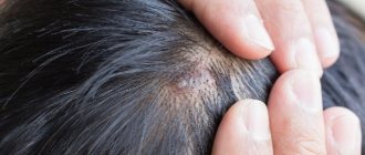
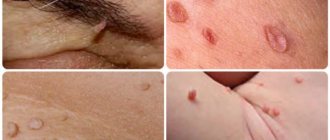
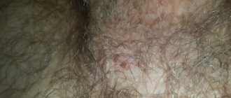
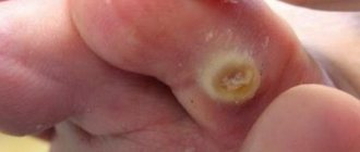
What does a wen look like?
Wen Quite a large number of people suffer from a similar disease. It is not always possible to detect a lipoma on your body, since when it appears, it is small in size and does not cause any inconvenience. It looks like a pea under the skin. A fatty tumor can be located anywhere in the body where adipose tissue is present, even inside the abdomen. The wen looks like a small soft lump on the body with clear boundaries. If you press on such a pea, it can move painlessly. Over time, the lipoma increases in size, which bothers the person. It also has an unpleasant cosmetic appearance. (Read here )
Basic facts about differences and common features
Let's try to summarize the information about the similarities and differences between atheromas and lipomas:
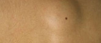
- The main difference by which the average person can distinguish between these tumors is mobility. The lipoma moves easily upon palpation, and the atheroma is fused to the skin.
- Lipomas can be located in different parts of the body, and atheromas can be located only where sebaceous glands are present.
- If you carefully examine the atheroma, you will see a small dot (usually black) on it; lipomas do not have this feature.
- Lipomas and atheromas are practically not dangerous from the point of view of malignancy.
- Multiple lipomas require surgical treatment, and a significant number of atheromas are treated exclusively conservatively (doctors identify the cause of total blockage of the glands and relieve it).
- Both lipomas and atheromas are distinguished by clear boundaries, a rounded shape and a tendency to grow. Both types of tumors have the potential to put pressure on nearby tissues, causing discomfort.
- Both types of tumors can create significant aesthetic inconveniences, spoiling the patient’s appearance.
Today, the only effective method of treating both lipomas and atheromas remains removal. The operation can be performed using a conventional scalpel, but modern clinics use safer and less invasive techniques - laser or radio wave removal.
Types of lipomas
According to the classification, lipomas are distinguished:
- perineural. These are neoplasms surrounding the nerves. They are always painful and difficult to remove;
- lumbosacral. Neoplasms are produced in the spinal canal or near the vertebrae. Often such tumors are associated with congenital underdevelopment of these areas;
- sheaths, tendons and synovium of the joint. Neoplasms are located in the named places;
- intermuscular. Such lipomas grow through the muscles. If they are not completely removed, they reappear;
- soft tissue myolipomas. Among them, subcutaneous neoplasms are rare;
- angiolipomas. They are most often formed in the kidneys, on the skin - rarely, as a rule, they are found in mature men and look like a painless dense knot under the skin of the limb;
- adenolipomas. Such neoplasms have the structure of a sweat gland, and therefore are difficult to distinguish from atheromas.
What is lipoma?
A lipoma is a benign growth that usually appears as a “lump” under the skin that is made up of fatty tissue. Lipoma is often called a “fat tumor,” but do not be alarmed—it has nothing to do with malignant tumors.
Almost nothing is known about why lipomas occur. It is believed that this is due to the malfunction of certain enzymes and a hereditary predisposition.
Most often, wen, oddly enough, appears where there is little fat under the skin: on the back and in the area of the shoulder girdle, on the outer side of the shoulder, thigh. This “bump” is soft and painless to the touch. If you press it with your finger, it easily “rolls” under the skin. Dimensions rarely exceed 5 cm.
There are also exotic localizations: around the nerve (in this case, the lipoma leads to pain), in the spinal canal, in the joints, in the thickness of the muscles.
Lipomas usually grow slowly and in most cases do not cause any problems other than cosmetic discomfort.
Laser method
Removing lipomas with a laser is the most effective, safe and painless. The advantages of the method include:
- lack of blood;
- minimal risk of infection;
- absence of pain;
- absence of scars;
- Duration of the procedure is 1 minute.
The laser beam helps to remove the wen from the surrounding tissues and at the same time “seals” the capillaries. If the lipoma is large, the surgeon will need to close the wound.
The removed lipoma is sent for mandatory histological examination. But if the size of the formation is very small, the fat will be completely destroyed by the laser beam.
According to statistics, an increasing number of people are using laser lipoma removal. Reviews indicate painlessness and the absence of a rehabilitation period.
READ ALSO: Purulent acne appeared after depilation, what to do?
Lipoma: video of its removal
Is it worth removing a lipoma, how dangerous is it and why does this tumor occur? These are the most common questions asked by people who have discovered a nodular lump under their skin.
In order to answer these questions, you need to consider all aspects of the disease: etiology, symptoms, prevention, direct removal of the lipoma, reviews of people who have already removed lipomas using various methods.
When should a lipoma be removed?
Surgery to remove the wen may be indicated in cases such as:
- Wen on a leg. It is imperative to get rid of such a lipoma, otherwise the leg may twist and cause tissue necrosis.
- The neoplasm compresses the organs. Sometimes lipomas form near any organs, which they can put pressure on and cause pain.
- Wen inside the organ. Such lipomas interfere with the normal functioning of the organ and sometimes degenerate into malignant tumors.
- The lipoma is growing.
Causes of neoplasms
Skin neoplasms appear due to the pathological proliferation of tissue cells that form tumor lesions of the skin. The reason for their occurrence may be:
- frequent skin damage;
- intense ultraviolet rays;
- genetic predisposition;
- chronic skin diseases;
- viruses (for example, the human papillomavirus provokes the appearance of warts and moles).
Most tumors require surgery to remove them, since injury or exposure to ultraviolet radiation increases the risk of benign tumors becoming malignant.
In addition, benign formations, being large in size, can disrupt the normal functioning of various organs, cause pain, squeezing nerve endings and blood vessels. In our clinic, at competitive prices, you can undergo the procedure of removing benign skin tumors and get rid of:
- warts;
- moles;
- birthmarks;
- papillomas;
- fibroids;
- condylomas;
- syringoma;
- adenoma;
- hemangiomas;
- lymphangiomas;
- lipomas and other formations.
Our best dermatologists, dermato-cosmetologists and oncologists will work with you.
ul
Causes and symptoms of development
At this stage of medical development, doctors cannot clearly name the cause of the appearance of fatty tumors. Lipomatosis (as you can see in the photo on the Internet) does not depend on the patient’s weight category and the amount of subcutaneous and internal fat in the body. Often, even with absolute starvation or anorexia, fatty tumors do not resolve on their own.
Scientists name a number of factors that provoke lipomatosis:
- genetic predisposition;
- disruption of metabolic processes in the body;
- hormonal changes - this especially applies to women during and after pregnancy and menopause;
- disturbances in the functioning of the hypothalamus, pituitary gland;
- pathological processes affecting the thyroid gland;
- alcoholism;
- malignant tumors.
The localization of lipomas is varied - legs, neck, head, limbs, thighs and buttocks, forehead and abdomen, shoulders. Wen that forms on internal organs is especially dangerous. They compress tissues and can provoke inflammatory processes.
Symptoms depend on the location of the wen. If the tumor is located in the mediastinum, signs characteristic of organ damage appear near the tumor.
Subcutaneous lipoma has the following characteristics:
- soft consistency;
- painless when pressed;
- lack of adhesion to the skin;
- lobulation of the structure;
- the size of the neoplasm is from 5 to 50 mm. And in rare cases, wen of gigantic size can be found.
Lipomastia in men is not a benign fatty pathology. The growth of breast tissue is not a classic wen, but may be a harbinger of the development of cancer.
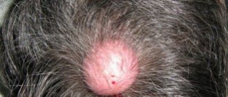
- Why does a wen appear on the head?
- Features of localization of papules on the sole
- How to remove condylomas with nitrogen
With subcutaneous growths, the patient's main complaint is a cosmetic defect. In internal localizations – the appearance of symptoms associated with disruption of the internal organs.
What is fibrolipoma and its causes
Fibrolipoma can appear at any age, gender does not matter. Typically there is no pain, but there are tumors that cause painful discomfort. There are formations from 2-10 cm or more, single and multiple. According to ICD-10, it has code D17, ... - after the decimal point a number is placed that specifies the location of the wen.
Fibrolipoma is a hereditary disease that is not characterized by a malignant course.
Localized in any part of the body. Classification of lipofibromatosis:
- soft. The consistency of fibroma depends on the predominance of components in the composition. Connective tissue, multiple cells are characteristic of soft tissue, area of distribution - neck, groin and subcutaneous folds;
- hard. The predominance of collagen fibers, a small content of cells gives a solid texture to the tumor. More often, mixed type tumors are observed, which contain the same amount of connective and collagen tissue.
Causes
It cannot be said that the causes of the disease have been fully studied. However, scientists say with certainty that genetics and fat metabolism in the body may be at play here. For example, a person may have a congenital predisposition to lipomatosis, that is, multiple distribution of formations throughout the body - lipoma on the neck, lipoma on the face, and so on.
There is a misconception that only obese people can develop a wen. This is not true. There are many cases of multiple lipomas in very thin people. This is affected by the appearance of leaky lipoproteins in the blood - these fats clog blood vessels from the inside, which leads to the appearance of lipomas in the neck or elsewhere in the body.
Lipoma on the back is one of the most common types of the disease, but older people often develop fatty tissue in the abdominal area. As a rule, a lipoma does not cause any significant harm, other than a cosmetic disadvantage.
Etiology of lipomatosis
The causes of lipomatosis are not yet reliably known. Scientists put forward at least two theories for the occurrence of lipomatosis, both of which are quite viable.
- According to the first theory, a person mechanically injures acne on the skin, which provokes a strong inflammatory process. That is, the duct of the sebaceous gland is blocked, a boil is formed, which, in turn, develops into a wen. This theory perfectly explains the reasons for the formation of a single lipoma, but does not explain the etiology of lipomatosis - multiple lipomas.
- The second theory for the appearance of lipomas is a violation of the biochemical process of fat metabolism, as a result of which the amount of adipose tissue increases. And the reasons for the violation of fat metabolism itself can be based on psychological and physiological stress (infectious diseases, hypothermia, climate change, etc.) In this case, it is important to fight the root cause of the disease, because even if the lipoma is effectively removed, reviews warn about the risk of relapse of the disease.
There is evidence that genetic factors play a significant role in the etiology of lipomatosis, but this theory does not yet have scientific evidence. Also, many scientists claim a relationship between lipomatosis and endocrine diseases and pancreatic pathologies. There is evidence of a hypothetical relationship between the formation of wen and alcoholism.
Soft tissue fibrolipoma is a spherical tumor-like formation that is benign in nature. A compaction with clear edges is located in the subcutaneous fat layer. Can be formed in singular or plural. Locations: back, chest, tendons, hand, etc. The lump does not provoke pain.
What is a “wen”?
In everyday life, the term “wen” usually refers to two types of formations on the skin. The first option is lipoma, a mesenchymal tumor that develops from adipose tissue cells. By its nature, lipoma is a benign tumor and does not pose a threat to health. Malignant transformation into liposarcoma is extremely unlikely. The second type of wen is atheroma. Unlike lipoma, atheroma is not a tumor. This is a retention cyst of the sebaceous gland, the result of blockage of the glandular ducts and impaired secretion drainage. Since there is no cell proliferation in atheroma, it cannot be called a tumor. This is a tumor-like formation that does not threaten health and cannot transform into a tumor.
Diagnostics
Examination methods:
- physical examination. The doctor probes the location of the fibrolipoma, checks its structure, density, and level of mobility. When located in hard-to-reach areas, hardware tests are used;
- X-ray. Used to study growths located deep under the skin. The rays make it possible to carefully study the structure of cells;
- Ultrasound of soft tissues. A diagnostic measure that allows you to determine the degree of echogenicity of fibrolipoma and determine the presence of a capsule;
- biopsy. A thin needle is used to sample the internal contents of the shell for a laboratory study of its composition and nature.
Diagnosis is carried out only if the formation is suspected of malignancy or before surgical removal.
Reasons for the formation of a wen
The reasons for the formation of lipoma are not fully understood. It is believed that a certain role is played by hereditary predisposition, trauma to the skin and subcutaneous tissue, as well as the age factor - lipomas more often appear in middle-aged people. Dietary features, obesity, endocrine and metabolic disorders do not increase the risk of developing lipoma.
The formation of atheroma can be promoted by both external and internal causes. Exogenous includes working at high temperatures, since this enhances the secretory function of the skin glands. Among the internal factors, it is necessary to highlight hyperhidrosis (increased sweating), metabolic disorders, endocrine diseases, and seborrheic dermatitis.
What is the danger of wen
Despite the fact that lipoma is a benign neoplasm, you must be careful about its presence on the body, since if the lipoma is encapsulated, it can be subject to mutation and become a beneficial place for the development of pathogenic microflora. After all, the antibodies that protect our body cannot get close to it and save us from the inflammatory process that begins in this place.
Some people really like to self-medicate. When this love of theirs spreads to the lipoma, and they begin to squeeze it out or pierce it at home with non-sterile objects, then there is a possibility of an inflammatory process. An even worse result of home treatments can be abnormal growth of wen tissue. Therefore, leading surgeons strongly recommend that you contact professionals with such questions, who will save you from problems without unnecessary and completely unnecessary complications.
News MirTesen
What danger does a lipoma on the stomach pose?
Gastric fibrolipoma is asymptomatic. It is harmless until it grows and compresses other organs. Quite often, the formation intersects with the pancreas, affecting it. Fibroma is a benign formation. Gastric fibrolipoma has a number of symptoms:
- Nausea, vomiting and other gastric symptoms.
- Changes in body weight towards decrease or increase.
- Pain in the epigastric region.
- Problems with appetite and absorption of nutrients.
People with an asthenic body type observe intensive growth of fibrolipoma due to the accumulation of adipose tissue. Difficulties in diagnosis arise due to vague symptoms.