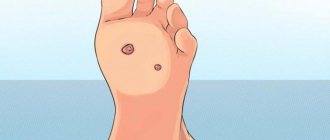
Keratoma is one of the types of benign formations that appear on human skin. Most of all they resemble freckles or age spots. Dermatologists consider abuse of sunbathing as one of the main causes of keratomas. Under the influence of ultraviolet radiation, epithelial cells become keratinized, which contributes to the formation of neoplasms.
The color of keratomas varies from flesh-colored to dark brown. Most often, these tumors appear on the arms, neck and back, much less often they appear on the legs.
Sometimes keratomas disappear on their own over time, but in some cases it is necessary to resort to removal.
Types of keratomas
Dermatologists distinguish several types of keratomas. For example, after forty years, so-called senile formations may appear - spots of a white or grayish tint, the diameter of which may increase over time. They occur most often on the neck, face and on the back of the hands. Sometimes these spots can become crusty and inflamed.
Content:
- Types of keratomas
- In what cases should a keratoma be removed?
- The essence of the radio wave method
- Advantages and disadvantages of radio wave removal of keratomas
- Contraindications to radio wave removal
- How is radio wave removal performed?
- How to behave after radio wave removal
- Possible complications
Solar keratomas appear most often in men with fair skin. They occur in those areas of the body that are most often exposed to sunlight. These neoplasms can be considered as harbingers of skin cancer, and therefore require mandatory medical consultation.
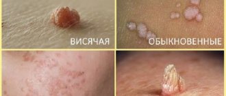
One of the most dangerous is seborrheic keratoma - a yellowish-brown spot that over time begins to peel and bleed, causing inflammation. These formations also require mandatory medical monitoring, as do horny keratomas, which are most prone to transform into malignant tumors. Follicular keratomas are the least common. Women are more susceptible to them than men. These neoplasms usually appear on the upper lip and scalp and are pinkish-gray nodules.
Why is keratoma dangerous and why is it necessary to remove it?
Keratoma itself does not pose a threat to human life and health. However, due to the fact that over time it is increasingly exposed to negative factors, such as mechanical damage and exposure to ultraviolet radiation, this benign neoplasm can degenerate into a malignant tumor.
Sometimes a keratoma can cause significant discomfort to a person. Itching, burning sensation, inflammation, bleeding, pain - all these are alarming signs. The appearance of these symptoms may indicate that the process of degeneration of the keratoma into a malignant neoplasm has begun.
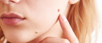
If you notice new moles, spots on your skin, or strange growths, be sure to visit a dermatologist-oncologist who can conduct research and determine whether the formations on the skin are malignant. Next, it is important to undergo regular medical examinations to prevent the occurrence of cancerous tumors.
Also, in a medical institution, you may be offered to remove keratomas on the face and other parts of the body and get rid of the need to regularly see a doctor.
In what cases should a keratoma be removed?
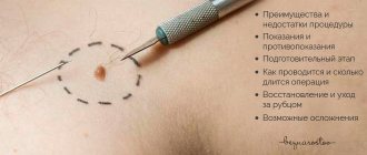
The decision to remove a keratoma should be made only after consultation with a doctor. In the event that the formation is located on an area of the skin hidden from view and does not cause either aesthetic or physical discomfort, removal can be delayed. However, in some cases, the formation must be removed without fail. For example, doctors recommend removing keratomas that rise above the skin, as a result of which they are often injured and subject to friction. A keratoma should also be removed if it begins to rapidly increase in size, begins to bleed, or becomes covered with cracks. People who spend a lot of time in the sun are advised to get rid of tumors.
The aesthetic aspect is also important. Often, keratomas located on open areas of the skin cause cosmetic inconvenience, causing complexes and self-doubt. In this case, the tumor can also be removed by first consulting a dermatologist.
There are several ways to remove keratomas. The tumor can be removed using a laser, liquid nitrogen, electric current, or surgical excision or radio wave removal. The final verdict regarding which removal method to use in each specific case is made by the doctor after conducting a visual examination and collecting an anamnesis.
Thus, small formations can be removed using cryodestruction with liquid nitrogen - the keratoma simply dies under the influence of low temperatures. The disadvantage of this method is the rather high probability of scarring and scar formation, as well as the inability to use it with reduced blood clotting. In turn, laser removal is quick and painless, eliminates bleeding, but also has a fairly wide range of contraindications.
Electrocoagulation, in other words, cauterization of neoplasm tissue with electric current, is a very effective method of getting rid of keratomas, but in most cases it leaves scars and is contraindicated for people with hypertension.
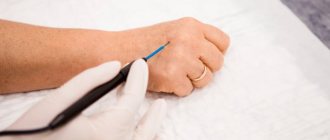
The most radical method of removing keratoma is surgical excision of the tumor with a scalpel. However, in this case, the appearance of scars of varying severity is inevitable, and in addition, the procedure is painful, although it is performed under local anesthesia.
Recently, the radio wave method of removing keratomas has become the most popular. It is considered the most advanced, very effective and at the same time almost painless.
The essence of the radio wave method
The radio wave surgery technique is based on the use of an “invisible scalpel” - high-frequency waves. This procedure is also known as radioknife. This method began to be used in dermatology and aesthetic medicine relatively recently and is considered one of the most gentle and at the same time most effective.
Thanks to its configuration, the “radio knife” concentrates high-frequency electromagnetic radiation in a small area, as a result of which it becomes possible to cut tissue with it, as if with an ordinary scalpel. Moreover, if the device has been correctly configured, the radiation does not affect healthy tissue, affecting only the area that has undergone pathological changes. Under the influence of radio waves, several processes simultaneously occur in the epidermis. First of all, microscopic cuts appear, and the pathologically changed cells themselves, under the influence of the knife, begin to generate heat and evaporate on their own. It is noteworthy that wounds under the influence of the same radio waves are immediately disinfected and coagulated.
Radio wave removal is most often used to remove single skin tumors with a small area. The radioknife makes it possible to remove keratomas in one procedure. Moreover, the procedure is non-contact and therefore sterile. Radio waves cut tissues and simultaneously cauterize them, which eliminates the development of bleeding. In total, removal of one keratoma using the radio wave method takes no more than three minutes, and the wound heals within a week.
Advantages and disadvantages of radio wave removal of keratomas
The method of radio wave surgery has recently become increasingly popular. This is quite natural, since this procedure for getting rid of tumors on the skin boasts a huge number of advantages compared to other methods.
First of all, the radioknife allows the surgeon to regulate the radiation power, thereby controlling the depth of penetration of the waves. As a result, skin formations are removed very carefully, without damaging surrounding tissues. As a result, unaesthetic scars do not appear at the site of removed tumors.
A tangible advantage of the radio wave method of removing keratomas is the absence of pain. Since the radiation is directed, it does not irritate nerve endings and does not provoke muscle contraction. Therefore, during the procedure the patient practically does not feel any discomfort.
Since the radioknife not only cuts, but also coagulates tissue, the removal process is bloodless. This eliminates the development of inflammatory processes and infection. In addition, the likelihood of developing postoperative edema and other complications is minimized.
Another undeniable advantage of this method is the ability to remove tumors located in hard-to-reach places. Also, the removed tissue can be sent for histological analysis to exclude the possibility that malignant degeneration has begun.
In fact, the radio wave method of removing keratomas has only one drawback: it is used only in cases where it is necessary to remove a formation of small diameter. If the area of the keratoma is large enough, other methods are usually used.
general description
In order to eliminate any neoplasms on the skin, many different techniques and methods are used, which can be done both in a specialist’s office and at home. Removal of keratomas can also be carried out by a whole group of methods, but the doctor must study the nature and causes of this skin condition. First of all, this is done in order to reduce the likelihood of recurrence of keratomas in the future, as well as to avoid other complications. Moreover, without special knowledge, a person can often confuse a keratoma with other skin tumors, so self-medication in this case can even harm the skin.
There are several main methods for removing keratomas - laser, liquid nitrogen, electrocoagulation, radioknife, chemical and surgical. Almost all of them are produced within the walls of specialized medical institutions, some require certain preparation of the patient and subsequent post-procedural treatment. There are recipes for eliminating these tumors at home - however, you need to remember that it is important to correctly determine the type of keratoma before such treatment, and this can only be done by a doctor. Therefore, if you have such a problem, you first need to contact a specialist and discuss the possibility of treating this tumor at home.
Contraindications to radio wave removal
Like any surgical intervention, radio wave removal of keratomas also has its contraindications, in which the use of this method is highly undesirable:
- You should refrain from using a radioknife if the patient has been diagnosed with diabetes.
- Radio wave removal of tumors for pregnant and lactating women is excluded.
- The procedure should be approached with extreme caution if the patient is diagnosed with cardiovascular failure.
- Removal of the keratoma using the radio wave method should be postponed to a later date if the patient has recently suffered from ARVI, influenza or other similar diseases.
Preparation for the procedure
Removal of keratoma is preceded by diagnosis. Without confirmation, you cannot begin any procedures.
You must proceed in the following order:
- Contact a dermatologist. The doctor will examine you and refer you for the necessary tests. If there is a suspicion of a malignant tumor, you will need to additionally contact an oncologist.
- Consult about contraindications for surgery: pregnancy, infectious diseases, HIV, poor immune system.
- Go through all diagnostic procedures; if the diagnosis is confirmed, then proceed to removal. Blood and urine tests, a coagulogram will be required, and in some cases a biopsy of the transformed tissue will be necessary.
After diagnostic measures are carried out, treatment is prescribed. The doctor will choose the method of tissue removal, but the patient’s preferences will be taken into account, because all methods lead to the same result. Another method of removal depends on the availability of the necessary equipment in the clinic.
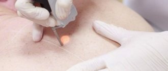
How is radio wave removal performed?
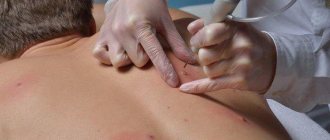
No preliminary preparation is required from the patient before removing the keratoma using the radio wave method. All preparatory work is carried out by the doctor. He must carefully examine the neoplasm, study its boundaries and depth of penetration into the skin. It is on the basis of this data that he makes a decision as to which radio waves need to be used in a given situation and adjusts the radio knife to the desired frequency.
The patient lies down on the couch. The area of skin that will be exposed is disinfected and anesthetized. It should be borne in mind that pain relief in this case is more of a “reinsurance” nature, because the procedure itself is practically painless. The frequency of the current used for manipulation is so high that it does not cause muscle contraction and does not stimulate nerve endings, as a result of which the patient does not experience any unpleasant sensations.
As anesthesia, either an injection of lidocaine or novocaine, or the application of special creams that are used to treat the area of treatment can be used. After five to ten minutes, the anesthesia begins to take effect, after which the doctor begins the procedure.
Externally, the radio knife looks like a thin metal rod connected to the device with a wire loop at the end. It is through this loop that radio waves influence the cells of formation. Also, in addition to the main loop electrode, a ball electrode can be used, which is usually used to level the wound and coagulate blood vessels to prevent the risk of bleeding.
Radio wave surgery is a non-contact method. In other words, there is no physical pressure on the tissue. As noted above, under the influence of a powerful radiation flux, a surge of thermal energy occurs and tissue heating occurs, as a result of which the cells simply evaporate.
Many patients who are not too knowledgeable about how keratoma removal is carried out using the radio wave method are afraid that they can get burned or receive an electric shock during the procedure. However, such worries are completely groundless. During surgery, the electrode does not come into contact with the skin; the doctor simply places it near the surgical field.
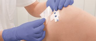
After removing the tumor, the doctor treats the wound with an antiseptic and covers it with a sterile gauze bandage.
How to behave after radio wave removal
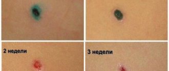
Removal of keratoma using the radio wave method is considered a simple operation that causes minimal harm to the skin. However, a small wound will appear at the site where the tumor was previously located.
She will heal in stages. First, a thin crust forms, which will fall off on its own over time. Under no circumstances should it be scratched. Also, for at least five days after surgery, the affected area of skin should not be kept wet.
Best materials of the month
- Coronaviruses: COVID-19
- Antibiotics for the prevention and treatment of COVID-19: how effective are they?
- The most common "office" diseases
- Does vodka kill coronavirus?
- How to stay alive on our roads?
Dermatologists recommend not using decorative or care cosmetics based on alcohol or other aggressive substances to care for the area of skin exposed to radio waves for at least six months after removal of the keratoma.
Also, you should not overuse sunbathing or visit a solarium.
Recommendations for care after removal of skin tumors:
- After the removal procedure, you should try to avoid being in the sun for a long time;
- If there is a change in color at the removal site or any other significant changes occur, you should immediately consult a doctor;
- When exposed to the sun, it is necessary to protect the place of removal from exposure to rays using special means;
- It is necessary to carefully palpate the removal site for the appearance of a compaction, and also visually inspect for any increase;
- Avoid going to the bathhouse, sauna, swimming pool, or solarium in the first days after the procedure;
- After removing moles, the affected area should not be wet for several days;
- Skin treatment should be carried out according to the doctor’s recommendations (treatment with an antiseptic should be done 3 times a day).
Specialists of the Absolut Med clinic
Chvyrova Tatyana Nikolaevna
Chief physician, director of the Clinic, participant in Russian and international scientific conferences for cosmetologists, dermatologists, allergists. Read more…
Dermatologist, cosmetologist
Deryugina Elena Yurievna
In 1987
Graduated from Vladivostok State Medical Institute. Experience in medicine for more than 29 years. She worked as a dermatologist for 10 years. In aesthetic medicine since 2000. Read more…
Dermatologist, cosmetologist. Doctor of the highest category