Moles that are located on a person’s skin do not need to be removed if they do not cause discomfort. The reason why a doctor will prescribe surgical removal of a mole is when the nevus is suspected of degenerating into a malignant formation, and also when the mole is subject to constant mechanical stress. With systematic trauma to the growth, the risk of infection and the development of melanoma increases. This is an oncological disease that is characterized by the rapid spread of metastases to human organs.
Mole - what is it?
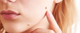
A mole is a collection of melanocytes that forms on the skin. Since melanocytes contain pigment, they are responsible for the color of moles from light brown to black.
Nevi are not only congenital. They can appear throughout a person's life.
Convex ones are formed if melanocytes are located deep in the skin layers. When the cells are located on the surface of the epidermis, flat moles appear.
Sometimes nevi can degenerate into a malignant neoplasm - melanoma. In such a case, there is a need for its urgent removal.
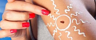
In order to promptly notice any pathological changes, moles of large sizes and unusual shapes should be constantly monitored. There are several indicators that a nevus does not pose a threat to human health:
- The size of the neoplasm is no more than 5 mm;
- Uniform color of the mole;
- Smooth surface and contours of the nevus;
- The presence of hairs growing from the body of the formation.
General characteristics of formations
A mole is a pigment formation of one of the shades of the brown palette. The base contains melanin and the melanocyte cells that produce it. Melanins are high molecular weight pigments. They are characterized by a heterogeneous structure and complex chemical composition. Pigments are found not only in moles, but also in all skin, hair, iris, inner ear and even some parts of the brain.
It is important not to confuse nevi and birthmarks. Moles do not form in the prenatal period, but as a person grows older and are closely related to his lifestyle. The cause of the formation of pigment spots can be a hereditary factor, exposure to ultraviolet radiation, or hormonal changes.
Each mole is a separate formation with a unique life cycle. At first it is a flat spot, which can increase and rise slightly above the skin as it grows and develops. The size and location of the formation depends on the level of melanocytes. If they are in the epidermis (top layer of skin), the mole will be flat. The deeper the melanocyte is immersed in the dermis, the higher the formation is located on the surface of the human body.
Convex nevi, which rise slightly above the skin, should not frighten their owners. This condition has no negative consequences and is considered a variant of the norm. The main thing is that the mole is miniature, round and uniform (in terms of color and structure) throughout its life cycle.
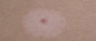
Can a mole disappear on its own and how safe is it? Yes, the formation can disappear without the influence of mechanical factors. This happens gradually and can alert a person. First, a white outline is formed around the formation, which resembles an orbit. This contour begins to increase and gradually covers the surface of the entire formation. The nevus disappears, and a small white spot remains in its place. Most often this occurs after severe sunburn. The disappearance of moles can also be a harbinger of the rare disease vitiligo.
If you notice changes in a mole (increase/decrease, uneven edges, change in shade or structure), be sure to consult a specialist.
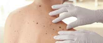
Indications and limitations for mole removal
Surgical removal of a nevus is prescribed if there are the following indications:
- Presence or suspicion of cancer;
- Nevi that have broken into several parts;
- Moles of large area or size.
Surgical removal of a mole, like any surgical procedure, has a number of limitations. These include:
- Herpetic infection in the active stage;
- The presence of inflammatory diseases;
- Infectious diseases.
Removal of nevi can be carried out after elimination of the clinical manifestations and causes of the listed pathological processes, that is, after the patient has recovered.
It is strictly forbidden to remove nevi on your own, as this can cause infection of the body and the development of oncology.
Surgical excision of a mole should only be performed in a clinical setting by a qualified specialist.
Contraindications
Removal may be contraindicated for moles whose malignancy process affects surrounding tissues or has spread deep into the body. Therefore, before the operation, a fragment of the formation is first excised and sent for histological analysis.
Other reasons why nevus removal is contraindicated:
- The presence of concomitant skin diseases, for example, acne, acne or viral rashes.
- Ultraviolet ray intolerance or photodermatosis. In the presence of such a disease, laser excision cannot be used. Otherwise, side effects will appear: swelling, redness of the skin, blisters.
Advantages and disadvantages of surgical removal of nevi
Despite the fact that surgical removal of nevi is the oldest method among all known, it has a number of undeniable advantages over others:
- Possibility of getting rid of deep and large moles;
- Safe excision of malignant neoplasms;
- The procedure is painless, since all manipulations are performed using anesthesia;
- Affordable price;
- One hundred percent removal of the nevus in one session (after excision, no neoplasm cells remain, which eliminates the reappearance of the mole);
- Possibility of conducting histological examination of remote formation;
- No contraindications.
Excision of a mole using the classical method is an effective and safe way to get rid of a nevus. However, despite this, it has several disadvantages:
- High likelihood of keloids and scars;
- Long period of rehabilitation;
- After surgery, you should not be in the sun.
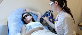
Laser destruction
How are moles removed surgically, which method is the safest? One of the most popular treatment methods is laser destruction. The doctor has the ability to control the depth of penetration of the beam, which eliminates the risk of injury to healthy tissue. The therapy has virtually no contraindications and lasts no more than 5 minutes.
When surgical removal of moles is contraindicated:
- pregnancy;
- lactation period;
- diabetes;
- immunodeficiency states;
- exacerbation of chronic diseases.
How is mole removal done? Several types of laser are used for treatment: carbon dioxide, erbium or pulsed. The beam evaporates fluid from the tissues, coagulates the vessels, leaving a crust at the site of the nevus, which disappears on its own after 14 days.
How is surgical removal of moles performed?
There are two methods for surgical removal of a mole: excision with cauterization (burning out using a special device) and excision followed by suturing.
The operation lasts from 40 to 60 minutes. The duration of the manipulation will depend on the individual characteristics and condition of the patient, as well as the size and location of the mole.
Excision of nevi without cosmetic sutures consists of the following steps:
- Treatment of the operated area with antiseptic drugs;
- Carrying out local anesthesia;
- Excision of the nevus using a scalpel at the skin level;
- Cauterizing the wound with a special device for cauterization or applying a drug for local use;
- Treatment of the wound with a local antibacterial agent;
- Applying a sterile dressing.
Upon completion of the operation, the doctor provides comprehensive recommendations for caring for the postoperative area and sends the resulting material for histological examination.
Excision of a mole with suturing is carried out in several stages:
- Treatment of the operated area with antiseptics;
- Use of local anesthesia;
- Excision of the nevus with the capture of surrounding healthy tissue;
- Treatment of the wound with antiseptic drugs;
- Cosmetic stitches. Depending on the depth of the formation, superficial, self-priming and deep sutures can be used;
- Treatment of the wound with a local antibiotic;
- Applying a patch and sterile dressing.
The removed material is also sent for histology to confirm the benignity of the neoplasm. If the nevus turns out to be malignant, then another intervention is performed. A week after the operation, the sutures are removed (if superficial sutures are used).
Recently, surgeons have been using special silicone absorbable patches, which make the scar left after excision less noticeable.
They are glued to the scar and worn for 15-20 days. In addition, if the patient is not satisfied with his appearance, plastic correction can be performed in the future.
Technique of the operation
The removal technique depends on the location and size of the formation. The mole must be excised within healthy tissue. The area of excision is determined by the doctor, warning the patient about it. For example, if the nevus is located on the face, the specialist must remove a minimum amount of epidermis. On other parts of the body, the result of the intervention will be less noticeable, so the doctor can capture more healthy tissue.
There are two main methods of surgical removal. One of them is cutting off the mole without suturing. The doctor uses a scalpel to cut off the growth slightly below the skin level. If bleeding occurs during the procedure, the wound is cauterized and a local antibiotic is applied. A bandage is applied to the affected area and skin care rules are explained. Most often, the surgical area is prohibited from getting wet or subjected to mechanical or ultraviolet influence. Based on the individual characteristics of the patient, the doctor draws up a list of necessary medications, care products and sets a date for the next visit to assess the body’s reaction.
The second method is removal with sutures. Most often, the method is used in the treatment of dark or flat moles. The doctor cleanses the skin, injects an anesthetic, and excises the formation and its surrounding surface.
During the operation, the deep layers of the skin are affected, so suturing is a mandatory procedure.
The healing rate of the area is 7-14 days and depends on the individual characteristics of the patient. The operated area should be cleaned periodically, protected from damage, and ointments or oral medications should be regularly applied.
The main thing is not to self-medicate, but strictly follow the doctor’s instructions. After some time, you need to make an appointment with a dermatologist again. He will evaluate the condition of your skin, possible side effects/complications, and your overall body response to surgery.
Rehabilitation period after mole removal
Surgical excision of nevi is associated with the longest period of rehabilitation. Depending on the structure, location and size of the nevus, the recovery period can last several weeks.
Throughout the rehabilitation period, the patient should adhere to the following recommendations:
- Do not scratch or tear off the crust that covers the postoperative wound;
- Do not sunbathe on the beach or in the solarium;
- Avoid getting water on the wound for a week after the intervention;
- When excising plantar nevi, do not subject the operated area to excessive stress. This will prevent possible bleeding;
- Carry out independent replacement of postoperative dressings only with the permission of the doctor;
- Follow hygienic skin care rules.
Also, rehabilitation after excision of a nevus involves cosmetic surgery to eliminate visible scars.
Scar restoration and care
Removing a mole is an operation that requires a recovery period. This means that the patient will be told about general wound care. A protective crust forms on the area of skin where the nevus was located within two days; it must not be injured or torn off. If its integrity is compromised, bleeding will occur, and an infection will enter the wound, which can lead to health complications. The protective crust will disappear in 10–15 days. After the crust falls off, a light red or pink spot is observed. This means that a new layer of skin has begun to form. The flesh tone will return in three to four weeks.
Possible complications after surgical excision of nevi
After surgical removal of a mole, the following side effects may occur:
- Itching, pain, discomfort at the surgical site;
- Allergic reaction to a drug for local anesthesia;
- Prolonged bleeding;
- The appearance of keloid scars;
- Swelling in the area of intervention.
An increase in body temperature may indicate the presence of an inflammatory process, so in this case you should consult a doctor.
Surgical removal of moles is one of the effective methods of getting rid of nevi, which helps prevent the possible development of cancer pathologies.
Our clinic employs qualified and experienced doctors who will help you efficiently and quickly get rid of not only nevi, but also other neoplasms.
Features of rehabilitation
Any surgical procedure is followed by a recovery period. Most often, the patient is given sick leave for at least 1 week.
The time frame may vary and depend on the characteristics of the procedure and the individual reaction of the body. Periodically, the patient must visit the doctor to remove the stitches and monitor the degree of tissue restoration. The bulk of rehabilitation takes place at home.
How exactly to care for your skin? For the first 4-5 days, contact of the wound with water should be avoided. But this does not negate general body hygiene and cleansing around the affected area. Before going outside, wear clothes that can cover the scar and be sure to use cream with an SPF filter. What is this for?
Ultraviolet radiation can disrupt the regeneration process and provoke the formation of age spots. The skin takes about 2-4 weeks to heal completely. Treat the wound daily and lubricate it with the products prescribed by the specialist. Most often these are medications with wound-healing, softening and antibacterial effects.
Surgical removal of nevi is a standard medical procedure. Immediately before the intervention, the patient should consult with a dermatologist and undergo epiluminescence dermatoscopy (helps to track changes in the mole). Do not under any circumstances try to get rid of education using folk remedies.
The nevus cannot be injured, and various medicinal infusions simply will not give results. Do self-examinations regularly. If the size/shape/shade of one of the moles is suspicious, contact a dermatologist immediately. Be attentive to your own body and healthy!