Small marks in the form of moles and birthmarks sometimes add charm, and sometimes cause a lot of inconvenience to their owners. Unfortunately, not every mole can be removed. And the main difficulty is not the postoperative scar, but safety for human health. The potential danger of a mole is that it can become malignant - evolve into a malignant tumor. And to eliminate the cosmetic effect, the patient is required to undergo several tests to exclude malignant degeneration of the tumor.
Before applying for mole removal, we recommend that you observe it. There are “calm” and “suspicious” moles. Only a specialist can make a final verdict regarding this or that formation, who will direct you to a series of tests and make the correct conclusion. But you can first determine how “calm” an unattractive mole is on your face or body. Our leading specialist Kirill Viktorovich Listratenkov has extensive practical experience in laser surgery, so we will be happy to help you remove your mole painlessly, atraumatically, and of course, safely.
“Will you take a test from the mole the day before?”
The only analysis that can reliably determine the nature of the formation is histological examination. The formation is removed entirely and sent for pathomorphological examination. No “plucking” is performed in advance before removing moles.
It is possible to perform a cytological examination in advance if there is discharge, ulceration or trauma to the formation. This study allows you to establish a preliminary diagnosis and is performed when it is difficult to make a preliminary diagnosis. Often, an external examination or dermatoscopy is sufficient to make a diagnosis. For example, in relation to fibroepithelial polyps (papillomas), keratomas, fibromas, viral warts, a fairly large group of nevi (intradermal, warty, non-pigmented, etc.).
For what symptoms should you make an appointment with a doctor?
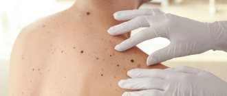
Pigmented formations usually do not cause problems for humans. Moles that are not prone to degeneration do not change in size or color. It is necessary to consult a doctor when suspicious changes appear on their part:
- The surface becomes glossy
- The shape changes (the edges are uneven, asymmetrical)
- Starts to grow
- Unpleasant sensations such as itching appeared
- Hair falling out from a mole
- The color has changed
- Became very soft or, on the contrary, dense
- Dry crusts form on the surface
- Starts to bleed and hurt
- Nodules have formed on the mole or around it
- Bubbles and small ulcers form on the surface
- Appearance of a halo
Sometimes nevus can be located in unpredictable places, in both men and women. If at the same time it is regularly subjected to any mechanical influences, then there is no need to wait until it manifests itself in an undesirable way.
Consultation with a doctor and timely removal can save health and life.
“Should I remove moles or not?”
There are formations on the skin that need to be removed because there is a high risk of cancer.
Mostly such formations are excised with mandatory histological examination of the surgical material. Benign, non-precancerous skin formations must be removed for cosmetic reasons, in case of unpleasant sensations (trauma, itching), if permanent damage to the formation or other discomfort occurs.
In any case, you should consult a dermatologist or oncologist about whether moles need to be removed. He should also recommend the optimal removal method.
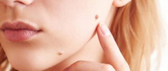
Is a mole on the chest dangerous?
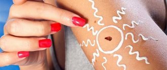
Most nevi are not dangerous and do not require treatment. If they are located on open areas of the body and are often exposed to sunlight, you can simply cover them with a bandage of gauze or bandage.
Pigmented nevi are at risk if they are damaged due to injury, rubbing, or exposure to ultraviolet radiation. At the same time, there is a danger of degeneration into one of the most dangerous forms of malignant tumors - melanoma.
The locations of birthmarks can be the most unexpected. Sometimes they are in places where there is a greater risk of injury. For example, the sole of the foot, palm, neck, waist, chest.
A mole can add sexuality to a woman, but it can also threaten her health. There can be a danger if the nevus is located in the areas where the bra adheres to the body. Regular friction can cause the degeneration of a mole. Sometimes nevi are convex, hanging formations that look like a papilla. The location of such a mole on the chest is a cosmetic defect and is dangerous because its integrity can be damaged, for example, when undressing. Red moles on the breast are not dangerous and mean hidden dangers in the body.
It is interesting that our ancestors considered moles on women’s breasts to be signs of fate, and not a threat to health.
This is understandable, since women tanned little, valued white skin, and exposed their bodies less in the summer. Importance was given to where the mole was located, on the left, right breast or in the middle. For example, a birthmark on the right promised a happy marriage for a woman and the birth of a child on time. On the left - it meant that the woman would not have a good personal life, although she would have many fans.
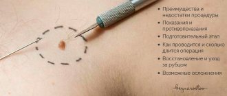
Ways to remove moles
- Surgical
All formations with minimal suspicion of malignancy are subject to excision within healthy tissue, with mandatory histological examination of the surgical material. Also, the surgical method is preferable for some large formations (more than 1.5 cm) from the point of view of the healing process and cosmetic effect.
- Minimally invasive: laser destruction, radio wave method
- Laser destruction
Most preferred for convex lesions, pedunculated lesions, seborrheic keratomas and warts, including large ones.
The advantage of the method is instant coagulation, without damage to adjacent tissues, without bleeding and instant formation of a crust on the wound. Laser destruction, unlike radioknife and electrocoagulation, does not conduct through the human body and is acceptable in patients with pacemakers and pacemakers.
During the laser destruction process, no ionizing radiation is generated and the impact is limited to the point of application, and is allowed in children and pregnant women. The method is practically non-contact, has a very high temperature at the point of application, and therefore eliminates the possibility of contracting infections such as HIV or hepatitis. The disadvantage of this method is that in most cases the removed material is not suitable for histological examination, so laser destruction is suitable for removing exclusively benign formations that do not raise doubts about the diagnosis.
- Radio wave method
It is a dissection of soft tissues in a non-traumatic manner using narrowly directed high-frequency radio waves, it allows you to make thin incisions with the adhesion of small vessels, sterilization of the surface - the high-frequency radio wave destroys microorganisms.
The method is suitable for removing small formations that require histological examination, since during removal the surgical material is minimally damaged or as an alternative to a scalpel when excising a formation. It has contraindications - pacemakers, epilepsy, glaucoma, diabetes, pregnancy, inflammatory processes in the acute stage.
Not suitable for removing “dry” formations - viral warts, formations with abundant keratosis.
Is it necessary to remove a nevus?
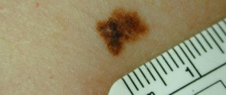
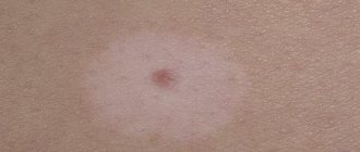
Most moles are not dangerous. And although they differ in appearance, they usually have the correct shape, uniform coloring and small size. Nevi are considered dangerous:
- Appearing in adults
- Diameter more than 1 cm
- Changing
You need to monitor such moles, and if you have any suspicions, consult a doctor. Moles can also be removed if they represent a cosmetic defect. This requires a preliminary consultation with a doctor or oncodermatologist. The nevus is removed by a doctor. Under no circumstances should you do this yourself, at home, without a preliminary examination and appropriate tools and equipment. Any injury or partial removal of a nevus can provoke its degeneration.
Ways to remove moles
Modern clinics offer the following methods for removing nevi:
- Surgical is the most common. It is used to excise large and deep formations, also when the process of degeneration into an oncological tumor has begun. The disadvantage of this method is the scars that remain after removal. It is undesirable when applied to the face.
- Cryodestruction is the removal of a nevus using liquid nitrogen. Nevus cells are destroyed by exposure to cold. This method has its pros and cons and is used very rarely. A mole treated with liquid nitrogen does not need to be removed; it dries out and forms a dry crust. There is no need for wound treatment or dressings. The disadvantage of this method is that during treatment of the mole with nitrogen, healthy cells may be damaged. It also does not guarantee complete removal of the formation. A repeat procedure may be required.
- Electrocoagulation is exposure to high-frequency current. Cell destruction occurs due to thermal effects. Next, the nevus can be sent for biopsy. However, after removal, scars also remain.
- Laser method - exposure to a laser beam. This method allows you to treat formations of various sizes without harming the surrounding healthy tissue. Removing small nevi leaves no traces, but large ones may leave discolored spots.
- Radiosurgery is a method that uses a radiocoagulator. The most effective way to remove nevi today. The device simultaneously cuts out bad tissue, cauterizes capillaries, and disinfects the wound. The removed nevus can then be tested for cancer.
Only a doctor can decide which method to use to successfully eliminate a dangerous mole.
Types and reasons
Moles (the medical term is nevi) on the top of the head and any part of the scalp are common. It’s just that the formations are usually hidden from view. The reasons for the appearance of the marks have not yet been identified with one hundred percent accuracy, which provides a strong argument in favor of the esoteric version of signs given from above at birth. Doctors believe that multiple accumulations of melanin in the upper part of the body and the expectant mother’s long exposure to the sun contribute to the appearance of formations in the child, although they cannot explain why this substance accumulates in a certain organ of the body.
Most owners of marks live with the understanding that moles on the head in the hair are dangerous, because a comb, shampoo, dyes and cosmetics constantly come into contact with them and can cause not only pain, but also serious damage. This is especially true for those who have many convex spherical nevi. Before deciding whether to remove them or not, you need to conduct a thorough study and determine what type of growth it is.
Nevi are divided into 2 types: congenital, forming already in the womb, and acquired - appearing a few days after birth or before the age of three due to prolonged exposure to the sun. This often happens to light-skinned immigrants who move to southern countries. The structure of formations is very diverse:
- vascular - located near the capillaries, scarlet or bluish in color;
- melanin - dark in color, appear in places of increased accumulation of the substance of the same name;
- convex - sometimes covered with villi, rising above the surface in the form of an elongated ball, easily damaged and most often removed on the recommendation of a doctor;
- flat - do not cause inconvenience, it is difficult to injure them, but can be potentially dangerous if a person is constantly under the burning sun;
- hanging - often formed from convex ones, which turn into a ball on a string of skin, and pose a threat, so they can be removed promptly in a hospital without any problems.
Is it possible to remove all moles with laser?
During the consultation, when a patient requests removal of a mole, the doctor carefully examines the mole and selects the most suitable removal method. If the mole has no lesions and dermatoscopy or siascopy does not reveal signs of atypia, removal for medical purposes is not required.
In this case, the removal of a mole is carried out only when the patient wishes it for aesthetic or comfort reasons, and in each case the method of removing a specific mole is selected individually. However, even unchanged moles are recommended to be removed if they are often injured by clothing, jewelry, shaving, and so on.
Moles are usually removed with a laser after administration of local analgesics. Laser mole removal is a quick procedure and the wound heals faster than surgical removal as there are no stitches, so a better cosmetic result is achieved. In addition, the likelihood of postoperative complications is much lower.