Thirdly, ultraviolet radiation could have a negative effect. Despite the fact that tanning makes the skin beautiful, it can also negatively affect human health. Due to systematic sunburn, new moles may appear and old ones may change, so it is not recommended to stay on the beach or in the solarium for too long without the protection of special products.
Fourthly, self-removal of a mole or the hair that grew from it could provoke redness. Doctors do not recommend trying to remove a nevus on your own using pharmaceutical drugs or traditional medicine. You will not be able to do this properly, since only the superficial manifestation of the neoplasm will be removed, and its base will still remain in the deep layers of the skin. It is also not recommended to pull out hairs from a mole, since in this case it will be damaged, and you will not be surprised if it becomes inflamed, red or itchy.
Fifthly, hormonal changes may manifest themselves in this way. In some cases, a change in the shade of a mole may indicate an imbalance of hormones in the body. This can happen during puberty, pregnancy, or menopause in women. If your case does not fit those listed, you may have a hormonal disease, for which you need to see a doctor as soon as possible.
Finally, the reason that a mole is inflamed, red or itchy may be degeneration into melanoma. This is the most dangerous reason for a change in the shade of a nevus; the advanced process of malignancy, that is, the transformation of a benign neoplasm into a malignant one (melanoma) can lead to death. In this case, you must definitely go to the oncology hospital for tests and consultation with a surgeon.
Causes of inflammation of birthmarks
The appearance of redness around the mole indicates the beginning of the inflammation process.
In most cases, it is a consequence of the following reasons:
- Mechanical impact. Redness around a mole often occurs due to friction with clothing or accidental hand contact, such as during bathing or scratching. Men often forget about them when shaving and can easily cut the top layer of skin, causing inflammation. In girls, the same problem concerns the groin area, legs and armpits. Damage and inflammation of hanging moles occur especially often due to their structure;
- Chemical damage. Inflammation of the mole in this case is a consequence of exposure to various chemicals, for example, detergent or reagents;
- Radiation exposure. Radiation is one of the main factors in the development of cancer, therefore, with prolonged exposure, a mole on the face or any other place can become inflamed and painful;
- Hormonal surges. Hormones have a strong influence on the body, so they are often the cause of a reddened birthmark;
- Sun rays. Ultraviolet radiation has a detrimental effect on moles not covered by clothing, which can cause them to become inflamed. Many people confuse this process with a banal sunburn, but in fact it may begin to degenerate into melanoma.
Less common reasons include:
- Bleeding into melanocytes (the cells that give color to the mole);
- Infection in the nevus due to injury.
Moles that require attention
Not every mole can develop into cancer, but some of them need to be closely monitored:
- Convex nevi. They become very inflamed when injured and can lead to cancer;
- Moles on the nipple. In girls who are breastfeeding, they are often damaged due to the nature of this process, so nevi can become inflamed and painful. For males, this problem is relevant in the summer, when many of them walk bare-chested all day in the open sun;
- On the arms and legs. The degeneration of nevi into malignant formations often occurs when they are localized on the extremities. These places are regularly injured, and the process of inflammation begins;
- Nevi on the face. You need to be careful with moles on your face, as they are constantly exposed to sunlight and come into contact with your hands, so it is extremely easy to injure them.
Watery bubble on a mole - No warts!
The appearance of moles in various parts of the body is associated with the division of melanocyte cells. When there is a large accumulation, they break out in the form of a nevus. They do not pose a health hazard if there are no factors influencing the process of degeneration into melanoma.
One of the reasons why a water bubble appears on a mole is ultraviolet radiation. Cases of degeneration are often observed in southern countries where the weather is sunny all year round.
In the northern regions, solar activity is lower, but there is no need to neglect preventive measures and sunbathe during dangerous hours. In childhood, sunburn increases the risk of developing skin cancer.
Ultraviolet radiation accelerates the aging of the skin with the appearance of age spots.
Sometimes in the depths of a mole there may be a dangerous type of fungus or larvae, which, under the influence of the sun, begin to actively multiply, forming a bubble on the mole. Changes in education occur for various reasons.
Features of the appearance of a bubble on a mole and the release of fluid from it
A watery mole is a sign of degeneration into a malignant formation that requires immediate treatment. You should not self-medicate; you need to seek help from specialists.
Symptoms
It is possible to respond in a timely manner to the process of inflammation of the birthmark that has begun quite quickly, since the external signs are quite bright and appear approximately 2-3 days after the influence of the irritating factor. The main symptoms are as follows:
- There is redness and swelling around the birthmark, which is very painful;
- Nevus can change its coloring. It may become brighter or fade;
- As inflammation develops, the contours of the mole may change. Most often, they become unclear, and the formation can itch and cause pain upon palpation.
Actions of the patient in case of inflammation of a mole
What to do if a mole becomes inflamed? When the first signs of inflammation appear, you should consult a dermatologist or oncodermatologist. The specialist will interview the patient to find out about any symptoms that are occurring and examine the affected nevus. In the absence of visible reasons that could trigger the onset of the inflammatory process, the doctor will advise you to donate blood for hormones. If the mole turns red and has been inflamed more than once before, then there is a suspicion that cancer has begun to develop. The patient is redirected to oncology to continue diagnostics. The oncologist may recommend the following actions:
- Taking a skin sample from the affected mole for analysis. This method will provide comprehensive information about the beginning of the process of rebirth or its absence. However, unnecessary trauma to the affected nevus may cause the situation to worsen;
- Using computer diagnostics or dermatoscopy to examine the skin layer;
- Mole removal.
Features of the degeneration of moles into a malignant formation
Melanoma or cancer poses a serious threat to human life and health. In medical practice, recovery is recorded only in 50 cases out of 100. Today, this type of cancer accounts for approximately 10% of the total statistics. Unfortunately, according to worldwide data, 40,000 people die from this disease every year. To prevent the development of a dangerous pathology, it is recommended to be attentive to the condition of the skin and, if abnormalities are detected, seek advice from a doctor.
A person should know the following symptoms that indicate the start of the rebirth process:
- The structure of the nevus begins to slowly change. One formation may contain several shades and colors at once. When palpated, compaction or excessive softness is noticeable. In this case, the condition is negative and requires immediate consultation with a doctor.
- The outlines lose clarity, and the formation begins to protrude above the surface of the skin. Additionally, it should be noted that a mole in its normal state should have symmetrical edges.
- The mole is covered with a crust, on which cracks appear over time. The formation may also ooze fluid and blood.
- Pigmentation changes the character and causes discomfort to a person.
- The nevus disappeared, and after that a new formation appeared in its place.
- Hair began to fall out around the mole.
A crust can also form on a mole if it has been injured. The situation arises in case of regular scratching. In this case, the formation of a crust is considered completely normal.
After a certain period of time, it will heal or become covered with pink skin. If the patient had a burn before, the crust becomes black. A person can get such a burn in a bathhouse or sauna. It is important to prevent the development of infection, because due to it the nature of the disease can be aggravated.
Causes of redness of a mole
You need to constantly monitor moles on your body. Redness of the nevus or the appearance of a halo around it is an alarming symptom that often appears during inflammation or the initial stage of oncology. In both cases, mole cells respond to health problems by activating or decreasing the production of melanin (coloring pigment).
In the worst case, inflammation of the nevus indicates the degeneration of the formation into a malignant tumor. But most often, redness appears as a result of trauma to the tissue of the mole and the penetration of dirt and pathogenic microbes into the wound. In rare cases, such a reaction can be triggered by taking medications during the treatment of acute forms of chronic diseases.
Possible complications and precautions
The main complication of a reddened mole is malignancy of the formation. Melanoma occurs. This tumor is malignant. It metastasizes quickly. All internal organs and bones are affected. Melanoma is formed from damaged birthmarks, ordinary moles. Treatment in the early stages of the disease increases the chances of survival several times. Neglected cases end in death. Once completely cured, melanoma may recur within a few years. The life expectancy of patients after recovery depends on the resistance of the body.
The disappointing prognosis and statistics confirm the need for timely diagnosis and treatment of the disease.
Other complications include bleeding, suppurative processes, and the addition of a bacterial infection. In these situations, you should immediately seek advice from a highly qualified specialist.
To protect yourself from serious illness, try to avoid intense tanning, do not visit the solarium, and take care of moles.
Signs of an infected mole
Infection in the nevus tissue can occur as a result of injury. Even minor damage, a light scratch, is a potential danger for the development of inflammation. The following signs indicate that pathogenic microflora begins to multiply in the mole or adjacent tissues:
- redness;
- swelling of the skin around the mole;
- itching and pain;
- bleeding or watery discharge.
If signs of infection appear, you should consult a doctor as soon as possible to prescribe treatment.
What to do if a mole turns red and inflamed
You should immediately consult a doctor (oncologist or dermatologist) for advice!
Pre-medical care for an inflamed mole should consist of keeping the affected area clean and dry. If blood or fluid is released from the nevus, it is necessary to treat it with hydrogen peroxide and cover it with a dry bandage.
If the doctor diagnoses the presence of an infection in the inflamed tissues, he may prescribe a course of oral antibiotics: Analgin, Nimesulide, etc. For local treatment of the infection, antibacterial ointments, for example, tetracycline, are used. Very severe inflammation may require treatment with intravenous antibiotics.
Mole removal
If there is a high probability of developing oncology in the nevus area, then it must be removed. It is also recommended to perform surgery if the mole becomes inflamed again or if it is located in a place where it is often injured.
Your doctor may remove the growth with a minor surgical procedure. But before it is carried out, it is necessary to undergo an examination, including a number of tests:
- general blood analysis;
- research to identify tumor markers;
- analysis of blood clotting indicators.
This examination is carried out to assess the general condition of the body and determine the cause of inflammation. Additionally, tests for drug reactions and allergy tests may be prescribed.
There are several ways to remove a mole:
- Laser surgery is considered one of the safest methods for removing nevi. Therefore, it is suitable even for sensitive skin. Allows you to eliminate a mole in one procedure. The ablation laser used for the operation is the most gentle tool, allowing you to gradually remove the stain layer by layer. The beam is directed to pigmented tissues, leaving healthy epidermis untouched. Performed under local anesthesia.
- Cryodestruction is a method of exposing tissue to low temperatures using liquid nitrogen. The cells of the mole die as a result of freezing, and a crust appears at the site of formation, under which the process of skin regeneration occurs. This is a rather extreme method, after which a long recovery period is required. Cryodestruction is usually not performed to remove moles on the face.
- The radio wave method is based on the use of a special tool in the form of a loop, which cuts off the build-up. And exposure to radio waves cauterizes the wound and prevents bleeding. This method is only suitable for small growths protruding above the surface of the skin.
- The surgical method is a proven and proven method for removing large moles. If there is a suspicion that the nevus is malignant, doctors recommend surgery.
Features of the appearance of a bubble on a mole and the release of fluid from it
A watery mole is a sign of degeneration into a malignant formation that requires immediate treatment. You should not self-medicate; you need to seek help from specialists.
Causes of watery moles
The appearance of moles in various parts of the body is associated with the division of melanocyte cells. When there is a large accumulation, they break out in the form of a nevus. They do not pose a health hazard if there are no factors influencing the process of degeneration into melanoma.
One of the reasons for the appearance of a water bubble on a mole is ultraviolet radiation. Cases of degeneration are often observed in southern countries where the weather is sunny all year round.
In the northern regions, solar activity is lower, but there is no need to neglect preventive measures and sunbathe during dangerous hours. In childhood, sunburn increases the risk of developing skin cancer.
Ultraviolet radiation accelerates the aging of the skin with the appearance of age spots.
Sometimes in the depths of a mole there may be a dangerous type of fungus or larvae, which, under the influence of the sun, begin to actively multiply, forming a bubble on the mole. Changes in education occur for various reasons.
What to do if fluid comes out of a mole
You can identify a water mole by regularly examining your body. If fluid is released from a mole, it is recommended to contact a clinic to diagnose the formation for malignancy.
Watery discharge is one of the alarming signs of degeneration. It is not recommended to self-medicate: this will cause pain relief, not healing.
Lack of proper treatment will contribute to the development of cancer.
During the examination, the doctor will find out the reasons for the changes. If cancer is suspected, the patient will have to undergo examination. First, material is taken for histological examination to determine the nature of the formation.
If oncology is confirmed, a series of tests are prescribed that reveal the spread of metastases affecting the lymphatic system and organs. Based on the test results, the doctor prescribes treatment.
It is recommended to remove a water mole.
Modern medicine has several methods of removal:
- Surgical intervention. A safe method for removing nevi in the presence of cancerous signs. Excision of the tumor and its roots along with the surrounding skin to prevent spread. Surgery removes large, flat growths and deep-growing ones.
- Cryodestruction – removal by freezing with liquid nitrogen. Exposure to nitrogen causes tissue necrosis, a bubble with watery content appears on the surface, which bursts on its own. A crust forms, after which it peels off leaving a pink spot. The disadvantage of the method is the inability to calculate the depth of nitrogen exposure. In the case of malignant formation, further development of melanoma is possible.
- Laser therapy helps remove small nevus. In case of metastasis, the method is ineffective - it is impossible to remove the tumor completely.
- The radio wave method is effective in removing small benign formations. A small tumor can only be removed at the first stage.
Are weeping moles dangerous?
If a mole on the surface of the skin begins to become weeping, this is a sign of abnormal behavior of a benign tumor; it may begin to transform into melanoma. Dangerous symptoms:
- fluid, ichor or blood oozes from under the formation;
- the formation of wet blisters on the surface of the formation and the area around it;
- changes in the size, shape and color of the nevus;
- inflammatory process with redness and swelling of the formation. The neglected condition leads to the release of pus with an unpleasant odor;
- long-healing wound;
- itching, burning, pain when touched;
- loss of hair from the birthmark, increase in the area of pigmentation.
If treatment is not started, characteristic symptoms may develop:
- increased fluid secretion;
- increased pain;
- rapid growth of nevus, change in skin color around;
- enlarged lymph nodes;
- the formation of ulcers on the surface, which can produce an unpleasant odor and pus;
- painful formations appear;
- bleeding;
- compacted structure of formations.
Melanoma grows by metastases, their signs:
- chronic obsessive cough;
- a sharp decrease in body weight;
- headache;
- muscle spasms.
The nevus begins to become wet due to the development of malignant melanoma, which manifests itself in different shades and asymmetrical shapes of nevi measuring more than 6 mm.
Timely treatment will help avoid serious complications that can lead to death.
The tumor can spread quickly, growing through several layers of the skin, traveling through the blood and lymphatic vessels. The liver, brain, and lungs are often affected.
Rules for caring for water moles
If a water blister forms on the nevus, do not self-medicate. The treatment is carried out by a doctor in the clinic.
Under no circumstances should you puncture a blister!
Amateur activity is fraught with the development of the process of transformation of a malignant tumor. If alarming signs appear, visit a dermatologist or oncologist who will prescribe the correct treatment.
Link to main publication
articles:
Loading…
Didn't find suitable advice?
or see all questions...
Source: https://ProMelanin.ru/rodinki/vodyanistaya.html
Prevention of inflammation
Typically, adults have no more than 40 moles on their body. People with lighter skin tend to have more spots. These can change as a person ages. Some become darker or lighter, and may increase slightly in size.
Most moles are harmless, but people should check them for changes and follow simple tips to prevent breakouts:
- Protect skin from direct sunlight;
- Avoid excessive tanning and sunburn;
- Be extremely careful when visiting a solarium;
- Use sunscreen;
- Wear clothes that do not rub moles;
- In case of trauma to nevus tissue, carry out timely disinfection.
Taking steps to protect your moles will help reduce your risk of developing inflammation and skin cancer.
A birthmark is a benign formation that consists of pigment cells. By its nature, it does not pose a health hazard. However, if you discover that the mole has swollen, reddened, or begun to itch, you should contact a dermatologist for examination. Dermatoscopy will determine the cause of the modification of the mole, on the basis of which the doctor will make a diagnosis and prescribe treatment. It is forbidden to treat or get rid of a nevus on your own; this will aggravate the situation, resulting in consequences.
How quickly does melanoma develop from a mole?
The transformation of a nevus into a cancerous formation can occur in different ways. The process depends on the stage of the disease and the type of tumor. Instant metastases are dangerous. Begins:
- growth of cancer (oncological) cells in the deep layers of the epidermis;
- their entry into the blood and lymph;
- penetration into the lungs, liver, kidneys;
- growth in these organs;
- complete damage to the body;
- death.
The growth phases of pigment cells are observed, along which melanoma develops from a mole. There are varieties:
- horizontal – damage to the upper layers of the skin occurs, lasting up to 10 years, but metastases do not appear;
- vertical – accompanied by the spread of cancer cells throughout the organs, can last two years, has an unfavorable prognosis;
- nodular - especially dangerous - characterized by deep spread within two months.
The first signs of melanoma
The patient can be assisted only when suspicious changes begin to be identified. The diagnosis, research, and referral for surgical treatment save a person’s life. The first signs of melanoma:
- increase in the height of the tumor;
- bleeding;
- the appearance of discharge;
- redness;
- burning, itching;
- swelling of tissues;
- softening of the nevus;
- the appearance of a crust;
- thickening;
- hair loss;
- expansion of pigmentation around the lesion.
With the further development of dangerous melanoma, the following are observed:
- significant change in size;
- the appearance of pain;
- enlarged lymph nodes;
- surface ulceration;
- formation of new foci;
- bleeding from places of pigmentation;
- liquid separation;
- skin thickening;
- the appearance of an earthy tint;
- signs of metastases are chronic cough, weight loss, cramps, headaches.
How to distinguish a mole from melanoma
To recognize which moles are dangerous and which are not dangerous, you need to know what they look like. A person with nevi, in order to eliminate terrible consequences, must constantly monitor the appearance of new formations and changes that occur. You can distinguish a mole from melanoma by its signs. Non-dangerous neoplasm:
- symmetrical;
- with smooth edges;
- uniform in color;
- with dimensions not exceeding 6 millimeters.
Features of dangerous melanoma that require seeking help from dermatologists:
- growth in a short time;
- pronounced asymmetry of shape;
- heterogeneity in color - the presence of inclusions of several shades;
- lack of clear boundaries - the contour line is blurred, jagged, and looks like a coastline on a geographical map;
- increased diameter over six millimeters;
- variability of any parameters - color, size, shape.
Why can a mole swell?
Traumatic factors that cause a swollen mole:
- Mechanical damage. Nevi do not affect a person’s life rhythm, but if they are poorly placed, there is a risk of injury. Such places include the neck, head, armpits, groin, legs and face. Items of clothing, shoes, and hair removal cause rubbing or tearing off of the mole. Children are more often exposed to mechanical damage.
- Chemical exposure. Products that consist of chemical components have a negative effect. Cosmetics containing excessive amounts of synthetics, hygiene products or household chemicals that contain abrasive substances.
In the absence of injury to the nevus, redness or swelling occurs due to the following factors:
- The beginning of the process of degeneration of a mole into a malignant tumor poses a danger to life. At the initial stage, melanoma can be treated without the risk of death. The condition is a timely visit to a doctor who will conduct an examination and identify the nature of the mole.
- Prolonged exposure to open sunlight provokes the appearance of new nevi or the transformation of old spots. It is not recommended to be in the sun in the summer from 10 am to 4 pm, when ultraviolet radiation is most aggressive.
- Hormonal imbalances appear in adolescence, during pregnancy, menopause, and lactation. Hormonal imbalance also occurs during treatment with hormonal drugs, abortion, and thyroid diseases.
- When you try to remove the formation yourself. There are means and methods of traditional medicine that promise to rid a person of the stain. Doctors say that resorting to self-removal is prohibited. With the help of such means, you can get rid of the upper layer of the nevus, while the base of the spot remains deep in the dermis.
- A hair was removed from the formation. Removing hair from the surface of the formation causes skin damage and increases the risk of infection. The onset of the inflammatory process is characterized by itching and swelling.
Features of the appearance of a bubble on a mole and the release of fluid from it. Water bubble on a mole
The appearance of moles in various parts of the body is associated with the division of melanocyte cells.
When there is a large accumulation, they break out in the form of a nevus. They do not pose a health hazard if there are no factors influencing the process of degeneration into melanoma. One of the reasons why a water bubble appears on a mole is ultraviolet radiation. Cases of degeneration are often observed in southern countries where the weather is sunny all year round.
In the northern regions, solar activity is lower, but there is no need to neglect preventive measures and sunbathe during dangerous hours. In childhood, sunburn increases the risk of developing skin cancer.
Ultraviolet radiation accelerates the aging of the skin with the appearance of age spots.
Sometimes in the depths of a mole there may be a dangerous type of fungus or larvae, which, under the influence of the sun, begin to actively multiply, forming a bubble on the mole. Changes in education occur for various reasons.
After injury or burn
A benign formation can be accidentally damaged if it is located in a dangerous place. On the head - accidentally cut with scissors when cutting, rip off with a comb.
On the neck - clothes or jewelry. Blisters under moles may appear in the facial area due to an allergic reaction to low-quality cosmetics.
It is possible to provoke discharge by pulling out hair from the surface of the birthmark.
It is possible to start the process of malignancy by getting rid of a water mole using traditional medicine.
In a sauna or bathhouse, a thermal burn of the nevus, which becomes covered with a dark, dry crust, is possible. As the disease progresses, fluid begins to leak from the mole. You can get a burn after visiting a solarium.
A watery bubble with white liquid appears on the surface. If there are many small vessels in the mole, the liquid in the bladder may be red.
It is dangerous for those with fair skin to be in the open sun: there is a risk of burning not only the skin, but also nevi located on the back and shoulders.
Sudden appearance of a blister
A blister may appear suddenly on a benign lesion due to heredity. Due to a genetic disorder, the skin is not able to adapt to the aggressive influence of external factors. The predisposition is passed on from generation to generation; in the presence of pathology, constant monitoring by a dermatologist and oncologist is recommended.
Dangerous moles: symptoms of melanoma
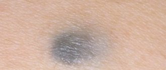
Cancerous pigmentation can vary greatly in its symptoms. Sometimes a person is able to adequately assess only some of the features. You should pay attention to what a dangerous mole looks like:
Uneven edges, but a fairly clear border with healthy tissue. Diameter – 10 mm.
Blue-black newly formed melanoma that has irregular borders. It arose from a dysplastic nevus (pink-brown area in the upper left corner). Size about 12 mm.
Oncological dysplastic nevus with black distant malignant extension, which was previously absent. It is only about 3 mm.
A malignant skin tumor consisting of three parts: a dark brown area on the left, a red area on the right and a light area on top. Size – about 15 mm.
When there is cause for concern
Although the last reason is the most serious and requires immediate medical attention, it occurs less frequently than the others. Therefore, there is no need to panic as soon as you notice a pimple on a mole on your face. Of course, it never hurts to see a dermatologist to either dispel your fears or take action on time.
In what cases do you really need to “sound the alarm”? Here are some of them:
- the birthmark has changed color and shape;
- the area became painful;
- itching is felt around the spot;
- bloody formations appeared;
- hair fell out around the inflamed area.
If these symptoms appear, you should see a doctor. If a pimple appears next to a nevus and looks as usual, there is most likely no reason to worry. But if you notice that acne appears on moles very often, this could theoretically indicate the development of melanoma - skin cancer.
However, only a specialist can diagnose this cancer by personally examining the suspicious area and conducting histological studies. If melanoma is nevertheless detected, then you can be glad that it is at an early stage, that is, you can get rid of it forever.
What can be done?
Most of us tend to fight acne on our own, without consulting a doctor or cosmetologist. There are a lot of procedures and remedies, both medical and folk, on how you can get rid of annoying acne. However, you should be careful.
https://www.youtube.com/watch?v=4q_FgHF7-II
If a pimple appears on the face or body, it is not recommended to squeeze it out yourself. Moreover, you can’t put pressure if he jumped up on a birthmark.
It is not uncommon to hear how someone ignored this advice and still squeezed out an unsightly blackhead. Result? Even more severe inflammation and spread of infection to healthy areas of the skin. There is a double danger here: infection and mechanical damage to the mole itself. And this is the first step towards developing cancer.
Therefore, it is impossible to crush acne on nevi. Instead, you should wait until they ripen on their own. And to protect yourself, they need to be disinfected. To do this, you can use simple tools available in every home. This is iodine or alcohol.
They will stop the development of the infection and prevent its spread into the tissues. These products will also dry out the pimple and relieve inflammation. Dip a cotton swab into the solution and wipe the growth with a pointed motion.
Repeat after a couple of hours several times a day.
Some people treat acne with hydrogen peroxide. It is undesirable to do this when it comes to a mole, since peroxide can damage its very structure.
Source: https://bolezn.info/kozha/osobennosti-poyavleniya-puzyrya-na-rodinke-i-vydeleniya-iz-nego-zhidkosti-vodyanoj-puzyr-na-rodinke.html
What to do if a mole is swollen
When a hanging nevus swells, it is forbidden to apply a cotton swab soaked in alcohol or iodine to the inflamed area. If itching and burning occurs on a swollen mole, you should avoid contact of the mole with external irritants and do not scratch. If severe itching is present, you can relieve the condition with a vinegar solution. To do this, soak a cotton swab in a weak vinegar solution and apply it to the itchy area for 5 minutes. If the mole is swollen and painful, it is not recommended to press on it while wetting it with a vinegar solution. After applying the tampon, lubricate the bladder with three percent hydrogen peroxide. This will reduce the risk of infection getting inside the growth.
Treatment and medical intervention
When contacting a dermatologist, a person will undergo a siascopy, during which the stage of the inflammatory process is determined. After examining the patient, the dermatologist sends him to an oncologist, who prescribes tests. Histological examination can detect melanoma. Based on the histology results, appropriate treatment is prescribed. If no danger is detected, the swollen nevus will be cauterized. If a pathological process inside the formation is detected, the doctor will prescribe removal of the mole, which is carried out using the following methods:
- Laser removal is based on cauterization of the nevus using a laser. The method is safe and does not leave a scar on the body.
- Cryodestruction. Involves the use of liquid nitrogen. Used for medium and small moles.
- Radio wave removal involves the use of high-frequency current, which removes the inflamed nevus layer by layer.
- The surgical method is based on the use of a scalpel. Before the procedure, the patient is given local anesthesia. The disadvantage of the operation is the scar. Surgical intervention is necessary for large nevi.
The operation lasts half an hour. The duration of the procedure depends on the location and size of the nevus. The doctor treats the skin area with a disinfectant, administers an anesthetic, and removes the formation. After excision, it is treated with a solution that will reduce the risk of infection and sutures are applied. The removed lesion is sent for histology. The patient is advised if a mole is swollen, what it means, and how to care for the wound.
If there is no need to remove the birthmark, the doctor will prescribe medication treatment. To relieve the inflammatory process, use calendula tincture or ointment, which contains an antibiotic. Wipe the swollen area with a cotton swab dipped in tincture or ointment. Carry out the procedure until the abscess disappears. An effective remedy for removing swelling is streptocide. Crush two tablets and sprinkle on the nevus. After 15 minutes, wipe the birthmark with hydrogen peroxide.
You can relieve swelling with flax oil, which heals tissue. Pour ten drops of oil onto a cotton swab and apply to the inflamed nevus. Carry out the procedure three times a day for no more than five days. Tincture of celandine helps reduce the blister. Apply a cotton swab soaked in the solution to the mole, and stick an adhesive plaster on top. After 15 minutes, remove the lotion. Perform the procedure three to four times every day for two weeks.
An effective remedy is a mixture of garlic and lemon juice. Apply the components one at a time no more than twice a day. An ointment made from hemp oil can relieve redness, itching and thickening. To prepare you will need 15 grams of chalk and 3 pieces of butter. The treatment is carried out for 10 days three times a day. You can add fig juice to the mixture.