The skin is a huge organ with a total area of up to 2.5 square meters. in an adult, performing many functions.
It protects the body from unfavorable environmental factors, ensures thermoregulation, respiration and other important processes.
The integument consists of three main layers - epidermis, dermis and subcutaneous fat.
All tissues are formed from cells that continually grow and die. Sometimes the coherence of their work is disrupted, the cells begin to divide randomly, forming a pathological tumor - a neoplasm. There are several dozen varieties, both absolutely harmless and hazardous to health, including:
- nevi, moles, birthmarks;
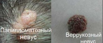
- warts and papillomas;
- dermatofibromas.
Each species differs not only in external characteristics, but also in the causes of its occurrence. Some need to be removed, while others are better just monitored. Some appear from birth, others develop throughout life.
If you learn to recognize them, you will be able to protect yourself from serious consequences, for example, infection or the degeneration of a benign form into a malignant one.
The main differences between moles
In medical practice, moles are called nevi; they can differ in size and color. In most cases, moles are red, black or brown in color, located at the same level with the skin or protrude slightly above its surface.
Moles, as a rule, appear on the human body from the age of 6 months; the formation process lasts a lifetime and occurs with varying degrees of intensity. The appearance of moles is the result of the accumulation of too much pigment in certain areas of the skin.
It should be noted that nevi themselves are absolutely harmless, but under the influence of certain factors they can degenerate into melanoma in a short time. Melanoma is dangerous due to the rapid development of metastases and death in approximately 80% of cases.
The causes of moles should be noted:
- genetic predisposition;
- hormonal changes;
- accumulation of melanin;
- prolonged exposure to the open sun.
Types of moles
Active growth of nevi is often observed during puberty and pregnancy, which is dictated by an increased amount of hormones produced by the pituitary gland.
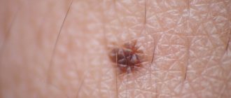
What do nevi look like?
Photo of nevus
Nevi are otherwise called moles. These are congenital or acquired pigmented neoplasms on the skin. They can be genetically determined or appear under the influence of various factors during life. Nevi are formed as a result of the production of melanin by skin cells. It accumulates in the upper layers of the epidermis and forms melanocytes - a coloring pigment.
In most cases, children are born with clear skin, without nevi. It is quite rare for babies to appear with congenital birthmarks on their bodies. As a rule, the first pigmented neoplasms form at 1-2 years of age. With age, the number of nevi on the body increases.
The number of nevi, their natural shade and size depend on genetic characteristics. This is the difference between nevi and papillomas. Typically, children inherit a general tendency to develop age spots from their parents. Identical nevi may also appear on the body of children, like one of their relatives.
In addition, prolonged and regular exposure to the sun can cause increased pigmentation. Also, nevi present on the skin can darken under the influence of ultraviolet radiation.
The nevus is dense and smooth to the touch. Its shape is quite symmetrical, and the edges are normally clear. The shade can vary from light brown to almost black.
When does a mole become dangerous?
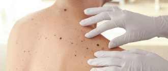
The appearance of moles on the skin is normal; even new nevi that appear in adulthood should not cause concern. Despite this, it is important not to forget that it is necessary to constantly monitor such neoplasms in order to prevent their degeneration.
Doctors advise paying attention to certain symptoms that indicate a possible danger: asymmetry, jagged edge, bleeding, color, change in size.
It is recommended to mentally divide the mole in half; if the halves look different, this indicates danger. The edges of a healthy nevus have smooth boundaries; the patient should suspect something is wrong if the neoplasm has jagged edges.
Another alarming symptom is bleeding; normally, nothing should come out of the mole, otherwise you need to contact a dermatologist or oncologist as quickly as possible for:
- consultations;
- taking tests.
The doctor will suggest taking a photo of the tumor to make it easier to monitor its dynamics. It is normal for a mole to have a uniform color, with no spots or inclusions of different colors. A person should be alerted to changes in the size of moles. Next, you need to consider how a mole differs from a papilloma (photo).
Removing formations
Every person has a certain number of moles on their skin. Sometimes there are few of them or they are located in hidden places, but do not cause concern to humans. But there are formations that cause a feeling of discomfort, appearing in visible areas, for example, on the face, neck, hands. Many people are indifferent to this, but there are also those who want to get rid of such skin formations. There are several modern methods for removing warts, papillomas, moles, condylomas and other unwanted growths.
Surgical intervention
The oldest method, known since ancient times. The operation is performed using a scalpel. Despite the apparent simplicity of the procedure, there are unpleasant side effects: pain, prolonged healing of the operated area, postoperative scars. In modern medicine, this method is practically not used.
Using a laser
This is a non-contact removal method using a medium power red laser, which can precisely adjust the laser beam. With its help, unnecessary cells are heated and evaporated, which makes it possible to cut off the unwanted formation. Due to the small area of exposure of the beam, neighboring tissues remain untouched by the radiation.
Laser removal has some obvious advantages:
- minimal pain;
- fast healing process;
- impossibility of infection during surgery;
- no scars.
The disadvantage of using a laser is the inability to take a tissue sample for histological examination. In addition, there is a technical problem with the laser operating mode. Its impulse is as fast and powerful as possible, which is preceded by a long charging process.
Radio wave method
The method is based on the effect of high-frequency electric current (100−300 kHz) on living tissue. In this case, there is no danger of damage to internal organs, and the current has the ability to remove cells from the skin. This effect is called diathermotomy, and the device that uses it is Surgitron.
Simply put, this is a high-frequency radio wave non-contact scalpel that can remove the smallest layers of skin. The cut boundaries are welded in the same way as when using a laser. The advantages of using Surgitron can be highlighted:
- no pain;
- speed of the process;
- impossibility of infection;
- no bleeding or other complications;
- fast healing;
- preservation of adjacent tissues;
- excellent cosmetic effect;
- possibility of conducting histological examination.
The market price of Surgitron is lower than a laser device, and accordingly, the procedure for removing any tumors is cheaper. The operation of both devices is based on the use of electromagnetic radiation, but with different frequencies. At the same time, the radio wave method is the most progressive and modern.
What is papilloma
Papillomas are also benign neoplasms, but their occurrence is dictated by the presence of the human papillomavirus (HPV) in the patient’s body. A wart can appear on any part of the body, not only on the skin, but also on the mucous membrane and internal organs.
The growths are a consequence of human papillomavirus infection; they are detected in approximately 80% of people in our country. Infection occurs through sexual and domestic contact. Papillomas, like nevi, can be of different sizes and shapes, but can be completely invisible.
The papilloma virus enters the body through the skin and mucous membrane, then begins to multiply, causing pathological changes of varying severity. Symptoms of infection and the warts themselves may not appear for a long time; the patient is unaware of his disease for many years.
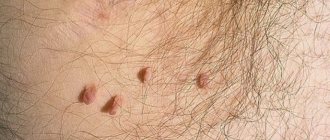
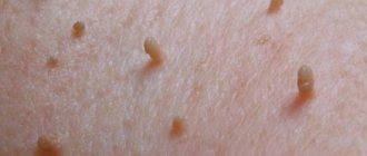
Papillomas on the human body
Papillomas grow when certain factors occur, primarily the following:
- alcoholism;
- decreased immunity;
- frequent infectious diseases;
- diseases of the digestive tract;
- depression and stress.
No less often, the impetus for the growth of warts is: casual sexual intercourse, prolonged use of certain medications (usually antibiotics), visiting public baths, swimming pools.
Papillomas can cause cancer; some types of human papillomavirus are especially dangerous; they are characterized by increased oncogenicity. Doctors say that it is these viruses that provoke cervical cancer; they are much more dangerous than moles.
According to statistics, hanging and large papillomas are most susceptible to degeneration. A person can protect himself if he leads a healthy lifestyle, strengthens his immune system, and eats rationally and properly. Among other things, you must not forget about the basic rules of personal hygiene.
Overview of treatment methods for growths
Treatment of papillomatosis involves analysis to determine the strain and oncogenicity of HPV, viral load, the use of antiviral agents and immunomodulators. Then the formations are removed in various ways.
Nevi are examined using a dermatoscope. Then they are removed using minimally invasive techniques. A histological examination of the biopsy specimen is required to exclude cancer.
Removal of moles and papillomas is carried out in the following ways:
- Evaporation using radio waves or lasers is a minimally invasive intervention. The blood coagulates, the risk of bleeding and infection is minimal.
- Diathermoelectrodestruction - the doctor places a loop electrode on the formation and cuts off the tumor.
- Cryotherapy using liquid nitrogen - the formation is frozen at low temperatures. Approved for use in the treatment of pregnant women and children.
- Surgical intervention is indicated for significant tumor sizes and malignant nature. There is a risk of bleeding and wound infection.
- The use of unconventional methods - attempts to get rid of it using castor oil, homemade ointments, and other traditional medicines.
If a mole begins to grow and its edges change, you should immediately consult a doctor! You should only get rid of tumors in medical institutions!
Dermato-oncologist surgeon, plastic surgeon. Doctor of Medical Sciences, Professor.
Professional experience includes work as a manager. Department of Laser Surgery Department of the LRC of the Ministry of Health of Russia, Head of the Department of the Moscow Regional Oncology Dispensary.
She has the skills to diagnose tumors of the skin and breast, advises on the problems of age-related changes, congenital and acquired defects of the skin and soft tissues of the face and body. Performs surgical interventions using laser, radiofrequency, and endoscopic techniques. Surgical treatment of oncological diseases of the skin, soft tissues, and mammary glands. Performs operations to remove tumors of the skin and mammary glands. Breast reconstruction operations. Aesthetic surgery: blepharoplasty (upper and lower eyelid surgery); face and neck plastic surgery; endoscopic plastic surgery of the forehead, eyebrows; correction of ear shape; changing breast shapes, reconstruction; abdominoplasty (figure correction). Aesthetic surgeries (with the exception of rhinoplasty).
Co-author of 3 methodological recommendations. Author of more than 50 published works.
What is the difference between a mole and a papilloma?
How to distinguish a mole from a papilloma? There are several criteria that help the common man to easily distinguish these tumors. First, you need to pay attention to the pigmentation; if a mole can be of any color, then a papilloma is always an exceptionally light color.
Another sign is the structure, papillomas have a loose structure, and moles are highly hard and dense, but there are cases when the neoplasms in question do not fit these criteria.
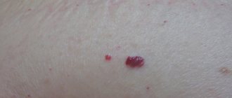
The difference between a mole and a papilloma is also in the presence of blood vessels; if moles do not have blood vessels, then papillomas will become their absolute opposite. Another difference is that moles can be inherited, but warts cannot.
Comparison of a mole with a papilloma
The difference between neoplasms is also in the place of localization; moles appear on almost any part of the skin; warts often appear:
- in places of maximum friction;
- on mucous membranes.
Healthy nevi are characterized by smooth edges and a symmetrical shape, in which they differ from papilloma; these characteristics are not characteristic of them. How to distinguish a mole from a papilloma (photos are available on the Internet) if they are very similar? In this case, you need to pay attention to the size; papillomas are always small in size, no more than one and a half cm. If the wart is damaged, it can begin to grow rapidly, reaching 6 cm in diameter.
Reasons for appearance
The main difference between a papilloma and a mole is the mechanism of its appearance.
The mechanism of mole formation
Moles can begin to form already during intrauterine development. They retain their shape and color throughout their lives. Only under the influence of provoking factors (sun rays, injuries) can they change shape and color . This is a sign of degeneration into melanoma. Such moles should be removed immediately, and under no circumstances should you do it yourself. In this case, the question of surgery is decided by oncologists.
Mechanism of papilloma formation
The main reason for the formation of papilloma is the entry of the HPV virus into the human body. Depending on the state of the immune system, the virus may not manifest itself, but when favorable conditions arise, it begins to actively multiply, which leads to the appearance of a neoplasm . There are capillaries inside the papilloma, and therefore its damage can cause bleeding and the formation of ulcers.
Note! Papillomas are of closed and open type. Open ones look like a kind of crater in which several pointed warts are located. Closed warts have a kind of thin stalk with which they are attached to the skin.
Diagnostic methods
Diagnosis of skin tumors can be carried out in several ways:
- colcoscopy;
- histological examination;
- cytological examination;
- PCR;
- visual inspection.
Diagnosis is usually made based on routine visual examination. If the doctor doubts the type of formation, or wants to determine the type of HPV virus, PCR may be required.