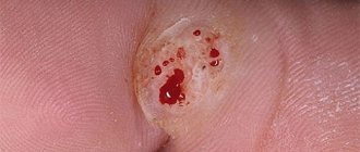
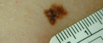
We all know what a mole is - it is a clearly defined spot of light brown or black color. Large moles are called birthmarks, and in scientific terminology - nevi. Birthmarks are common in fair-skinned people and do not cause them any particular concern. But if you find similar changes on your eyeball, you will immediately ask yourself the question: can there be a nevus on the eye or not? The answer is that moles form not only on the skin, but also on the eyeballs. A nevus in the eye can occur in a child, adult, or elderly person. But don’t panic – most often the neoplasm is benign.
To find out the cause of a nevus, it is necessary to understand what the color of a person’s hair, skin and eyes depends on. Their individual shade is given by the specialized pigment melanin, evenly dispersed throughout the cells. In the tissues of people with fair skin, the amount of melanin is minimal - when there is an excess of it in a certain area, a pigment spot is formed. Among the reasons, heredity comes to the fore - if at least one of your parents has a birthmark, do not be surprised if you find it in yourself. Age-related and hormonal changes are the second reason for the formation of spots: adolescence, hormonal fluctuations in men, in women - pregnancy, menopause or menopause. When exposed to ultraviolet radiation, infection or injury, a nevus may also appear. Are there large moles in the eyeball? Are they capable of growing and impairing vision?
The answer is that neoplasms can grow and negatively affect vision, but it is extremely rare - according to statistics, only 1 out of 500 people have a birthmark that progresses into a malignant tumor.
Why does such a tumor appear?
Today, the main cause of nevus on the conjunctiva and eyeball, as well as on other parts of the body, is a large amount of melanin that accumulates on a small fragment of skin and is responsible for the shade of the eyes, skin and hair. Nevi can also form under the influence of other factors, but the main one is disruption of the functioning of the endocrine system and hormonal fluctuations.
Today, there are a number of factors that provoke the occurrence of conjunctival nevi. These include:
- pregnancy (especially in later stages);
- stress and nervous disorders;
- exposure to various radiations on the body (including UV rays);
- infectious diseases;
- menopause;
- natural aging of tissues;
- taking contraceptives;
- inflammatory processes on the skin.
IMPORTANT! Another reason for the appearance of nevi of the conjunctiva and eyeball is genetic predisposition. It is heredity that becomes the main cause of nevi on the eye of young children.
Is it possible to remove a choroidal nevus from the eye?
Removal of a pigmented mole of this type is carried out only in extreme cases, when an ophthalmologist has examined the health of the patient’s organ of vision and determined the presence of malignant cells in the eyeball located inside the nevus. As a rule, such neoplasms begin to sharply increase in size, become darker, and sometimes even provoke an inflammatory process in the eyeball. In this case, the ophthalmologist, in close collaboration with the oncologist, performs electrical excision of an area of eye tissue whose cells contain a large amount of melanin.
At the same time, the surgeon performs plastic surgery of the eyeball. It is also possible to get rid of choroidal nevus using special medical equipment that allows eye microsurgery to be performed with the evaporation of areas of the surface epithelium of the organ of vision that have been excessively saturated with dark pigment. If a benign neoplasm of this type is too large and surgical intervention is contraindicated so as not to injure important elements of the eye, then doctors use laser therapy. Using a beam, the surface layer of the epithelium, which contains cells saturated with melanin, is removed.
Which treatment method to choose is determined solely by the attending oncologist or ophthalmologist-surgeon who will perform surgery to remove the choroidal nevus. The therapeutic process for this neoplasm on the organ of vision is individual in nature and is developed separately for a specific clinical situation. All manipulations on the eye to remove a birthmark from the surface of the pupil or its circumference are performed using local anesthesia and in an operating room. The period for complete healing of the eyeball lasts no longer than 3 days, and the rehabilitation period does not exceed 3-5 days.
Forms of the disease
Modern experts distinguish two types of ocular nevi:
- conjunctival moles;
- nevi of the eyeball.
Conjunctival nevi
The conjunctiva (iris) of the eye is the transparent mucous tissue.
Neoplasms on it can arise both from the outside and from the inside.
Depending on which cells take part in the formation and growth of the nevus, iris pathologies are divided into:
- Vascular. They occur in places where small blood vessels accumulate on the mucous membrane and have a red or pink tint.
- Pigmented. This form of pathology is characterized by a dark color, which is explained by the large amount of melanin in the tissues.
- Cyst nevi , as a rule, they are colorless, and in appearance they resemble small air bubbles. Cyst-like moles appear in places where lymphatic vessels connect.
Sometimes moles on the iris can change under the influence of hormones. In addition, some nevi may lighten over time and even fade completely.
Moles on the eyeball
Just like moles of the iris, nevi of the eyeball can be divided into several types depending on their variability:
- Stationary. This type of mole does not change its color or size. As a rule, no serious problems arise with such neoplasms. They are not dangerous to health and do not require surgical intervention, although examinations by an ophthalmologist should not be ignored.
- Progressive. This group of moles has the ability to change their shape, shade and size. Progressive nevi have a yellow border, which in some cases can cause worsening vision or a decrease in viewing angle.
IMPORTANT! If there is the slightest change in the appearance of the nevus or if a discomfort occurs in the eye, you should immediately consult a specialist.
Mole on the iris of the eye
One of the components of the vascular layer of the eye is the iris. It is rich in pigment, which gives the eyes an individual color. Vascular nevus is red with a light tint, has the appearance of a limited spot, prone to growth. A pigmented mole is located, as it were, inside the iris; if the colors match, it is very difficult to see it. Cystic spots at first glance resemble raw egg white, with limited contours. This version of the nevus is the most harmless and is prone to resorption.
Many people are concerned about the question - how to treat a mole in the eye? Can I lose my vision if the surgery goes wrong?
To treat a nevus, it is better to go to a specialized clinic with trusted specialists. The treatment method mainly depends on the nature of the disease itself - malignant variants capable of proliferation do not require delay. The appearance of a mole that covers the pupil is especially dangerous, as it will lead to loss of vision. However, an experienced ophthalmologist prescribes surgical treatment based on the patient’s age - for example, for a child, removal of a nevus is indicated only if it grows and has a negative effect on vision. Superficial moles on the mucous membrane of the conjunctiva or growing on the cornea are removed quite simply - by electroextraction with plastic surgery of the affected area or microsurgical operation. Hard-to-reach birthmarks that are localized deeply and can involve the retina in the pathological process are treated with laser correction. All of these methods are minimally invasive, which minimizes the destruction of healthy eye tissue.
Attention! Timely diagnosis of growing nevi allows you to prevent precancerous conditions and preserve your vision!
The location of moles is most often chaotic; for more information about the most common locations of moles, see our next video:
Localization and appearance
Nevi can be located on both the external and internal sides. Most often they are localized at the edge of the inner eyelid in the corner of the eye, at the border of the conjunctiva, in the area of the lacrimal caruncle, or in the semilunar fold.
In some exceptional cases, moles appear on the inside of the eyelid or near the pupil, but they do not affect vision.
New growths on the eye can have different shapes and colors. There are flat and convex moles in shape. The hue of nevi can vary from light pink or yellow to blue-black. The color of the mole directly depends on the number and type of cells from which the nevus is formed.
Sometimes colorless neoplasms occur. This is explained by the fact that the cells that form it do not contain pigmentation. As a rule, a conjunctival nevus has clearly defined boundaries, a flat shape and a velvety surface.
Choroidal nevus
This type of mole differs from a conjunctival nevus in that its localization area is the surface of the pupil, the fundus and the equator of the eye. Most often, choroidal nevi occur in the form of a single birthmark on one or both eyes. Also, medical statistics show that the simultaneous appearance of moles of this type in both eyes at once is extremely rare. If this happens, then in 93% of cases the etiology of choroidal nevus is hereditary, and its treatment will most likely result in a relapse of the disease.
By itself, it does not rise above the general level of the eye, and does not have characteristic features or pronounced contours. This is a common brown spot formed due to the saturation of healthy eye cells with the pigment melanin. The diameter of a choroidal nevus ranges from 2 to 4 mm. Excessive pigmentation of this type does not threaten the patient’s health and is absolutely safe, but still requires careful monitoring and observation by both a dermatologist and an ophthalmologist.
Symptoms
Iris nevus usually begins to appear in the first 10-20 days of life. At this stage, local irritation or small pigment spots are observed.
Single, maximally limited, flat or slightly convex epithelial neoplasms appear, which move without problems along the surface of the sclera. Quite often at this stage, cystic cavities form within the boundaries of the nevus.
Depending on the individual characteristics of the body, pigment spots may have different shades, and in some nevi there may be no pigmentation at all.
Pigmentation color ranges from light brown and tan to dark chocolate and blue-black. In adolescence, the nevus may change in size and become more pronounced.
IMPORTANT! Like other nevi, a mole on the iris can be benign or malignant.
A malignant neoplasm on the conjunctiva has the following clinical manifestations:
- unusual location of the formation: the border of the eyelid or the arches of the iris.
- Pigmentation extending to the cornea.
- Sharp growth and change in the shade of pigment spots.
- The appearance and development of vascularization (with the exception of puberty).
Causes
A mole can appear in infants, adults and the elderly. In this case, the ocular nevus can be localized on the iris, on the white or on the cornea. In some cases, the mole is not visible and can only be detected using an ophthalmological microscope.
As a rule, a birthmark is visible from the side. It has a darker pigment composition. Therefore, the nevus is very noticeable in people with light eyes. To understand why it is dangerous, you need to understand the reasons for the formation:
Congenital feature
Then the pupil and white will have a nevus from the first days of the child’s life. In newborns, this spot is often small in size. But over time, the eye changes color and it grows. Moreover, its increase is very intense.
| Congenital nevus does not pose a threat to vision. It does not interfere with visual function and does not affect the functioning of capillaries. |
Acquired nevus
In this case, the mole appears throughout life. And this can happen at any moment. After all, the conjunctiva and the eye are exposed to different influences throughout life. Moreover, such effects can be directed not only directly to the conjunctival region. This is caused by severe stress, hormonal changes, pregnancy, and so on. In any of the above cases, there is a possibility of a birthmark appearing on the eye.
How is it diagnosed?
During the initial examination of the nevus of the iris and the eyeball (the white of the eye), the specialist must pay attention to the following complaints of the patient:
- constant discomfort and sensation of the presence of a foreign body in the eye.
- Cosmetic changes (defects).
- A feeling of “sand” poured into the eyes, which does not leave the patient for a long time.
- Hemorrhage into the mucous membrane.
After listening to the patient’s complaints, the ophthalmologist must examine the eye. He studies the condition of the iris, cornea and mucous membrane. Then, using a special slit lamp, he checks the transparency of the shells.
At the second stage of diagnosing the disease, the following instrumental techniques are used:
- Ophthalmoscopy is a method of studying the retina, small vessels and the condition of the optic nerve by reflecting light from the fundus of the eye. This procedure is carried out using an ophthalmoscope on a dilated pupil.
- Ultrasound of the eye makes it possible to study the condition of the deep components of the eye structure. This technique is used to control the growth of nevus.
- Duplex color scanning allows you to study the structure of the eye and its general blood circulation.
- Angiography, which helps determine the presence of retinal angioma. During the procedure, a fluorescent dye is used, which accumulates in the places where moles appear.
In addition, if necessary, the attending physician may additionally prescribe CT and MRI. Thanks to these diagnostic methods, the malignancy of the neoplasm is confirmed or refuted.
Stationary and progressive nevus
| The appearance of a mole in the eye does not mean a risk of developing cancer. This sign can be completely safe. Moreover, the nevus does not affect visual acuity or eye function in any way. |
Nevus is not considered a disease. This is an education that every person has. It’s just that moles are usually located in other anatomical areas. For example, on the back, arms, and so on.
In this case, the nevus can grow over time. Such formations are called progressive. They change size and shape, gradually increasing. Often, growth occurs at a very rapid pace. So, in one year a mole doubles in size.
If any other spot begins to grow, this indicates a potential cancer threat. But this principle does not work regarding nevus in the eye. It's important to clarify the situation. An increase in the size of the spot does not mean that the mole is turning into a malignant formation. The risk of developing cancer remains throughout the entire period of existence of the nevus in the eye. Therefore, when a mole appears, it is necessary to consult a doctor, undergo an examination and monitor the dynamics of its condition.
Treatment
If the neoplasm is stationary, then treatment is not required. The main thing is to visit an ophthalmologist once a year to prevent the growth and development of a nevus. If the mole begins to grow and change color, treatment must be started immediately. Today, medicine uses several methods for treating nevus. These include:
- microsurgery;
- laser therapy;
- cryodestruction;
- brachytherapy.
Microsurgical removal
This treatment method is suitable for treating all types of nevus. Most often used to combat large and malignant tumors. If a mole creates discomfort, pain occurs, and experts suspect melanoma, then it is recommended to remove the nevus surgically.
During the operation, local anesthesia is used, so there is no need to be afraid of pain. The main advantage of this treatment method is its relatively low cost, compared to other methods of combating nevi.
Laser therapy
Quite often, neoplasms on the eye are associated exclusively with cosmetic defects. But at the same time, everyone forgets that such a pathology can cause loss of vision, and, consequently, significantly worsen the quality of life.
It is for this reason that before surgery it is necessary to undergo a complete eye examination and consult with an oncologist. And if necessary, make a histology of the contents of the nevus. Laser therapy allows treatment without plastic surgery or sutures.
Using a laser, excision of the tumor is carried out within tissues not affected by pathology.
This technique has become widespread due to its effectiveness, safety, and the high speed of patient recovery. And the healing of postoperative wounds occurs with an excellent cosmetic effect and does not leave scars.
Cryodestruction
Today, a very popular method for removing nevi using liquid nitrogen is cryodestruction. The method is based on exposing moles to extreme cold, so that it is enough to completely destroy any organic matter.
Brachytherapy
This method of removing nevi is very simple, but safe. After the brachytherapy procedure, the patient does not feel pain, and it has no consequences or complications.
The main advantage of this procedure is that in one session the body is exposed to several types of radio waves that remove nevi and trigger tissue regeneration. Today, brachytherapy is used as part of a comprehensive treatment or as an independent procedure.
Conjunctival nevus
This is a type of mole on the surface of the eye. They are always especially noticeable, since their localization is not the iris of the pupil, where the pigment spot is not always visible, but the corner of the eye, located closer to the bridge of the nose. A conjunctival nevus is an epithelial growth that can have a regular geometric shape in the form of a sphere, or take the form of a cone. This benign neoplasm does not impair the quality of vision, as it is located at a sufficient distance from the pupil. A conjunctival nevus differs from a choroidal mole in that it is too large and the tumor itself can reach a size of 2-3 mm.
The danger of this type of mole is that its cells quite often degenerate into a cancerous tumor. Therefore, if a foreign epithelial growth is detected located in the inner corner of the eye on the side of the bridge of the nose, you should immediately consult a dermatologist and ophthalmologist. In 30% of cases, a conjunctival nevus develops as a normal neoplasm that does not threaten human life, but after some time, under the influence of unfavorable environmental conditions, the birthmark can acquire all the symptoms of cancer.
Birthmarks on the choroid
In this case, the neoplasm will be located at the junction of the pigment membrane and blood vessels. Such nevi are pigmented or vascular in nature. The difficulty with this type of birthmark is that it is absolutely invisible to others.
This makes diagnosis difficult. Typically, choroidal nevi occur in 2% of the population. At risk are mainly teenagers who are experiencing a period of significant hormonal changes.
A mole in the choroid is small and dark. Rarely, it may be completely devoid of pigmentation. In this case, it will be impossible to determine the presence of a birthmark until a certain point.
The thing is that choroidal nevus, as a rule, is progressive. As the spot grows, it can cause visual disturbances and deterioration. In addition, the person will complain of a feeling of a foreign object in the eye. If such symptoms occur, you should definitely consult an ophthalmologist.
Particular importance should be attached to treatment. To do this, the doctor selects the most appropriate methods, taking into account all the characteristics of the patient’s health condition. Eye microsurgery is often prescribed.
Diagnosis of formations
Detection of a birthmark is carried out using special equipment. After all, it is necessary not only to study the cornea or pupil. The doctor must look inside the eyeball, examine the fundus, and so on. Therefore, various techniques are used:
Ultrasound diagnostics
It is a well-known ultrasound examination. Only the patient's eye is the object. Using this technique, it is possible to identify accumulations of melanin in certain parts of the eye. This test is very accurate and provides a complete picture of what is happening inside the visual organs. There will be no hidden areas, which is very important for further treatment of nevus;
Angiography
This is a type of x-ray examination. It is necessary for studying and assessing the condition of blood vessels. As stated above, a nevus can form directly on a collection of blood vessels in the eye.
During the examination, it is recommended to use both methods. They complement each other perfectly and give an accurate picture of the development of the birthmark. A lot of information can be obtained by studying the nevus over several years.


