In the modern world, people have to lead an active lifestyle. In this regard, they are daily overwhelmed with emotions, which are: stress, excitement or joy. This does not go away without a trace, and manifests itself in the form of an imprint on the body of various spots, warts or moles. Their owners have a question: How to protect themselves from this scourge?
Unfortunately, it is impossible to avoid this completely. Whether we want it or not.
Why does it appear
Moles on the leg and the reasons for their appearance are as follows:
- The influence of ultraviolet rays. With prolonged and frequent exposure to open sunlight from 10 am to 4 pm, hanging formations may form.
- Heredity. If one of the parents has several hanging moles on the neck, a similar phenomenon can be inherited by the child.
- Hormonal imbalances. Pigment formations can form during pregnancy, puberty, a stressful situation, menopause, or termination of pregnancy.
- A pedunculated mole can occur as a result of an insect bite, as a result of which an infection has entered the body through an unhealed wound.
- When the protective crust is injured by an insect bite, the process of activation of melanocytes begins.
- Diseases of the endocrine system provoke the formation of hanging growths.
Is a mole on a leg dangerous?
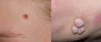
Pedicled nevi are the most dangerous type of moles. The danger is based on the constant mechanical impact on the convex formation, as a result of which the risk of degeneration of a benign growth into a malignant one increases. Another danger is the similarity of the growth on the leg with papilloma. Papilloma is a viral disease caused by the human papilloma virus. Whether a pedunculated mole is dangerous or not can be determined when an examination is carried out, during which the presence or absence of pathology is confirmed.
Locations
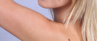
Pedicled formations often appear in women on an open area of the body, resulting in aesthetic discomfort. Common places where hanging nevi are located:
- In the intimate area. The groin location of the growth is less dangerous because it is not exposed to ultraviolet rays and does not cause discomfort. It is necessary to observe the size and color of the nevus. Be careful when removing unwanted hair in the intimate area. If it begins to grow and become inflamed, you should consult a doctor.
- In the armpit. A mole on a leg under the arm causes inconvenience. Carefully remove hair in this area. If you accidentally cut off a mole, the risk of inflammation increases. Sweat glands are located under the armpit; sweat is intensely released, which causes the spread of bacteria. When a nevus is damaged, bacteria are able to penetrate the wound.
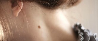
- On the neck. The danger comes from moles located in the collar area. A large formation on the neck is subject to constant contact with clothing or jewelry and is in direct sunlight. This can provoke a violation of their integrity, which will lead to the process of degeneration into a malignant convex formation.
Injury to the nevus will lead to heavy bleeding. If damaged, the area must be treated with a disinfectant and lubricated with brilliant green. To avoid re-injury, you should consult a surgeon for surgery to remove the growth.
When to see a doctor
Regardless of the reason for the appearance of a mole on a leg, it is necessary to know the symptoms that may indicate that the process of transforming a nevus into a malignant formation has begun. These include:
- Rapid increase in the size of a raised mole.
- Discharge of blood or clear fluid from the formation.
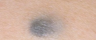
- Change of shade. When degenerated, the nevus may turn red or black.
- Itching and burning in the area of the growth.
- The surface of the mole is covered with small nodules.
- The appearance of small spots around the mole.
- A dry, shiny crust appeared on the surface.
- The raised mole began to peel off.
- A white spot appeared around the formation.
The above symptoms may be a signal of an inflammatory process, caused by a hormonal imbalance, prolonged exposure to the sun in the summer, or thyroid disease. To determine the cause of the appearance of a mole on a leg, you need to visit a dermatologist or oncodermatologist. After the examination and the necessary tests, the doctor will determine treatment tactics or prescribe removal of the raised mole.
Diagnostics
An experienced doctor, upon visual examination, can easily distinguish a hanging mole, since they have a characteristic appearance, color and shape.
If there is a suspicion of malignancy, a fine-needle biopsy is prescribed. This diagnostic method is intended to determine the presence of pathologically altered cells.
The patient is also required to donate blood and urine. Laboratory testing helps determine the presence of papillomavirus, which most often causes the appearance of various growths on the skin. But if it was he who caused their formation, the treatment will be ineffective.
If the color or size of a mole changes, the patient is referred to an oncologist for examination.
Removal methods
To get rid of a pathological or awkwardly located mole, you can choose one of the following methods:
- Removal by radio wave. The thermal effect is able to remove the nevus without contacting it. The method is safe and painless, which can be taken on spots located on different parts of the body. Layer-by-layer removal of the nevus leads to its reduction and disappearance. After the operation, a protective crust will remain, which is not recommended to be peeled off. After two weeks it will dry out and fall off.
- Surgical removal. It is advisable to use surgical removal if the nevus is large or malignant. The doctor uses a scalpel to remove the nevus and its deep part. The sutures that are placed after removal are removed after a week. The disadvantage of this method is the presence of scars and scars, so you should not use this method to remove moles located on a visible area of the body.
- Laser removal. It is a popular but expensive method. The laser action is directed at the base of the convex nevus, during which layer-by-layer disappearance occurs. You can remove small or medium growths with a laser. The advantage of the method is the absence of contraindications and possible penetration of infection into the wound.
- Electrocoagulation method. The build-up is removed using the thermal effect of electric current. The operation is safe and painless. The patient is given a local anesthetic to prevent pain during surgery. The advantage of the method is cauterization of the wound, which prevents infection.
- Cryodestruction method. The removal process uses liquid nitrogen. The operation must be performed by an experienced doctor, since liquid nitrogen can cause an inflammatory process if it gets into a healthy area of the skin.
In addition to medical methods, it is possible to get rid of a nevus on the stem yourself. These methods include:
- Three times a day, cauterize the formation with iodine solution.
- In the morning and at night, lubricate the nevus with a swab dipped in celandine juice. When lubricating, do not violate the boundary of the formation. Celandine juice can be used in pure form or mixed with Vaseline in equal proportions.
- Bandage the stalk of the nevus using your own hair. The method is aimed at gradually stopping the flow of blood to the nevus, as a result of which it will begin to die. After the bandaged mole falls off, you should consult a dermatologist. The doctor will examine the area for any pathological tissue changes.
Diagnostic measures ↑
Diagnosis of hanging moles involves recognizing a malignant formation. For this purpose it is prescribed:
- fluorescent microscopy is a method of histological examination carried out using a special microscope;
- computer diagnostics – makes a differential comparison with similar formations.
- blood test - checks the presence of tumor markers that confirm the existing malignancy.
Luminescence microscopy is one of the diagnostic methods
Doctors recommend removing hanging moles because of their shape and location.
It is not recommended to eliminate them yourself: this can cause inflammation or serve as an impetus for the formation of a malignant tumor.
Currently, the removal procedure is carried out in many medical clinics and beauty salons. When prescribing surgical excision of a mole, a doctor is guided by external data and its location. A specialist will advise and recommend a removal method suitable for each individual case. There are several elimination methods that can be used if necessary.
Drug treatment of moles
How to protect a pedunculated mole from degenerating into cancer
To avoid the conversion process, some rules must be followed. These include:
- Take careful care of the area of skin where the raised nevus is located to avoid drying out.
- Avoid prolonged exposure to the summer sun on the mole.
- Protect from mechanical impact.
- Do not allow household chemicals and cosmetics that contain aggressive components to come into contact with the growth.
- Visit a dermatologist every 6 months to examine and examine the hanging formation.
If you follow the above rules, you don’t have to worry about the growth degenerating into a malignant tumor.
What is the danger?
Growths of this type are benign in nature, but cases of degeneration of the formation into malignant are not uncommon. You should immediately contact a specialist when the following signs appear:
- intensive growth of education;
- painful sensations, itching;
- color change or blackening;
- if there was a pattern on the nevus that disappeared over time;
- the appearance of peeling;
- the appearance of a halo of light or dark color;
- nevus thickening;
- blood is released;
- symptoms of inflammation are present.
The above signs indicate that the formation may have become malignant.
The main threat of hanging moles is injury and complications.
There is also a danger in cases where a mole has come off. In this case, an inflammatory process or bleeding occurs. It is urgent to stop the bleeding using peroxide, then treat the damaged area of skin with brilliant green. Apply a sterile napkin or an antibacterial patch to the wound. After this, you should definitely see a specialist.