In a healthy state, the female genital organs have a uniform light pink color. Any deviations from the norm are a sign of various pathological processes: from improper functioning of the sebaceous glands to dangerous viral or fungal diseases. Thus, white dots on the labia minora are a symptom that accompanies several diverse gynecological diseases. This is a serious problem that requires urgent consultation with a specialist.
Rashes on the labia: what are they?
A rash on the genitals is a symptomatic sign belonging to the group of genital lesions.
Moreover, such a manifestation does not necessarily characterize infection with a sexually transmitted infection, and sometimes even turns out to be a variant of the norm.
To determine the diagnosis, it is not enough to simply note that small white dots have appeared on the labia.
Not only the color and size of the elements are important, but also their density, fullness and other characteristics.
In fact, upon examination, a specialist differentiates such a point as a spot, pustule, vesicle, nodule, etc.
Only based on the totality of all the signs can one preliminarily assess which disease the rash corresponds to.
Therefore, it is so important to seek medical help if suspicious rashes appear in the genital area.
It does not matter which doctor will conduct the initial examination.
As a rule, with a similar problem they are referred to a dermatologist-venereologist or gynecologist.
In any case, the patient has the right to decide for herself where to go: to a skin and venous dispensary, a medical center or to a private doctor.
The final diagnosis is established after all the necessary procedures: questioning, examination of the patient, laboratory and instrumental studies.
Dermatological classification distinguishes between primary and secondary elements of the rash.
The first (cavitary/non-cavitary) appear directly on the skin and mucous membranes, the second develop from the first.
A specialist who represents all the features and patterns of formation formations can assess the type of not only existing formations, but also the state preceding it.
Primary elements include:
- Spots are localized, well-defined changes in skin color. These include roseola and erythema. Primary spots can turn into pigmented scales.
- Blisters are convex formations without a cavity. An example would be a rash at the site of an insect bite.
- Nodules (papules) are small elevations without a cavity. Usually they pass without a trace, but they can turn into scales, thickening of the skin, cracking, etc., and merge into plaques.
- Tubercles are dense elevations up to a centimeter in diameter, prone to scarring and atrophy. They can transform into scales, ulcers, and crusts.
- Nodes are subcutaneous elevations without a cavity, with a diameter of 1 cm.
- Bubbles (vesicles) are small elevations with a cavity filled with liquid (serous, serous-purulent or bloody contents). They may burst with the formation of oozing, crusts, pigment spots, and scales.
- Bubbles (bullas) are the same as bubbles, but with a larger diameter.
- Pustules (pustules) are raised areas with purulent contents. There are follicular, which form in the hair follicle, and non-follicular.
Prevention
Advice from dermato-cosmetologists on the prevention of depigmented disease:
- Protecting skin from ultraviolet radiation. Always use creams with SPF protection of at least 30, and in the spring-summer period 50 to prevent the appearance of vitiligo in other parts of the body.
- Do not sunbathe or be in direct sunlight for more than an hour a day. Or wear clothing that will completely cover the affected areas.
- Eat foods containing copper and zinc.
- Use intimate hygiene products that are right for you;
- Take immunostimulants and antioxidants periodically.
White dots inside the labia: normal options
Normal manifestations include papular rashes in the following conditions:
- micropapillomatosis of the vulva;
- pilar and epidermal cysts;
- seborrheic cysts (Fordyce granules).
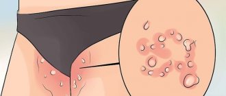
Multiple soft, pinkish-white dots on the labia minora (less commonly in the area of the labia majora) with symmetrical linear localization are a characteristic manifestation of vulvar micropapillomatosis.
This condition does not pose a health hazard, does not cause any complications, is not contagious and does not require treatment.
This cosmetic defect occurs in approximately 5% of women, appearing during puberty.
The causes of micropapillomatous dots have not been established to date.
There is an assumption about their connection with hormonal disorders.
Micropapillomas should be carefully differentiated from genital papillomas.
Diagnostic criteria are symmetrical localization, soft consistency and lack of blanching in the acetic acid test.
The most common cystic skin lesion is the epidermal cyst.
Such a cyst is formed when a closed cavity in the skin is filled with sebum and horny masses.
Cysts are most common in people with acne and seborrhea.
Despite the fact that such formations are considered only a cosmetic defect.
When their thin walls rupture, complications that require treatment.
A small epidermal cyst filled with keratin is called a milium.
Milia look like single or multiple white dots under the skin of the labia, face, and limbs.
They develop from hair follicles, sweat and sebaceous glands.
Fordyce granules are small visible sebaceous glands.
Despite the second name - seborrheic cysts, they are not actually cysts, and they have a very dubious relationship to seborrhea.
The reason for their appearance is considered to be the congenital location of the sebaceous glands closer than usual to the upper layer of the skin.
During puberty, the glands enlarge and produce more sebum, which makes them visible.
White-yellow granules are localized in the area of the labia majora.
When pressed, a small amount of content can sometimes be released from them, but more often such manipulation leads to pinpoint bleeding.
How to get rid of it?
If light-colored vaginal discharge appears, there is no need to panic. First of all, you should assess your condition. If leucorrhoea does not have an unpleasant odor and is not accompanied by burning, itching, pain, ulcers, or rashes, it is simply a waste product of the body or a reaction to detergents.
On a note! White plaque from the labia is removed with water. Soap or gel can aggravate the problem. To remove the substance from hard-to-reach places, use a wet cotton swab.
It is advisable to stop preventive douching for a while and think about changing hygiene products. Eating healthy foods and avoiding alcohol will also have a beneficial effect on the health of your microflora.
If unpleasant accompanying symptoms occur, you should not self-medicate. Unreasonable use of medications will worsen the condition. Only a doctor after the necessary examinations can prescribe adequate therapy. The use of folk remedies in treatment is possible only with the permission of a specialist.
White dots on the labia: pathology
Pathological conditions with white rashes can be infectious (bacterial, fungal, viral), allergic, hormonal, or autoimmune in nature.
A number of factors contribute to the development of the disease:
- violation of intimate hygiene rules;
- damage to the skin when shaving the perigenital area;
- tight underwear made of synthetic materials;
- stress, overheating, hypothermia;
- weakening of the body’s defenses after an illness, during pregnancy, when taking appropriate medications, etc.
The list of diseases that manifest themselves as white spots under the labia includes the following conditions:
- allergies;
- molluscum contagiosum;
- lichen sclerosus;
- candidiasis vulvitis;
- Bowen's disease;
- Fox-Fordyce disease;
- folliculitis;
- genital warts (anogenital warts);
- genital herpes;
- bartholinitis;
- leukoplakia;
- psoriasis;
- red ringworm, etc.
An allergic reaction in the form of a whitish rash may appear after contact with an allergen (synthetic underwear, semen, hygiene product).
A variant of the condition is spontaneous urticaria, in which pale pink rashes gradually change color to white.
Molluscum contagiosum is a viral disease that is transmitted through close skin-to-skin contact, including during sexual intercourse.
That is why in adults this disease (the causative agent is a type 2 virus, while in children it is type 1), according to WHO recommendations, is considered a sexually transmitted infection.
The pathology manifests itself as dense, flesh-pinkish, light-colored papules, from which, when pressed, a curdled, white content is released.
In HIV-infected people, the elements of the rash are prone to suppuration due to the addition of a bacterial infection.
Lichen sclerosus, otherwise called “white spot disease,” is characterized by the appearance of distinct light spots, papules, etc.
The exact cause of the pathology is unknown; an autoimmune mechanism of development is assumed.
With candidal vulvitis (thrush), the white dots are not elements of the rash, but fragments of a characteristic cheesy discharge.
Additional signs of the disease include swelling, redness, severe itching, which tends to intensify after showering, sexual intercourse, etc.
The cause of the disease is fungi of the genus Candida.
With such a rather rare precancerous pathology as Bowen's disease, pockets of red spots or white flaky plaques form on the skin.
As the lesions progress, they develop into squamous cell carcinoma.
When the ducts of the sweat glands are blocked, including in the perineum, symmetrical rashes appear, dense, up to 3 mm in diameter, from red to light in color.
This condition is called Fox-Fordyce disease.
With this disease, patients also complain of intense itching and hair loss in the affected area.
The disease is quite difficult to treat, but after menopause it often regresses spontaneously.
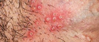
Infectious inflammation of the hair follicle is manifested by the formation of white, white-yellow, red pustules at the base of the hair.
With ostiofolliculitis, only the mouth of the follicle is affected and very small yellowish-white pustular dots, riddled with hair, are formed.
The cause of anogenital warts is the human papillomavirus.
Despite their common cauliflower-shaped form, condylomas can also appear as light papules or grayish-white spots.
Transparent-white multiple blisters on a red base, which after opening turn into whitish-gray erosive dots with a red rim, are a symptom of a sexually transmitted infection such as genital herpes.
The disease manifests itself with itching, pain in the affected areas, and is characterized by a chronic relapsing course.
When the lumen of the Bartholin glands, located in pairs at the base of the labia majora, is blocked, the outflow of secretions is disrupted and a cyst forms.
If such a cyst becomes infected and suppurates, bartholinitis develops.
Precancerous conditions include leukoplakia, in which a dystrophic change in the vulvar mucosa occurs in the form of white small spots and plaques.
Psoriatic plaques are pink-red elements covered with white-silver scales.
The autoimmune disease lichen planus is characterized by the appearance of papules of different colors depending on the form of the disease.
When the vulva is affected, the rash forms a peculiar grayish-white mesh pattern.
When the disease is intense, it causes erosions and scars.
Features of the disease
The color of human skin and hair is determined genetically and depends on the ratio of red, blue, yellow and brown pigments. The lack of some natural dyes leads to dischromic spots.
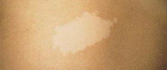
Vitiligo (also called dog) is a disease characterized by the development of primary depigmented white spots without peeling or atrophy. The spots tend to cluster and grow slowly. White lesions on the periphery are surrounded by a weakly pigmented corolla.
This pathology occurs due to the loss or decrease in the functional activity of melanocytes (cells containing the substance melanin).
This is an exclusively acquired disease. It is characterized by clearly limited areas of depigmentation of various sizes and shapes. Occasionally, vitiligo also affects the hair follicles, in which the pigment disappears and the hair becomes discolored. The first foci of the disease appear at a young or very young age.
The etiology and pathogenesis of this disease is not fully understood. There are several theories that explain why this phenomenon occurs:
- The theory of melanocyte destruction. In theory, researchers are of the opinion that vitiligo occurs as an autoimmune disease or due to a violation of the redox status of melanin. Some scientists associate the disease with a neurogenic etiology.
- The second theory says that there is a violation of the inhibition of pigment cells or a violation of their adhesion.
Predisposing factors for the appearance:
- dysfunction of the nervous system;
- constant stress;
- chronic inflammation of the urogenital area;
- diseases of internal organs;
- intoxication together with liver disease;
- destruction and physiological disorders of gland secretion;
- deficiency of copper, selenium and zinc ions;
- decline in the status of the immune system;
- occasionally – hereditary predisposition.
White spots on the labia: diagnosis and analysis
If white spots are detected in the genital area, a thorough diagnosis of the condition is carried out and only then treatment is prescribed.
A particularly important point is the differentiation of a normal state from a pathological one.
Diagnostic steps include:
- interviewing the patient (how long ago the rash appeared, is there any itching, were there any “suspicious” sexual contacts, what concomitant diseases are there, etc.);
- general and gynecological examinations with assessment of the type of rash (papules, vesicles, nodules, etc.), identification of elements of the rash in other parts of the body;
- inspection of elements under the light of a Wood's lamp;
- performing a test with acetic acid;
- carrying out laboratory tests (general tests, examination of scrapings from the surface of rashes, smears from the genital organs, etc.).
Laboratory analyzes are carried out using dark-field microscopy, PCR, culture, etc.
If necessary, samples are taken from the affected areas (biopsy) for subsequent histological examination.
Reasons for the development of the pathological process
Leukoplakia is a reaction of the mucous membrane to irritants. The following factors lead to its appearance:
- neuroendocrine diseases (disorders of the endocrine glands: hypothyroidism, polycystic ovary syndrome, diabetes mellitus, obesity, etc.);
- chronic inflammatory processes of the internal and external genital organs (herpes, HPV);
- cervical dysplasia;
- damage to the external genitalia;
- neglect of personal hygiene rules;
- stress and psycho-emotional overload;
- decreased immunity;
- vitamin deficiency (especially vitamin A deficiency);
- insolation;
- bad habits.