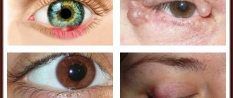
Causes of pathology
As already mentioned, a lipoma can appear anywhere, even on the buttock. Fat in such a delicate place causes some discomfort, usually while sitting. It is especially unpleasant when the tumor is localized in the anus. If such a tumor grows, it can close the anus, thereby complicating the act of defecation.
To date, scientists have not been able to determine the specific causes of the pathological proliferation of fat cells. However, it has been established that there are a number of factors contributing to the formation and growth of wen:
- genetic predisposition – autosomal dominant type of inheritance;
- metabolic dysfunction – in particular, lipid (fat) metabolism disorders;
- disruptions of the endocrine system, such as thyroid hypothyroidism and pancreatic pathologies;
- liver diseases, diabetes mellitus;
- injuries, prolonged excessive compression of tissues;
- infections that cause problems with the hypothalamus;
- period of menopause and puberty;
- addiction to alcohol and smoking.
It has also been noted that lipoma more often forms in people who have crossed the 30-year mark. It is important that the wen will never disappear on its own; how to get rid of the problem and whether it is advisable to eliminate it in principle will be determined by the doctor.
Signs of pathology
An increase in growth may be accompanied by itching and redness. The growth of hygroma causes the patient to complain of the following symptoms:
- itching in the area of the tumor;
- when bending or moving the wrist, the hand begins to hurt (acute pain syndrome);
- the appearance of dense cartilage;
- roughening and darkening of the skin;
- filling the thickened cone inside the phalanx with fluid.
Patients often complain of numbness in the palm, which leads to the inability to move their fingers. The cause of numbness is the growth of a tumor, which compresses blood vessels and nerve fibers in the area affected by the pathology.
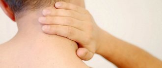
Wen (lipoma) on the butt
The cause of lipoma on the buttock is unknown. The lump can cause serious discomfort and disturb a person's condition. It usually hurts when pressure is applied to it, and given the area, this impact is frequent. Doctors recommend removing such a lump, since complications can develop due to it, for example, an abscess due to constant pressure on the surrounding tissues and their injury. Surgery is needed even if there is no pain in the lump on the buttock, since the tissue of the buttock is still damaged. Lipoma is not a malignant neoplasm and does not pose a threat to human health. Sometimes circumstances are not in the patient’s favor when a person tries to remove a wen on his own. The infection enters the wound and produces changes in the behavior of the growth, which begins rapid growth and development.
Opening tumors at home is strictly prohibited. Without knowing the structure and nature of the tumor, you can doom yourself to irreversible processes in the disease. Only a specialist, through research and analysis, can determine whether the formation is malignant. How to treat and get rid of tumors must be discussed with a surgeon, whose knowledge and experience will help you find the appropriate method for each individual case.
Manifestations and forms of the disease
In more than 90% of cases, lipoma is a single painless neoplasm, the size of which does not exceed 10 cm in diameter. The presence of unfavorable factors provokes the growth of the wen and in some cases it can reach 20 centimeters. This form of the disease is called nodular.
Multiple lesions (lipomatosis) are somewhat less common and usually develop in people with endocrine dysfunction and neurofibromatosis.
In this case, the pathology is called diffuse; it can spread not only to the superficial parts of the body, but also to the internal organs.
The histological features of wen determine their classification:
- classic lipoma is represented exclusively by adipose tissue - localized in the subcutaneous space, soft, painless (the most common type);
- fibrolipoma – characterized by the accumulation of fatty and connective (fibrous) tissues. The neoplasm is quite dense to the touch and does not hurt;
- myolipoma - not only fat cells, but also muscle cells are involved in the formation process. This tumor can be distinguished by its bumpy surface;
- myxolipoma - develops in the mucous subcutaneous layer with its gradual filling with adipose tissue, capable of producing mucin. Most often localized on the genitals, anus, and mammary glands;
- Angliolipoma - as a rule, this is a large-scale neoplasm growing with blood vessels;
- myelolipoma - is a combination of mature fat cells and hematopoietic tissue. Locations: retroperitoneal tissue, adrenal glands, subcutaneous connective tissue of the small pelvis.
READ ALSO: Is it possible to wipe your face with alcohol to remove oiliness and acne?
The mechanism of lipoma development, in principle, does not depend on its location, but the symptoms largely determine the location of the tumor.
Lump under the skin on a muscle
- Lipoma and fibrolipoma are the most common types of lumps under the skin.
- Atheroma is a lump under the skin, which they also like to call a wen.
- Hygroma is a lump under the skin associated with muscle tendons and joints.
- A lateral neck cyst is a common type of lump under the skin in this area.
- A lump under the skin due to damage to the lymph nodes (lymphoma, lymphadenitis, lymphadenopathy, cancer metastases).
- A lump under the skin due to bone overgrowth (osteoma).
- A malignant lump under the skin that looks like a sarcoma.
- Lumps under the skin on the arms and legs due to damage to the joints.
- A lump under the skin in and around the breast area.
- A lump under the skin on the arm, leg, neck, back. Why does it hurt?
- Comments to “Why did a lump appear under the skin on the arm, legs, neck, body? " : 8
- Ball-shaped lump under the skin
- Lump on an arm or leg
- On the face
- Lump in the groin, thighs and buttocks
- Seal on the joint
- Small ball under the skin
- Pimples have turned into hard balls
- Lump under the skin after vaccination
- articles
- Why did a lump appear under the skin: the main reasons
- How to treat lumps under the skin quickly and effectively
Often people note that a lump has appeared under the skin, a thickening or protrusion in different parts of the body.
This can be a symptom of many diseases, including cancer, which is why you should treat this problem with extreme caution. Diseases that are accompanied by the formation of lumps under the skin:
Also, bumps under the skin of certain varieties like to appear
Lipoma and fibrolipoma are the most common types of lumps under the skin
Lipoma is a benign formation that occurs in the subcutaneous adipose tissue, which is why people often hear its names: wen or fatty tumor. The lipoma is soft to the touch, painless and removable.
If the tumor contains dense fibrous tissue, it is called fibrolipoma, and is more dense to the touch. A lump under the skin such as a lipoma often appears on the arms, legs, back, abdomen, and in the mammary gland.
Their appearance is facilitated by injuries and heredity.
Atheroma is a lump under the skin, which they also like to call a wen.
Atheroma is a benign skin tumor that occurs due to disruption of the ducts of the sebaceous glands. There are two main types: epidermal cyst and sebaceous cyst. A lump under the skin, such as an epidermal cyst, has a blocked duct of the sebaceous gland in the form of a brown or black dot.
Likes to appear on the back, back of the neck, face, stomach, and other places on the body. A lump under the skin that looks like a sebaceous gland cyst does not have a blocked duct and looks like part of a sphere covered with normal epidermis. Associated with hair growth, appears on the head.
Both types of atheroma have a well-palpable capsule that feels like a ball.
Hygroma – a lump under the skin associated with muscle tendons and joints
Hygroma, or tendon ganglion, is a tumor-like formation, the cause of which is injury, past tendovaginitis (inflammatory process in the synovial membrane of the tendon), bursitis (inflammation of the joint capsule).
Diagnosis of hygroma in the early stages is difficult. Because, being small in size, it does not cause pain. A lump under the skin like a hygroma likes to appear on the hands near the joints, on the legs near the feet.
It feels like a ball to the touch and usually does not hurt.
Lateral neck cyst is a common type of lump under the skin in this area.
A lateral neck cyst is a lump under the skin that is associated with intrauterine developmental disorders. It may not manifest itself for a long time. However, after neck injuries or infectious diseases (sore throat, acute respiratory infections, flu), it begins to stretch and grow. It looks like a rounded protrusion above the skin of the neck, in which elasticity can be noted to the touch, like a ball.
Lump under the skin due to damage to the lymph nodes (lymphoma, lymphadenitis, lymphadenopathy, cancer metastases)
Lymph nodes are fortresses on the path of the spread of infections and malignant tumors. If microbes or cancer turn out to be stronger, they take over these fortresses, making them their own. Lymph nodes are located in the folds and folds of the neck, in the armpits, in the groin, and on the folds of the arms and legs.
The mildest variant of lymph node damage is called lymphadenopathy, and a slight enlargement of the lymph nodes occurs. They may feel like small bumps under the skin that hurt. In the case of lymphadenitis, the lymph node enlarges significantly, becomes a very painful lump under the skin, and may turn red and open with the release of pus.
In the case of metastases and damage to the lymph nodes by lymphoma, they increase to 1.5-2 cm or more, and practically do not hurt.
A lump under the skin due to bone overgrowth (osteoma)
Osteoma is a tumor arising from the bones. A hard lump under the skin (a growth on the outer surface of a bone) appears on people's bones in the head, arms, legs, and pelvis.
Osteomas can be transmitted genetically from parents to children, and also occur as a result of injuries, various diseases, such as syphilis, rheumatism, gout. Statistically, it occurs more often in men.
X-ray examination helps in diagnosis.
Malignant lump under the skin of the type sarcoma
Sarcoma is a group of tumors with aggressive growth (malignant). These diseases can arise from connective tissue (bone, cartilage, fat), grow from the walls of blood and lymph vessels and muscle fibers.
If left untreated, they are accompanied by rapid progressive growth and the appearance of metastases. A lump under the skin, such as a sarcoma, does not hurt, is relatively hard to the touch, has unclear contours, is covered with reddened skin, and occurs anywhere on the body, arms and legs.
Metastases of internal organ cancer look approximately the same.
Lumps under the skin on the arms and legs due to joint damage
Rheumatoid arthritis is a systemic connective tissue disease that affects small joints. It appears as hard bumps in the areas of the affected joints with redness and pain. Typically, bumps appear under the skin of the joints on the arms in the area of the hands.
In the absence of adequate treatment, the disease quickly progresses and leads to disability in patients. Osteoarthritis mainly affects the large joints of the lower extremities due to injuries, endocrine diseases, and obesity. In advanced cases, hard lumps under the skin appear around the joints.
Pain appears in the affected joints, swelling, and rarely redness.
Gout is a disease associated with metabolic disorders, as a result of which uric acid is deposited in soft tissues in the form of specific hard lumps under the skin that hurt - tophi, the size of which varies from a few millimeters to two centimeters.
Finally, the cause of the appearance of lumps under the skin around the joints of the arms and legs is determined by a surgeon, rheumatologist, or orthopedic traumatologist.
Source: https://BoliGolovnie.ru/varikoz/shishka-pod-kozhej-na-myshce.html
Treatment of boils
At the initial stage, it is quite easy to get rid of a boil. For this purpose, special ultraviolet lamps, external anti-inflammatory drugs and ointments are used, which provoke the rapid opening of a purulent growth.
If the boil does not “come out” for a long time, then it is cut surgically, after which the patient is prescribed a course of treatment with antiseptics and antibacterial ointments.
Sometimes the ulcers open on their own. In this case, you need to rinse the tailbone area yourself with hydrogen peroxide. After this, you should purchase Levomekol ointment and apply bandages with it until the wound is completely healed.
Symptoms
At the beginning of its development, a lipoma is detected only by palpation - usually a person feels a small bump on the body on his own. Often this neoplasm is soft, pliable, and painless. The pathology has a slow course, the formation process can last for years. With all this, the wen does not always increase in size - throughout life it can be the size of a pea and not show itself at all.
In another situation (usually in the presence of unfavorable factors), the lipoma grows rapidly, compresses nerve and vascular tissues, causes significant discomfort to the person, and provokes pain and numbness. In this case, no ointments, lotions or treatment with folk remedies will help - there is only one solution to the problem - surgical removal.
Specific symptoms of lipoma depending on location:
- neoplasm on the kidney - dull, aching pain of a constant nature, radiating to the lower back;
- large wen on the neck - the pathology is accompanied by complications in breathing and the swallowing process;
- tumor in the brain - can cause hallucinations, decreased vision, headaches;
- spinal lipoma - causes minor pain, as it progresses, weakness, numbness of the limbs, and dysfunction of the pelvic organs are possible.
Sometimes it is impossible to identify a pathological growth under the skin of the anus and buttocks on your own (with lipomatosis of internal organs); it is discovered during the diagnosis of other diseases requiring CT, X-ray or MRI studies.
Formation of lumps under the skin and on the hands: types of lumps, causes, symptoms and recommended treatment
Typically, bumps can appear on any part of the body. That is, lumps can grow on the face, legs and arms, buttocks, back or abdomen. Most often, the growth of a neoplasm under the skin can only be noticed after a certain period of time, when the lump reaches a large size.
Lumps grow especially slowly on the scalp, where it is quite difficult to notice a lump if its growth is not accompanied by pain. Most often, this is how the growth of lumps proceeds without pain and corresponding symptoms, which are benign neoplasms under or on the skin.
If lumps or lumps cause pain and discomfort , then most likely the problem is simply a consequence of an infection that has entered under the skin layer, for example, through the hair pores.
As soon as an infection gets under the skin, it begins to rapidly develop in a certain place and this may be accompanied by ordinary pain or an increase in local or general body temperature. Most often, infectious bumps change color at the site of inflammation and become red or burgundy.
You may also experience headaches, malaise, or weakness. The most interesting thing is that such bumps, with proper treatment, can be cured in just a few days.
The most dangerous lumps that appear under the skin are malignant tumors . You can notice them yourself or feel them. They are actually no different from benign neoplasms, so if such a lump is found on the body, it is better to immediately contact a specialist to remove the lump.
Lipomas (fat)
Often, after people find lumps on their body, they immediately run to the doctors, and that’s right, but you shouldn’t worry right away, because the lumps may just be lipomas. These are a kind of fatty formations that are benign neoplasms and therefore cannot cause any harm to the body or human health.
Such fatty tissues are distinguished by the presence of clear boundaries and the absence of pain and discomfort. In addition, when a lipoma occurs, the color of the skin in the affected areas does not change.
These bumps can appear on any part of the body, but are most often found on the neck, head, back, arms and legs.
Wen can be removed by conventional surgery, but if they do not interfere, many people simply do not touch them.
Lipomas can only cause discomfort in rare cases when their growth does not stop and the wen simply begins to put pressure on organs or muscles, which leads to pain during movement.
Atheroma
A disease such as atheroma is often confused with ordinary wen among people.
Therefore, when atheroma occurs, people may simply forget about the appearance of a lump under the skin and not consult a doctor, although in fact this disease is more serious and in some cases can even cause complications for the body. Atheroma is not a wen, but a cyst .
There is a whole list of differences by which you can distinguish atheroma from lipoma. First of all, it is worth checking the area of skin where fold atheroma develops. If the skin does not gather into folds, then it is no longer a wen.
Atheroma is a tumor that forms when the duct of the sebaceous glands is blocked. As a result, sebum begins to accumulate, which can lead to the formation of pus or simply become inflamed. It is impossible to cure atheroma with simple folk remedies, and the only way to remove it is surgery.
Hygroma
On the wrists it can often appear in the form of a hygroma bump. This neoplasm does not cause any harm to human health, except for spoiling the appearance, although most often such a ball under the skin is simply invisible.
Hygroma can be removed surgically or disappear on its own in the event of a blow, but no harmful consequences will follow, since such a lump is just an accumulation of fluid that is located between the fibers of the tendons.
Nodules on the joints
Often, when the disease occurs, lumps or so-called small nodules may appear on the joints. Each disease has a different type of nodules on the joints.
So, for example, if rheumatoid arthritis develops, then it is quite possible that a rheumatoid nodule .
Lumps, which are called Heberd's and Bouchard's nodes, may appear on the fingers if deforming osteoarthritis develops. Most often, such nodules develop to medium size.
Gouty nodes, or as they are also called tophi, can be significant in size. Such bumps can appear in people who have suffered from gout for several years, as a result of which the accumulation of salts begins.
It is also worth mentioning the subcutaneous lump that forms on the joints of the big toe. The growth of such a bump is accompanied by hallux valgus , or more precisely, the finger simply begins to bend due to an increase in the bone. This, in turn, causes great discomfort while walking and when choosing shoes.
Hernia
One of the most famous formations under the skin is a hernia. Many people know what dangers a hernia can pose and why it occurs. Mostly, a hernia can appear in the navel area or under the skin in the groin. A hernia may be simple and not cause discomfort, but it can also be accompanied by painful symptoms.
No matter how strange it may sound, it is often possible to set the hernia back by simply pressing with a finger.
More precisely, a hernia is formed with the help of internal organs, which, under pressure or heavy loads, are simply squeezed out , so they can be easily repaired, but it is better, of course, to consult a specialist.
Typically, a hernia can be caused by a load that puts too much pressure on the abdominal area. Also, a hernia can appear even during coughing or vomiting, because they contribute to an increase in local pressure.
Lumps in the breast (in the mammary gland)
Breast lumps in women can be scary, but in fact, almost everyone has experienced this problem.
Every woman has experienced the appearance of lumps or lumps in her breasts, and most often this problem appears during her period.
Small bumps or lumps may appear due to the action of hormones on the mammary glands , which simply change temporarily. After menstruation passes or just begins, such lumps in the breast immediately subside.
There are possible cases when lumps remain even after menstruation and this indicates that their occurrence lies in other reasons, so if after menstruation the lumps do not go away, then it is best to consult a mammologist or gynecologist. Most often, such tumors turn out to be benign, and therefore they can be easily removed using conservative treatment or surgery.
Reasons for urgent consultation with a doctor:
- the node quickly increases in size;
- pain in the chest area appears regardless of the stage of the cycle;
- the neoplasm does not have smooth contours and clear boundaries;
- ulcers or skin deformities appear;
- The lymph nodes in the armpits are enlarged.
If such symptoms appear after your period has ended, it is best to immediately contact a mammologist or oncologist.
Erythema nodosum
If the nodes on the buttocks cause pain upon palpation, it is possible that you had to deal with erythema nodosum. This is an inflammatory process of subcutaneous fat, which most often manifests itself in the fairer sex. There are many factors that can lead to the development of erythema nodosum on the buttocks. Secondary inflammation can develop as a complication of certain infectious diseases (leprosy, tuberculosis, tonsillitis, scarlet fever, etc.).
Many women have to deal with erythema nodosum of allergic origin. In this case, painful nodes on the buttocks appear after antibacterial therapy, the use of certain contraceptives or certain sulfa drugs. Only a doctor can accurately determine the cause of the inflammatory process. The following methods are used for this:
- biopsy of the inflamed area;
- general urine analysis;
- general blood analysis.
In addition to the soreness of the node, the development of the disease will also be indicated by symptoms of general intoxication of the body: an increase in body temperature to low-grade levels, chills, drowsiness, nausea.
Compresses with Dimexide will help relieve inflammation
Treatment of erythema nodosum begins with eliminating the causes of the pathological process. If the disease is allergic in nature, the patient will have to limit contact with the irritant (stop taking medications that caused the side effect). Antibiotics and non-steroidal anti-inflammatory drugs help relieve inflammation. Physiotherapeutic procedures such as ultraviolet irradiation, UHF, heat therapy, and magnetic therapy also show good results.
To speed up the recovery process of the diseased area, local medications can be used: anti-inflammatory and corticosteroid ointments, compresses with Dimexide.
How to treat
Medications
Medicines can treat bumps near the hand or on the curve of the hand. The inflammatory process leads to unpleasant symptoms, therefore, it must be stopped. Often the patient is prescribed antibacterial drugs (for purulent inflammation). If thickenings, nodules, compactions and bumps were removed by a surgeon, then the use of medications will prevent infectious blood poisoning. To eliminate the inflammatory process, experts prescribe:
- antihistamines, for example Clemastine;
- corticosteroids, for example, Diprosalik;
- NSAIDs - Ibuprofen, Nimesil, Nurofen.
Physiotherapeutic treatment
Magnetic therapy is often used in the treatment of hygroma. Physiotherapeutic procedures have a positive effect and help remove unpleasant tumors. To eliminate formation, the following methods are used:
- Exposure to ultrasound. Enrichment of affected tissues with oxygen promotes their regeneration.
- Magnetotherapy. The procedure lasts 20 minutes, reducing the inflammatory process in bone and cartilage tissues.
- UHF therapy. Ultra-high frequencies stop inflammation, improving local blood circulation, deeply warming tissues and stimulating restoration processes.
- Baths with salt and soda. The impact affects adhesions in the hand. The course includes up to 30 procedures.
Diagnostics and treatment methods
Visual examination and palpation of the affected area is the first thing the doctor does when diagnosing the disease. After this, in order to confirm or refute malignancy, a cytological analysis and blood sampling are performed, as well as CT, MRI, X-ray or ultrasound.
Having established a diagnosis, the doctor determines the method of treatment - conservative (medication) or surgical. Also, for small benign tumors, it is possible to carry out treatment with folk remedies, but first coordinate your actions with your doctor.
The reasons for surgical intervention are:
- soreness;
- intensive growth;
- weeping and inflammation of the lipoma.
In this situation, self-medication is prohibited.
Treatment of bruise
If a bump appears on your butt between the buttocks as a result of a fall or a strong blow, then first of all you need to rule out a fracture. To do this, you need to take a photo and consult with a specialist. If no fracture is found, the doctor most often prescribes the following treatment:
- A cold compress should be applied to the lump daily.
- The patient should completely avoid heavy exercise. It is best to lie down for a while (it is recommended to rest on your stomach).
- Massage and water procedures are completely excluded.
- It is necessary to apply special anesthetic ointments to the site of the injury 2-3 times a day.
- It is recommended to use an orthopedic pillow.
READ ALSO: Folk remedies for acne - healthy lifestyle recipes
Doctors also advise smearing the affected area with Dolobene gel and using ichthyol suppositories 1-2 times a day.
If the inflammation does not subside, then the lump is opened. After this you will have to take a course of antibiotics.
What to remember
- To cure a bump on your head, figure out what it is and why it appeared.
- Treat the bruise immediately with a cold compress and then with ointment.
- Treat lumps from insect bites with antihistamines or adsorbents.
- Treat boils on the head with antiseptics and anti-inflammatory drugs.
- If the lump is not caused by a bruise or bite, consult a general practitioner.
- If a lump forms on your newborn's head, contact your pediatrician.
See you in the next article!
Conservative methods
If the tumor does not exceed 5 cm in diameter and does not bother the patient in any way, the doctor may decide to get rid of it with the help of medications, the action of which is aimed at breaking down fat deposits.
The drug Diprospan is often used. It is injected into the fat capsule through a puncture, and it is important to observe the permissible dosage; an excess of the drug causes side effects.
The effect of the medicine is slow - over several months the lipoma will gradually resolve until it disappears completely. Therefore, this method is not suitable for those people who want to quickly fix the problem.
At home, you can use the well-known Vishnevsky ointment. The drug contains tar and castor oil, they improve blood flow to the affected area and draw the contents of the lipoma out. Use the ointment in the form of a compress (three-day course) - apply to a cotton pad, apply to the wen and secure with a bandage. Leave for 12 hours. In addition, the medicine has a regenerating effect.
Ichthyol ointment has the same properties. However, before using this or that product, be sure to consult your doctor. It should also be understood that such treatment is fraught with frequent relapses.
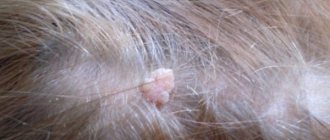
Bumps on the head in the hair: reasons for their appearance – Izvilina
29.12.2019
May 4, 2018
Sometimes, in the scalp you can find formations that feel like hard lumps to the touch. These are bumps. They should be classified as tumors, and the variety should be classified only according to the primary causes of occurrence, and based on the further course of the disease.
Less commonly than on the scalp, a lump can form on an open area of the scalp. The location of the formation is often on the back of the head. Before taking any action to treat the problem, it is necessary to correctly diagnose the cell division disorder.
Very often, women do not notice such a problem in the early stages of its development.
Types of tumors under the scalp
If we are talking about a relatively soft tumor that develops rapidly and is large in size, then most likely it is a hemangioma.
This type of formation is quite specific in nature, therefore it is less common than others, and is a consequence of problems with the activity of the circulatory system.
This tumor is unique in that it often has its own blood supply system, therefore, upon studying it in detail, a vascular network can be seen on the skin of the lump.
- Allergic bumps are not such a common occurrence, but still occur with some frequency in women suffering from various types of allergies. It doesn’t matter at all what causes the immune reaction: some food product or, for example, household chemicals. Such formations bring the greatest discomfort compared to all of the above, since one of their key symptoms is severe itching.
- Lumps that are formed due to a serious disorder in the division of connective tissue cells are called fibromas and sarcofibromas. Based on the name of the latter, one can draw conclusions about the malignant nature of the tumor. Most often, these formations can appear on the head of an adult woman, reaching impressive sizes. Their multiple appearance is also typical. The factors that provoke the growth of such cones do not depend on the person. This may be a fact of hereditary predisposition, diabetes mellitus or disruption of the endocrine system. A malignant variant of a connective tissue tumor, without proper therapy, can be fatal.
- When there is a history of damage to adipose tissue, a woman, especially one in the “30+” age category, may find bumps on her head – lipomas. Disorders of lipid metabolism in the body, as well as age-related hormonal problems can trigger the appearance of these lumps. It is noteworthy that this type of tumor can develop not only on the head, but also on any other part of the body. The formation is significantly softer to the touch than the connective tissue described above.
- There is another type of growth on the head in the form of bumps, which is associated with adipose tissue, since it has a capsule inside that is directly filled with fat. We are talking about atheroma. The problem can arise when a woman experiences blockage of one of the sebaceous glands. The lump has a smooth surface to the touch. Upon examination, you will notice that the skin color in this place is much yellower than elsewhere. Since this type of tumor is visually very similar to the one described above, establishing an accurate diagnosis can only be entrusted to a specialist. It is important to remember that if this problem is not solved by timely removal, then atheroma can cause discomfort and pain.
- Sometimes, when we talk about bumps on the head of women, we can talk about the formation of an ordinary wen. This tumor, unlike all of the above, forms above the surface of the human skin. The cause may be any hormonal problems of the woman, or chronic exposure of the body to all kinds of stress. Despite the fact that such bumps appear above the skin, one cannot talk about their infectious nature.
Causes of hair bumps on the head
Doctors have predetermined factors that can cause a bump to appear on the head, if we are not talking about some kind of neoplasm. It is noteworthy that this can apply not only to women of different ages, but also to men.
The most common factor is injuries received in the course of life. A common everyday bruise of the back of the head due to a blow to some blunt object provokes swelling accompanied by pain.
Lumps that arise for this reason go away after some time without additional medical care.
And in order to independently speed up the process of resorption of the tubercle, you should resort to using a cold compress.
Sometimes, the reason why a lump forms may be an insect bite. Since all of them, when biting, inject a small portion of poison under the skin, this can provoke a serious allergic reaction, accompanied not just by redness of the skin at the site of the bite, but by a whole lump.
Traditional methods for solving the problem
If we are talking about lumps, which are neoplasms, the question of using folk remedies may not be relevant.
At a minimum, because a woman will not be able to independently determine the nature of the tumor and its quality.
It is better to entrust the treatment of such formations to specialists who, in most cases, will prefer to resort to surgical intervention, which will get rid of the problem once and for all.
When the lump arises due to a bruise, and you want to get rid of it as soon as possible, it makes sense to think about resorting to the help of traditional methods of treatment.
The most common method of dealing with bumps from bruises is compresses with medicinal plants such as golden mustache and aloe. For the procedure, one sheet cut lengthwise is enough. It is applied directly to the place on the skin where the problem is localized.
Since the previous method is designed for additional fixation of the compress using an insulating bandage, it will not be convenient to use it on all areas of the head. There is another opportunity to speed up the process of lump resorption. To do this you will need regular food salt. It is dissolved in water, and lotions are made from the resulting solution.
Preparations for the treatment of bumps on the head
If a woman is faced with the problem of bumps of an allergic nature, she needs to resort to the help of antihistamines.
By suppressing the action of a substance that is released in the body under the influence of an allergen, the medicine will avoid further development of the immune reaction, as well as speed up the process of resorption of the lump.
An antiallergic agent should be selected based on individual susceptibility to the ratio of its components.
In the case of bruises, the active ingredients of ointments such as “Troxevasin” or “Rescuer” will help to remove the bump. The only disadvantage of using ointments may be the inconvenience of applying the drug to the scalp.
When mentioning various tumors, there can be no talk about their drug treatment, since, often, such therapy is not always able to even simply reduce the tumor in volume. What can we say about getting rid of it completely.
A surgical intervention performed by a professional doctor will allow you to forget about the problem of bumps on your head without fear of relapse. Regardless of the nature of the tumor, its size and the degree of complexity of the operation, the patient will be prescribed a course of antibiotics.
The edges of the postoperative wound should be treated with antiseptics, periodically making sterile dressings. This will prevent various infections from entering the vulnerable area.
With the development of modern medicine, many, both private and public medical institutions, can offer their patients to remove bumps on the head using a laser.
This is acceptable when the size of the formation is not too large, and its benignity is confirmed diagnostically.
The laser procedure, of course, will cost an order of magnitude more than a classic operation, but at the same time, trauma in the process is significantly reduced, allowing the amount of time required for rehabilitation to be reduced.
Source:
Bumps on the head: causes, symptoms and treatment:
Why do bumps appear on the head and how to get rid of them? It is these issues that this article will be devoted to.
General information
Surely every person has ever noticed that bumps periodically appear on his head. In most cases, they rarely bother their owner, but people try to get rid of them as quickly as possible. However, doctors are not so categorical in this regard.
Experts believe that if bumps on the head do not bother you or cause any discomfort, then they should not be removed or treated. Although in some cases (in case of swelling, redness, itching, increased body temperature, etc.) it is necessary to consult a doctor.
Causes and symptoms
Bumps on the head can appear for various reasons. And in order to get rid of them, you should definitely determine why you have such a benign neoplasm. To do this, it is recommended to consult a doctor. However, if you do not have such an opportunity, then we will help you identify the true cause of the appearance of cones in this article.
Wen or wart
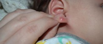
A hard lump on the head may be a wen or a wart. Such neoplasms appear not under the skin, but on it. They can be localized not only on the head, but also on the neck and behind the ears.
Pimples
If the bumps on your head hurt and itch, it could be regular acne or marks from insect bites. As a rule, after a few days such formations disappear on their own.
Injury
It is no secret that after a severe injury to the head, a lump may form on it. To prevent such swelling from occurring, it is recommended to apply a cold compress to the injury site immediately after injury.
Allergic reactions
Often, bumps on the head appear due to exposure to any allergens. To eliminate this phenomenon, you should review your diet, as well as change shampoo, conditioner, hair dye and other cosmetics that may cause an allergic reaction.
Treatment of cones
Not all bumps that appear on the head can be treated independently at home. For example, it is better to get rid of a formation such as hemangioma with the help of an experienced specialist.
If the bumps do not hurt and do not bother you in any way, then there is no need to treat them.
In the event that such a deviation is unpleasant for you, and you still decide to get rid of it, then there are several traditional methods that are actively used for therapy.
- Golden mustache. The tincture of this plant has been used for a long time and has been successfully used to treat cones. To do this, soak a cotton swab in the tincture, and then apply it to the swelling and leave for some time (6-8 minutes). By the way, this drug can also be taken orally in the amount of 10 drops twice a day.
- Kalanchoe leaves. Fresh leaves (without skin) of this plant must be applied to the cones, and then covered with film and a cotton pad. This compress should be secured with a bandage and left for 3 hours. It is advisable to carry out this procedure up to 3 times a day.
How to get rid of insect bites?
If bumps on your head appear as a result of insect bites, then it is recommended to use antihistamines (gels, ointments and aerosols) to eliminate them. Such drugs quickly relieve puffiness, swelling and tissue irritation. As for folk remedies, salt and soda solutions are good for insect bites.
Treatment of atheromas and lipomas
In traditional medicine, there are several ways to treat such formations. The main method is surgical removal, which is performed under local anesthesia. Laser excision is also a fairly popular way to eliminate various formations.
However, this procedure is much more expensive than minor surgery. If you need to get rid of a wen, then the third method is used for this.
Source: https://fiz-disp.ru/diagnostika/shishki-na-golove-v-volosah-prichiny-poyavleniya.html
Features of hemorrhoidal cones
The appearance of the described bumps is typical for both women and men. Women may develop hemorrhoids during pregnancy due to congestion of the venous system. In addition, a pregnant mother can be exhausted by the symptom of the described pathology in the form of constipation.
As mentioned above, hemorrhoidal cones in adults can be either internal or external. Internal formations are located directly on the walls of the rectum and are prone to prolapse from the anus and bleeding during bowel movements. The external bump is due to the presence of the outer skin. It is this tumor that has already fallen out, the so-called tail, that is susceptible to thrombosis to a greater extent than the internal one. Characteristic features of hemorrhoidal cones may include acute pain when they fall out and bleeding.
Surgery
In addition to medical indicators, quite often people remove lipomas located on open areas of the body, lyceum, neck, chest (this is done mainly for aesthetic reasons). Today, there are several methods for surgical removal of wen:
Classic excision is performed under anesthesia. An incision is made with a scalpel, through which the capsule and its contents are removed. Plus - there are no relapses. The downside is tissue scarring.
Endoscopy is a minimally invasive intervention. An incision is made (up to 1 cm), flexible equipment is inserted under the skin, with the help of which the wen is destroyed and removed. Plus - no traces. The downside is frequent relapses.
Laser destruction - a laser beam locally affects the problem without affecting healthy tissue. Plus – bloodlessness, minimal rehabilitation period, absence of scars. Minus - it is not used for large lipomas.
Liposuction, or puncture-aspiration method, uses special equipment to evacuate fat contents (vacuum pumping). Plus - fast healing, no scars. The downside is that due to the fact that the capsule remains in place and is not always completely cleaned, relapses often occur.
You can also get rid of lipomas using electrocoagulation and radio wave destruction.
How to safely remove a wen on the head: effective methods
Lipomas are benign, non-dangerous formations that usually bother a person only in terms of aesthetic discomfort. They can appear on any part of the body, sometimes appearing on the scalp. This is explained by the large number of sebaceous glands in a given location. Bumps are difficult to hide, and removal of the wen on the head is important.
Which doctor should I go to for a wen on my head?
A tumor on the head cannot always be covered with hair and often grows in height and width. If the lump is located behind the ears, it may cause discomfort when wearing glasses.
The formation under the hair makes it difficult to comb; on the forehead near the eyes, eyebrows can be easily damaged.
Owners of wen do not think about removal until they feel discomfort. Sometimes they try to cure a wen on the head using folk methods.
In a calm state it does not threaten health; self-treatment can lead to inflammation. The best solution would be to contact a specialist. Doctors who can help you get rid of an unsightly lump:
- Therapist. He will conduct an initial examination of the scalp and refer you for research.
- Dermatologist. Examine the condition of the skin.
- Ultrasound specialist, radiologist. Conduct hardware research. X-ray, ultrasound or computed tomography will determine whether the tumor is dangerous, determine its contents, and the condition of the tissues next to the formation.
- Oncologist. If a malignant nature is suspected, an oncologist takes over and performs a biopsy.
- Surgeon. He will examine the lipoma and choose the appropriate method of removal.
Is it necessary to delete
Typically, patients ask to get rid of an ugly wen on their head. A harmless lump from a medical point of view, it looks unsightly and interferes with daily procedures: creating a hairstyle, wearing glasses and hats.
However, there are cases when the doctor strongly recommends removing the wen. These symptoms include:
- Pain is felt when pressing.
- The skin became inflamed.
- Itching appeared.
- The tumor has burst or is leaking.
- It is growing rapidly.
- The swelling has changed color.
- Headaches started.
- Weakness appeared for no reason.
- Mental disorders arose.
Lipomas that protrude along the nasal line, mid-scalp, and cranial suture line are often examined with a CT scan or MRI for connections to neural tissue to avoid complications during the removal process.
Methods for safe removal of wen
You can safely get rid of a tumor on your head only by contacting a specialist. At best, independent methods of removal may result in zero results, and at worst, urgent medical attention will be required.
You cannot try to break through, cauterize, cut, puncture, or try to administer medications with a syringe. There is a risk of infection and the growth of new or malignant tumors.
The doctor may advise not to remove the lipoma, but to try to cause it to resolve. Only “young” formations can be cured. Medications will help soften the wen.
For this, Vishnevsky ointment, Ichthyol ointment, and Dimexide solution are used. Injections of lipolytics will reduce, but not eliminate.
Surgery
There is a classic surgical method, endoscopic excision. In the first case, the skin at the site of formation is dissected, the adipose tissue is cut out along with the capsule, and healthy tissue is sutured. If necessary, a drainage is placed to drain the fluid.
Endoscopic excision involves making an incision on the side of the lump. The surgeon can examine the position and condition of the adjacent tissues. This removal of a lipoma on the head will hide the scar.
The operation is performed under local or general anesthesia in a hospital. Previously, when removing from the scalp, patients were shaved bald to prevent hair from getting into the seam. Now the surgeon shaves a small area around the tumor.
The surgical method is suitable for getting rid of large specimens and does not cause relapses.
The main disadvantage is the formation of scars, the relatively long operation time and the need for a rehabilitation period if the suture becomes inflamed.
Laser
The method will avoid scars and scars. The skin incision on large formations is made not with a laser, but with a scalpel, since the surgical instrument makes a more precise and smaller incision. The laser cauterizes the tissue inside the incision and cuts off blood vessels.
Do you need advice from an experienced doctor? Get a doctor's consultation online. Ask your question right now.
Ask a free question
Removing a large wen on the head with a laser will eliminate relapses; the operation lasts several minutes and is performed under local anesthesia or without it. A distinctive feature is rapid healing, absence of traces of intervention, minimal possibility of infection. Most often, this method is used to get rid of fatty deposits on the face and swelling in children.
Small-sized formations are completely decomposed under the action of a laser.
other methods
Other treatment methods include liposuction, radio wave exposure, and cryodestruction on the head.
Liposuction
The doctor injects a thinning substance into the lump through a puncture. After some time, the contents are removed using a needle. In some cases, the thinning fluid is injected several times.
After the operation, there are almost no marks left on the head; the downside is the high probability of the tumor reappearing, since the capsule remains intact. The skin at the intervention site remains flabby, and the wen cavity remains. Liposuction is usually used on giant tumors to avoid large cuts in the skin.
Radio waves
Radio wave removal has the same advantages as laser removal. The operation is painless, no rehabilitation is required, and there are no scars left. The surgeon operates with a radio wave knife, which simultaneously cuts out the tumor and cauterizes adjacent vessels. A scratch remains at the site of the cut, which quickly disappears. The method is suitable for cones up to three centimeters in size.
Cryodestruction
Lipoma on the head is cured with liquid nitrogen. A special feature is the preservation of fat accumulation in tissues. After the operation, it decreases in size and gradually disappears.
Is the removal procedure painful?
Surgical excision of the wen is performed under local or general anesthesia. The surgeon injects the surgical site with an anesthetic liquid, which makes the manipulation painless. After the anesthesia wears off, the suture may ache, but does not cause severe discomfort.
Absolutely painless methods are laser, radiological exposure and cryodestruction. During liposuction, the patient does not experience any pain; he only experiences an unpleasant sensation during the puncture.
How to avoid relapse and healing rules
Relapses occur if the capsule remains on the head under the skin. Only a surgical method can eliminate the risk of the lump reappearing: using a scalpel, laser or radio waves. Liposuction and drug methods do not provide an absolute guarantee. This also applies to home methods of getting rid of education.
We can talk about the stage of rehabilitation after surgery only in the case of classic tumor removal, when the tissue is cut and stitched. There is a possibility of infection, therefore, after disposal, it is necessary to avoid damaging the operated area and monitor the condition of the skin on it.
There is no special prevention for the formation of wen on the head. General guidelines include advice to keep your body healthy and avoid injury.
The article has been reviewed by the site editors
Source: https://VashaDerma.ru/zhiroviki/na-golove-udalenie
What treatment to take
Some patients, before turning to a surgeon to solve their problem, try traditional methods for getting rid of lipoma. Self-treatment of a wen often does not bring any results, and sometimes, on the contrary, it provokes rapid growth of the tumor. By examining the condition of the tumor and determining its size, the attending physician himself decides on the method of removal. And since subcutaneous growths usually develop very slowly, it is often not recommended to touch them at all. Especially if the seal is located in a closed area of the body. In such cases, the doctor prescribes observation with regular ultrasound and histology studies.
Which doctor should you contact?
If a child has a lump on his head, contact his pediatrician . If this is an adult, see a therapist .
Of course, sometimes you may need the help of another doctor to treat a bump on your head:
- in the case of an allergic lump - an allergist;
- in case of an infectious disease with enlarged lymph nodes in the head area - an infectious disease specialist;
- in case of neoplasms - a dermatologist or oncologist.
In any case, contact your therapist first. He will conduct an initial diagnosis and, if necessary, prescribe a consultation with a specialist in another field that you need.
Recipes from traditional healers
Non-traditional treatment methods can be used for relatively small subcutaneous tumors and only in consultation with a doctor. They will certainly help soften and reduce the size of the tumor and perhaps the wen will become almost invisible. However, after some time it will definitely appear again, since the capsule and its contents remained in place.
- Kalanchoe - soak a gauze cloth with the juice of the plant and apply it to the affected area. You can also use the pulp.
- Golden mustache - slightly crush a leaf of the plant, apply it to the lipoma, wrap it in plastic and apply a warm bandage, leave for 12 hours. Coltsfoot is used in the same way.
- Baked onion + laundry soap – grind the ingredients until smooth. The resulting paste is applied to a cloth and applied to the wen, secured with a bandage, and left overnight.
- Black pepper + alcohol – a gauze cloth is soaked in alcohol, sprinkled with ground black pepper and a compress is applied to the lipoma for half an hour.
- Red clay + kefir - mix the ingredients so that you get a soft dough. Make a flat cake and apply it to the wen, cover with cling film and put an adhesive plaster on top.
- Garlic + vegetable oil – a small head is cleaned, the cloves are passed through a press, approximately the same amount of oil is added. Treat the affected area with massage movements three times a day.
- Lamb fat - you need to melt it in a water bath, let it cool a little and apply it to the fat with massage movements.
READ ALSO: Acne from birth control pills: causes and methods for eliminating side effects
You are unlikely to be able to eliminate the problem forever using folk remedies. And self-medication can sometimes even cause harm. Remember, if therapy with folk remedies for a month has not given the desired results, it is advisable to reconsider the treatment tactics, of course, together with your doctor.
Treatment of furunculosis
Fortunately, this disease can be treated with conservative methods. Typically, a doctor prescribes for a patient with a lump on the butt between the buttocks:
- A laxative diet, which involves the inclusion of fresh fruits and vegetables high in dietary fiber in the diet. Thanks to this, the peristalsis of the intestinal tract is normalized and carbohydrate metabolism is restored, the violation of which can precisely cause the appearance of furunculosis.
- Antioxidants (usually vitamin C).
- Superficial physiotherapy.
If furunculosis is pronounced, then the specialist may additionally prescribe Amoxicillin (penicillin of synthetic origin).
Much less often, a lump between the buttocks in men and women becomes a symptom of osteomyelitis of the coccyx, presacral teratoma or spina bifida.
Lump on the inner thigh in women photo
Hemangioma
When a benign tumor, which is a proliferation of vascular tissue, forms, a protruding seal with jagged edges appears on the inner side of the thigh. The color of the neoplasm can vary from red to bluish. The appearance of a dense lump is accompanied by pain and swelling in the hip joint, and manifestations of lameness. Hemangioma is treated through surgery.
Fibroma
A subcutaneous neoplasm formed by fibrous and connective tissues is localized in the upper layers of the skin. This compaction resembles a dense oval or round bump rising above the surface of the dermis. Dimensions can reach up to 3 cm in diameter, color changes are observed only when the tumor enlarges. Fibrous growths on the inner thigh are most often soft, may be pedunculated or adhere tightly to the surface of the skin.
The appearance of fibroma is not accompanied by pain and discomfort (possible only in the case of mechanical damage or irritation of the tumor), but in the absence of proper therapy, it is prone to degeneration into a malignant neoplasm. Such seals must be removed surgically.
Lipoma
A soft, mobile subcutaneous formation consisting of fat cells is localized close to the surface of the dermis. Both single and small multiple wen can form on the inner side of the thigh. A painful lump on the inner side of the thigh can be benign in nature, but over time, without proper treatment, it can transform into a malignant tumor. You can get rid of lipoma by surgical removal of the tumor.
Atheroma
When the outflow of sebaceous secretion is disrupted and subsequent blockage of the sebaceous glands, a cystic sac-like seal is formed - atheroma. Pain in the lesion is observed with significant tumor growth, as well as with inflammation of the neoplasm tissue. Removal of atheromas is carried out by surgical or laser excision of damaged skin surfaces.
Hygroma
A hard lump on the inner side of the thigh, clearly noticeable upon palpation, indicates the formation of a subcutaneous tumor-like neoplasm filled with mucous or serous contents. A stationary ball of elastic, dense consistency with a smooth surface begins to develop in the periarticular bursa or muscle tendons.
Usually, hygroma does not hurt or cause discomfort. The neoplasm is susceptible to mechanical trauma, which creates a risk of infection of nearby tissues. Therefore, doctors recommend removing the lump by puncturing the contents of the lump or surgical excision of the affected tissue.
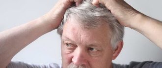
Tumor on the head in the form of a lump: types, why it appeared, symptoms, consequences, treatment
The appearance of a bump on the scalp is not immediately noticed; only when it reaches a fairly decent size does it reveal itself. Tumors differ depending on the type. They can be soft or hard to the touch. The location can be different: on the back of the head, on the forehead, on the temporal part.
Kinds
There are several types of cones:
- Lipoma, its other name is wen. The lump grows quite slowly, there is no pain during palpation, there is a mass in the form of fat inside.
- Papilloma is a benign formation. It can be on a leg or flat. On palpation, as a rule, there is no pain. The surface is loose and can be long or oblong. In older people, warts are often flat, brown, gray or even black.
- Hemangioma is a benign formation that occurs in children immediately from birth. Its surface is red, loose, loose, painless.
- Osteoma is a benign neoplasm that develops from the bone tissue of the skull; accordingly, it is hard to the touch and does not hurt.
- Atheroma is a cyst that begins to grow in place of the sebaceous gland, making it difficult for its secretion to escape. Inside the neoplasm there is sebaceous content, upon palpation pain occurs, and it tends to periodically become inflamed.
- Trichoepithelioma is a benign, small tumor. It develops at the site of the hair follicle. Soft to the touch, painless. The reason for its formation is hereditary predisposition. Multiple formations are often encountered.
- Fibroma is a benign type of tumor that develops from connective tissue and is similar in appearance to a wart. Doesn't hurt when touched.
- Sarcofibroma is a cancerous neoplasm that grows quite quickly from connective tissue, the boundaries are unclear. When palpated, it resembles a knot in the skin covering.
- A furuncle is a purulent formation.
Causes
The reasons are divided into internal and external.
Trauma, bruise
This factor is considered the most common in medical practice. After the blow, swelling occurs in the form of a lump. It is often painful on palpation. A simple bump does not require treatment and goes away on its own; to quickly disappear, you need to apply a cold compress immediately after the blow.
However, if after the blow a person loses consciousness or develops nausea or dizziness, this may indicate a closed head injury. In this case, the brain tissue is directly damaged, and the lump is just an external sign of a dangerous indicator.
If you experience the above symptoms, you should immediately consult a doctor to make sure there is no severe brain damage or bleeding. An MRI or CT scan will be necessary to determine how serious the injury is.
A bite of an insect
Manifests itself as an allergic reaction. The size of the resulting lump can vary from 5 mm to several centimeters, it depends on the severity of the allergy. In this case, anti-allergy medications will help; an examination by an allergist is advisable.
Atheroma
A lump that is painful to the touch develops due to a blockage of the sebaceous gland, making it difficult for its contents to exit. It grows quickly, often being larger than a chicken egg. Most often seen in the back of the head.
If there is damage to the integrity of the skin, there is a high risk of bacteria getting inside, which causes the development of purulent inflammation. In this case, the body temperature increases, and a tugging pain appears in the area of the lump. It is treated exclusively by surgery.
Hemangioma
The formation occurs as a result of an abnormality in the structure of the blood vessels located under the skin. Forms a bulge that looks like a button.
With its rapid growth, it invades healthy cells, which makes it very dangerous. They are most often located behind the ears and in the eye area. If they appear, you should immediately visit a doctor.
Fibroma (sarcofibroma)
A benign tumor that is hard to the touch. However, in order to make sure that this tumor is really a fibroma and not a malignant formation, it is necessary to undergo tests and seek advice from an oncologist.
The reasons for its occurrence are still not fully understood. Some believe that it can appear in anyone, others believe that it is a hereditary pathology, and others believe that it is due to various skin lesions.
Furuncle
A tumor with a reddish tint and a white-greenish purulent head. Purulent formation occurs due to infrequent hair washing, hypothermia, use of low-quality cosmetics, as well as when bacteria are introduced through minor skin lesions (often due to careless combing).
Lipoma
Formed as a result of repeated trauma, it does not cause any discomfort and is an accumulation of fat cells in one place. Lipoma is absolutely safe, but it becomes necessary to consult a doctor if it is large in size, as well as if it is swollen and compresses blood vessels. It is formed due to pathologies in adipose tissue, poor heredity and metabolic disorders.
Wart
When large in size, it resembles a lump and is often found on the scalp. Usually, after an examination, a good doctor can accurately indicate the cause of its occurrence, most often it is a sharp decrease in immunity.
Medications can be used for therapy; burning with laser, liquid nitrogen; surgical removal.
Symptoms
Symptoms are determined depending on the type of formation.
Injury
Painful sensations appear when pressed, redness and swelling occur. With more serious injuries, which indicate a closed craniocerebral injury, the following symptoms occur:
- Loss of consciousness occurs immediately after the injury. At this time, the patient does not respond to external stimuli and does not feel pain.
- Pain in different parts of the head - begins immediately after regaining consciousness.
- Nausea and vomiting do not give a feeling of relief.
- Dizziness.
- Hematoma - most often occurs with fractures of the bone frame of the skull. You can often observe the ear and near the eyes.
- The face and neck become red.
- Amnesia - a person does not remember events that happened before the injury (occasionally there are cases when a person forgets events that happened after the injury).
- Development of convulsive syndrome.
- Increased sweating.
If the vessels of the brain are damaged, then hemorrhage in the membranes is possible. This situation is manifested by the following symptoms:
- Sudden pain .
- Photophobia is pain in the eyes in bright light.
- Vomiting and nausea that do not make you feel better.
- Loss of consciousness.
- The muscles of the back of the head are tense, it is impossible to bring the chin to the chest.
Insect bites
With insect bites, sometimes there are no symptoms, but if they are present, immediately after the bite a person may experience itching, swelling, pain, and an increase in temperature. All these symptoms can last up to several days, the pain gradually decreases.
The lump itself is an allergy to insect venom. On palpation, this formation is solid, marked in the center with a red dot, as if pricked by a needle (the site of an insect bite).
Fibroma
Fibroma is a hard formation to the touch that is painless. The danger appears when it occurs on the scalp, as it can be injured when combed and become infected.
Diagnostics
Each tumor is diagnosed differently.
For traumatic brain injury, X-rays, MRI and CT are used, and angiography is often performed (examination of brain vessels with the introduction of contrast agents).
Insect bites are diagnosed based on clinical manifestations. Treated by an allergist.
Confirmation of atheroma begins with examination and palpation of the atheroma itself and nearby lymph nodes. Next, Doppler ultrasound, ultrasound of the cyst, CT scan, and head x-ray are prescribed. The most accurate method is histological examination.
Hemangioma is diagnosed in the following ways:
- general blood
- Ultrasound of the brain.
- Endoscopic studies.
- Magnetic resonance imaging.
- Multislice computed tomography.
The fibroid is examined by a doctor. Additionally, histological and cytological tests are prescribed.
Diagnosis of hemangioma is carried out by a dermatologist. For this purpose, ultrasound, biopsy and cytological examination are prescribed.
Confirming the presence of warts involves scraping and examining them using a microscope. If a malignant tumor is suspected, a biopsy is prescribed.
Treatment
Treatment for the resulting lumps depends on the type and cause.
First aid for injury is to apply a cold compress to the injury site (frozen foods are often used for this). Keep for about 10 minutes. Preparations in the form of ointments and gels are also used:
- Troxevasin – strengthens vascular walls, relieves swelling. Rub in gently twice a day.
- Troxerutin - effectively eliminates swelling; it is forbidden to apply to injuries where the integrity of the skin is compromised.
- Heparin ointment - relieves pain and resolves blood clots.
- Rescuer - antiseptic effect.
If insect bites occur, treat with soap and cold; if symptoms persist, use anti-allergenic agents. If necessary, consult an allergist or dermatologist.
For lipoma, self-treatment is not carried out; the help of a surgeon is required. Most often it is removed surgically, with a laser, or using a special substance that resolves fat (injected into the wen itself).
For atheroma, the use of various ointments and folk remedies turns out to be ineffective; they are best used for faster healing after removal or self-opening of the tumor. Removal of atheroma is carried out surgically, laser and radio wave methods.
Treatment for fibroids is primarily aimed at eliminating the cause. Next, the tumor is removed using a laser, surgery, cryodestruction (using low temperatures) or radio wave method.
There are several ways to remove a hemangioma:
- Application of liquid nitrogen (no traces are left).
- Microwave cryodestruction – for deep facial tumor localization.
- Radiation therapy – for damage near the eye.
Therapy is carried out exclusively under the supervision of a doctor, since the risk of transformation into a malignant tumor is high.
The boil is removed by a surgeon on an outpatient basis, after which antibacterial and physical therapy is prescribed.
The most effective way to remove a wart is to remove:
- Current therapy is not the most effective method; several sessions are often necessary.
- Freezing with nitrogen is expensive and the surgery leaves visible scars. Long-term postoperative care is also necessary.
- Laser burning is the most widely used procedure and is carried out quickly. No traces remain and similar neoplasms no longer appear.
- Surgical intervention.
Consequences and complications
Whether there will be consequences depends on the extent, type of damage and timeliness of treatment.
Minor bruises usually resolve without complications. Severe ones may in the future affect vision, motor function, mental activity and regular headaches.
Insect bites are complicated by allergic reactions, and with a tick bite, infection with encephalitis and borreliosis is possible.
Boils can provoke meningitis, encephalitis, and cerebral vascular thrombosis. Lipoma can lead to mental and sleep disturbances.
Atheroma provokes the development of infection, which in turn causes relapses, phlegmon - the destruction of the purulent capsule and the spread of pus through the subcutaneous layer and into deeper layers. The most dangerous thing is transformation into a malignant tumor.
Fibroids also carry a risk of cancer. Hemangioma leads to ulcers, blood vessel diseases, external and internal hemorrhages.
In most cases, bumps are not very dangerous, especially painless ones. However, it is always better to consult a doctor to find out the cause and nature of the tumor, thereby eliminating possible complications.
Source: https://nevralgia.ru/opuholi/golovy-v-vide-shishki/