Possible causes of formation
If a ball appears in your earlobe, you should pay attention to its size, color and other additional symptoms that will tell you what it might be.
Usually formations appear on the ear as a result of:
- Injuries. This happens after an ear piercing, when pathogenic flora begins to multiply in the resulting wound. As a result, swelling occurs, accompanied by inflammation, pain, increased body temperature, and the appearance of purulent exudate inside the tissue at the puncture site. To treat such a tumor, anti-inflammatory drugs, sometimes antibacterial ones, are used.
- Insect bites. If a child or adult, after a walk, has a swelling (maybe several) in the ear area that itches but does not hurt, you can suspect an insect bite. To eliminate the tumor, nothing is often used - it goes away on its own within 1-2 days. If the tumor turns red and a rash appears around it, the victim should be given an antihistamine and a special remedy for insect bites should be applied locally.
- Pore blockages. Such balls often appear and disappear after a while, looking like a pimple or boil. They are caused by weakened immunity, lack of hygiene, metabolic disorders, lack of necessary substances in the body, and infection.
- Epidermoid cyst. When epidermal cells begin to multiply excessively, a cystic formation forms under the skin on the lobe. Only a doctor can determine the exact cause of its origin after conducting a series of studies. Such a lump often festeres and hurts, and can increase over time. According to some signs, the cystic formation is similar in appearance to atheroma.
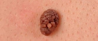
- Atheromas. Wen on the earlobe is quite common and looks like bumps or balls of a dense structure.
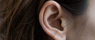
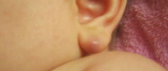
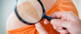
A few photos:
A doctor can determine why tumors appear on the hearing organs - inside or outside. Sometimes this may be associated with a malignant process - cancer of the skin or another organ.
Painless lump
A painless lump in your earlobe is most likely:
- A consequence of scar changes. If the ear was injured, a scar could form in this place. Most often, this situation occurs after an unsuccessful piercing and the bump is noticed completely by accident. It does not cause any discomfort and does not pose a threat to health.
- Wen (lipoma). This tumor can occur in different parts of our body (wherever there are fat cells), and it is a collection of fat cells. Lipoma has a soft, mobile structure and does not pose a threat to health. However, such a tumor can increase in size, and in rare cases, become inflamed.
- Atheroma. This is another type of benign tumor that occurs due to blockage of the sebaceous gland duct. Such a neoplasm looks like a superficially located dense lump with an elastic structure and clear contours. In the central part, a point can be seen - the place of blockage of the gland. The tumor is mobile and painless, but can become inflamed.
Most often, a painless moving ball on the ear is atheroma. Although such a tumor does not pose a threat to health, doctors advise removing it to avoid suppuration.
A lump has appeared and it hurts
The pain of a lump that appears on the earlobe indicates the development of an inflammatory process. A similar problem can happen as a result of:
- Complications after piercing. If the ear was recently pierced (especially at home), an infection could enter the wound and lead to the formation of an inflammatory process.
- Inflammation of a benign tumor. Lipomas become inflamed extremely rarely, but atheromas can cause a lot of trouble for a person. If the inflammatory process develops, the skin becomes red and swollen, as well as hot to the touch. Body temperature may increase. A festering atheroma can burst on its own, which is accompanied by the release of pus mixed with sebum-like contents.
- Formation of a subcutaneous pimple. Insufficient hygiene, hypothermia, decreased immunity and other factors can lead to the appearance of the most common pimple on the earlobe. It looks like a lump under the skin, is painful, and is covered in red skin (may be warm to the touch).
A painful lump on the earlobe is most often completely benign. You can suspect possible malignancy by:
- Itching.
- Burning.
- The appearance of an ulcer.
- Bleeding.
- Serous, mucous or purulent discharge.
In any case, the appearance of an incomprehensible tumor in the ear area that causes discomfort should be considered as a reason for an unscheduled visit for medical help.
Any inflammatory processes in this area are considered potentially dangerous, as they are located close to the brain.
Inflammation of the hair follicle
If a ball has formed on your earlobe and it hurts, this may be due to the fact that the skin glands are clogged and pus has begun to accumulate in them. Externally, this formation looks like a red pimple raised on the skin, sometimes with pus inside.
As long as the ball does not cause any particular discomfort, it should be treated like a pimple or herpes, depending on its appearance. But if a pimple on your earlobe begins to feel noticeably painful, you should contact a medical facility to prevent widespread inflammation, suppuration and the spread of infection.
Medical treatment of a lump in the earlobe
If a ball appears in your earlobe and it hurts and increases in size, you must urgently contact a medical facility for qualified help. There are a number of traditional treatment methods:
- Pimples on the earlobe are treated on an outpatient basis using anti-inflammatory and antihistamine medications.
- Inflamed purulent lipomas are opened with a scalpel, the contents are squeezed out, and the affected area is treated with antiseptic agents. Removal is carried out in a hospital setting under local anesthetic (anesthesia). It is possible to apply sutures at the removal sites, which dissolve over time without causing cosmetic discomfort to its owner. After the operation, the patient is prescribed antibiotics and antipyretic drugs.
- Introduction of a special solution into the seal, which “squeezes” the contents of the ball out. After the injection, the lipoma gradually resolves.
- Laser removal - cauterization of the compacted area with a laser beam. This method is especially effective in the initial stages of lipoma ball development. In more advanced cases, as well as in cases of suppuration, laser removal is not effective.
- Cauterization by radio waves. The effect of radio waves on the contents of the seal promotes cauterization (evaporation). The advantages of this method are painlessness, high efficiency, and the absence of skin defects in the form of scars, cicatrices, and sutures.
When a ball has formed on your earlobe, a qualified specialist will help you choose a method for its effective and painless removal. If necessary, he will prescribe a series of tests and smears, and after the procedure, he will prescribe medications to prevent inflammation at the removal site, decongestants, and also follow a number of measures to prevent recurrence of the lipoma.
Factors contributing to lipoma formation
Experts say that the formation of lipomas is provoked by a violation of metabolic (or hormonal) processes. There are a number of factors that can provoke the formation of a ball on the earlobe:
- Non-compliance (insufficiently thorough) with personal hygiene rules,
- Incorrect earlobe piercing
- Hypothermia,
- Excessive exposure to sunlight,
- Diabetes,
- Dermatological diseases of the ear,
- Injuries in the ear area,
- Increased sweating
- Use of low-quality cosmetics,
- The presence of pimples and acne on the skin.
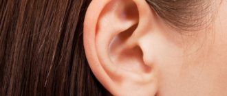
What can a lobe tell you about the owner’s health?
Is it really possible to somehow determine by the appearance of the left earlobe (or right) a person’s diseases or predisposition to them?
In any case, such attempts are being made. It is believed that the normal color of the lobe is pink, it feels smooth to the touch, and should not have bumps, pimples or folds. Normally, it is not thin, soft.
If it is observed that the earlobe:
- pale, thinned, hardened - this indicates a decrease in human immunity, exhaustion;
- too thick - may indicate obesity, mental lethargy;
- has a diagonal fold - a likely sign of stroke, heart attack, cardiovascular disease;
- includes multiple folds - possible presence of diabetes, atherosclerosis;
- unevenly “filled”, as if lumpy - a person may have cancer;
- has pimples - you need to pay attention to which organ these pimples are “projected”, problems are possible there.
According to observations, if a person has a square and elongated earlobe, he has a large supply of energy and vitality, but he may have a tendency to aggression.
Someone who has a long, pointed lobe has a lot of energy, is very hard-working, and smart.
The luckiest ones are those who have large ears with large, thickened lobes. It is believed that long-livers have precisely such ears.
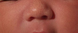
Manifestations in children
Most often, a ball on the earlobe appears in people aged 25-50 years, but the possibility of its formation in children cannot be ruled out. Children encounter this problem before the onset of puberty; from a medical point of view, we are talking about atheroma of the auricle. If you notice such a formation in a child, you should immediately consult a doctor, since in a child’s body such lumps can reach large sizes.
If a ball has formed near the girl’s earlobe and it hurts, the reason may be related to ear piercing. In this case, it is recommended to wear silver earrings rather than gold ones for some time.
From a medical point of view, the appearance of atheroma does not pose any danger, however, this phenomenon may indicate infection with AIDS. In some cases, a wen appears after unprotected sexual intercourse and is accompanied by deterioration of health, the appearance of acne and herpes. In such cases, atheroma is localized not only in the ear area, but spreads throughout the body, in the locations of the sebaceous glands. In addition to consulting with an infectious disease doctor, it is necessary to undergo appropriate tests.
According to doctors, the appearance of atheroma may be associated with:
- Hypothermia;
- Prolonged exposure to the sun;
- Improper hygiene;
- Injuries to the earlobes;
- Unsuccessful ear piercing;
- Acne;
- Hereditary predisposition;
- Hormonal imbalance caused by disruption of the endocrine system;
- Seborrhea;
- Wearing jewelry that is made from low-quality materials;
- Long stay in a hot, dusty room.
A common disease is atheroma
Structure of atheroma
Atheroma is nothing more than a blockage of the sebaceous glands. When the gland is blocked, a secretion is produced inside it, leading to an increase in the size of the tumor. It looks like a hard ball or lump under or inside the lobe, elastic, mobile and painless if there is no inflammation.
From the inside, the clot looks like a cyst, filled with yellow or white exudate and covered with epithelium. Several formations may appear at the same time, and inflammation of the lymph nodes occurs. If such a ball has formed inside the lobe, you should consult a doctor for its treatment (possibly removal).
Typically, a cyst occurs against the background of:
- genetic predisposition;
- hormonal disorders associated with diseases of the endocrine organs;
- metabolic disorders;
- acne;
- damage to the root part of the hair, which leads to blockage of the pores and suppuration in them;
- seborrhea;
- hyperhidrosis;
- inaccurate puncture of the lobe (may appear from earrings made of low-quality material);
- irregular or improper skin care;
- long stay in not very good conditions.
If the wen hurts when pressed, this may indicate an inflammatory process that has begun in it. It often begins with hypothermia or infection, as a result of trying to eliminate the tumor yourself, with dirty hands or other tools.
Manifestation of the disease
Atheroma is a benign formation of small size (initially). In the absence of proper therapy, the hard ball under the skin can increase to 50 mm in diameter. An ordinary non-inflammatory atheroma is often round in shape, smooth in surface, and mobile.
Inside it, the excretory duct is enlarged and swollen, the capsule is filled with exudate. When the wen becomes infected, the clinical picture changes.
A person complains about:
- the appearance of painful symptoms;
- hyperemia;
- increased body temperature;
- discharge from the center of the formation with purulent or bloody impurities.
If the capsule spontaneously opens, all the accumulated secretion comes out. Only a doctor can tell you how to treat a disease. Unfortunately, people turn to it very rarely, more often in critical situations, preferring to self-medicate.
However, such a negligent attitude may result in further surgery, a long period of rehabilitation, and repeated relapses.
Main signs and symptoms
The resulting wen in most cases is a benign neoplasm. In this case, a small compaction initially appears, but if it is not treated promptly, it may increase. Redness of the skin near the wen indicates the beginning of the inflammatory process and does not exclude the possibility of pus appearing in it. The ball has clearly defined boundaries, a smooth surface and a round shape. The excretory duct, which is located in the center of the compaction, can increase in size and fill with a porridge-like mass consisting of epithelial cells and secretions.
The appearance of pain in the area of the tumor indicates infection of the ball. The following features may be signs of an inflammatory process:
- Painful when pressed.
- A significant increase in body temperature in humans.
- When you press on the ball, ichor with impurities of pus may be released from it.
Main symptoms:
- Significant deterioration in the blood supply to the ear, which is caused by pathological growth of fatty tissue.
- Changes in the color of the skin on the earlobe. There may be some shine, tightness and severe swelling.
- When a ball appears on the earlobe, there is a high probability of developing otitis media.
- A person may experience a deterioration in health and the appearance of fever.
If the ball hurts, then you should not delay a visit to a specialist, as there is a high probability of developing ulcers, which can lead to meningitis.
Minor symptoms
Increased body temperature. Hyperthermia is observed in those patients in whom the atheroma has festered. Since the focus of the pathology is located extremely close to the brain, the syndrome of general intoxication will be intensely expressed.
Feeling of stuffy ear. The inflammatory process spreads extremely quickly to the ear canal, which leads to hearing impairment.
The danger of atheroma is that the cystic formation in the lobe can perforate. That is, the contents of the cyst independently break through the skin and flow out. An ulceration forms at this site, which is quite difficult to treat.
Like any tumor process, atheroma requires timely detection and initiation of treatment. It would be most advisable to contact an oncologist, since this type of pathology should be treated by this specialist. This is due to the fact that benign processes must be differentiated from malignant ones.
Otitis
Otitis is an inflammatory disease of the middle ear. Most often found in children. Adults get sick less often - about 20% of all cases. This disease can occur in chronic and acute forms.
Symptoms of otitis media include:
- Severe ear pain.
- Increased body temperature.
- General malaise.
- Noise in ears.
- Ear congestion.
- Hearing problems.
Somewhere on the third day of the disease, purulent contents begin to emerge from the ear cavity. The patient's condition improves somewhat and the pain gradually subsides. In severe cases, the pus cannot come out due to a certain density of the eardrum. Then a hole must be made in it. This disease cannot be treated independently, as it leads to serious consequences:
- Brain abscess.
- Meningitis.
- Sepsis.
The disease can also become chronic, which is difficult and takes a long time to treat. Relapses in this case can occur throughout life.
How to determine if there is an infection in a lump
When a tumor in the earlobe hurts when pressed, this does not at all indicate that an infectious-inflammatory process is developing inside. The presence of a pathological process is indicated by the following symptoms:
- the skin at the site of infection acquires a reddish tint;
- ichor and even blood may appear at the site of the neoplasm;
- discharge of pus;
- a sharp jump in body temperature - above 38°C.
If a wen in the earlobe is accompanied by the symptoms described above, then we can talk about serious problems that require immediate monitoring by specialists.
And remember, it is strictly forbidden to try to squeeze out the contents of a lipoma on your own, to extract it in any other way, or even to cauterize it with alcohol tinctures or apply compresses! This is fraught with aggravation of the current state of the pathology.
Signs of infection of the ball in the earlobe
- Redness in the area of infection, swelling,
- The appearance of painful sensations when touched,
- The appearance of blood and ichor at the site of compaction,
- Breakthrough of purulent contents to the outside,
- A sharp increase in body temperature above 38 [Symbol]C.
If any of the above signs appear, you should immediately contact a medical facility. It is strictly forbidden to squeeze out the contents of the lipoma yourself, do cauterization with alcohol-containing products, or apply compresses.
Medical diagnosis
The primary diagnosis is examination by palpation. The essence of this method is that a specialist uses his fingers to palpate the tumor, finds its duct and determines the level of density. All these measures can help prescribe correct and complete treatment in the future.
If, after an examination, a specialist says that it is necessary to remove the wen, a histological examination may be required. It is with the help of this method that the cause of the occurrence and its level of hardness are determined.
Which doctor should I contact for treatment?
If a lump appears in your earlobe, you can consult:
- To the therapist.
- To a dermatologist.
- Otolaryngologist.
Each of these specialists will be able to assess the situation and draw conclusions about the need for further more detailed diagnosis and/or targeted treatment. But if we are talking about a targeted consultation, then a dermatologist is more likely to come to the rescue.
A patient with such a problem may be prescribed conservative treatment, recommended to monitor the lump, or referred to see a surgeon.
Not such a simple part of the body
Already in ancient times, since the times of Avicenna and Hippocrates, the small lobe was not deprived of the attention of the Aesculapians. Taking a closer look at this formation, the sages drew conclusions about the patient’s state of health and even made attempts to predict his future diagnoses.
Some doctors of oriental medicine even now very closely monitor the appearance of the auricles of patients. Healers consider them an important organ, by influencing which they can identify the disease and cure a person. This is what the following areas do:
- auriculodiagnosis, which allows you to diagnose a person by examining the auricle and influencing its reflexogenic points;
- auriculotherapy - acupuncture, acupuncture, through which healing occurs.
Why is it important to visit a doctor?
More often, the appearance of a tumor on the earlobe is caused by blockage of the sebaceous glands, which leads to the formation of a wen. The latter, even in the case of a secondary infection, creates only discomfort for the person and does not pose a serious threat to life.
However, sometimes a neoplasm arising from the epithelium or fatty tissue becomes malignant. And it is possible to determine the type of tumor if a histological examination of the contents of the capsule is performed.
Therefore, when painful balls appear on the ears, and the skin in the problem area turns red, you need to seek medical help.
Grown or not
Lobes look different for everyone: they come in large and small sizes, varying shapes, and “attach” to the head in different ways. Based on their shape, they can be divided as follows:
- Free-hanging - when they are smoothly rounded at the point of attachment to the scalp and seem to hang, having a semicircular, oblong, square or pointed shape.
- Grown. This is what they are called if they do not hang down, but seem to be fixed to the scalp, without “room for self-expression.” They are also small in size.
Lobe types are genetically determined. What they will be like in a child depends on the relationship between the dominant and recessive genes of the parents.
Traditional therapies
A ball of pus located behind the earlobe can be opened by a surgeon using a scalpel. In this case, all contents of the formation are removed. The abscess on top is treated with antibacterial agents. If the ball is too large, the specialist may stitch the patient.
The cyst or atheroma is also removed through surgery. It is undesirable to leave such a neoplasm, since over time an inflammation process or a malignant tumor may develop.
To carry out the procedure, you need to use local anesthesia. The capsule is opened and all its contents are removed. After this, the surgeon cuts out the capsule directly. Instead of a classic operation, the doctor can perform laser removal of the ball on the patient. Despite the high cost of the method, it ensures the absence of cosmetic defects after its implementation.
Those who delay going to the hospital should be prepared for the fact that two operations will be needed. During the first, the doctor needs to remove the wen itself and send it for examination, which will confirm that the neoplasm is indeed benign. Only after this the surgeon has the right to remove the capsule itself.
Most often, fragments of all removed tumors are sent for histological analysis.
How else can you treat a lump near the earlobe?
Conservative treatment methods
A traumatic tumor or pimple does not require special treatment. Almost all cases resolve on their own. You can carry out local procedures that will locally improve blood flow and accelerate tissue regeneration.
But for atheroma, surgical treatment is indicated. Thanks to the development of medicine, various variations of surgical interventions are used to successfully treat the disease at different stages of progression.
The most primitive is simple peeling of the atheroma, which is carried out through a skin incision. It is important to remember that during such an operation it is advisable not to damage the tumor capsule in order to avoid recurrence and purulent-septic complications.
This method is used in cases where the atheroma is of significant size. This method of eliminating pathology has one significant drawback - scar formation. That is, patients need to undergo plastic tissue correction after such treatment in order to eliminate a rough and unsightly scar.
Modern methods of removing atheroma
More common is laser removal of atheroma. This type of surgery is well suited for minor operations, as it does not cause as much damage to surrounding tissue as with conventional treatment.
The duration of such an intervention usually does not exceed several minutes. One of the advantages of laser therapy is that the risk of relapse is minimized in the future.
In addition, treatment can be carried out using a radio wave installation. This method of treatment is used extremely rarely, and only in the early stages, when the size of the atheroma is still small. This method of tumor removal is used more often in other localizations of the process.
Surgical treatment of a pimple or abscess is radically different. All interventions that are carried out in this situation are aimed at creating an artificial opening for the outflow of purulent exudate. This can be achieved with a regular puncture.
Removal of atheroma using radio waves
This is the newest existing method for removing a ball in the lobe and is considered the most gentle.
In order to eliminate atheroma, high frequency radio waves are used. The energy of high-frequency radiation is concentrated on a special electrode, which heats the tissue, “evaporating” the cells.
Despite this, the surrounding tissues are not heated and are not damaged. The procedure is carried out under local anesthesia, during which all the contents from the capsule are evaporated.
If such an aesthetically unpleasant formation appears, you should not hesitate to go to the doctor, because the problem may worsen. To protect yourself from the appearance of a ball in the lobe, take care of your hygiene and eat right, emphasizes estet-portal.com.
Treatment with folk remedies
If a ball has formed on or inside your earlobe, it does not cause discomfort or pain, you can get rid of it at home yourself. For the treatment of lipoma the following is used:
- Compresses from aloe leaves 2 times a day for a month. To make a compress, stop watering the plant for 10 days, then cut off the most massive leaves, wash them thoroughly under water and place them in the refrigerator for 14 days. This is necessary so that the leaves begin to produce biologically active substances that promote tissue regeneration. Then the leaf is cut and the pulp is applied to the area affected by the lipoma, secured with a paper plaster. After 30 days, the contents of the ball will come out, and this area must be treated with hydrogen peroxide.
- Compresses from the houseplant Golden Mustache. The leaf of the plant is cut with a sharp knife, washed thoroughly, kneaded slightly (to increase the release of juice) and applied to the affected area. The compress is covered with a gauze bandage and secured with a bandage. To get results, applying a compress every day at night for 7 days is enough.
- Application of Vishnevsky's liniment to the area of compaction, which helps to draw out purulent contents and rapid healing of the wound.
- Garlic oil. Young juicy garlic (2 heads) must be crushed until mushy, add vegetable oil to form a thick paste. Massage the garlic oil into the seal area daily (preferably at night). For the ball to completely dissolve, it will take about 10–15 procedures.
- Ointment "Star". Local application of ointment to the area of the ball on the earlobe 2 times a day for 14 days. The ointment contains essential oils and flavonoids, which successfully warm and draw out purulent contents.
- Apply tea tree essential oil to the affected area with a ball 3 times a day until the lipoma is completely absorbed. Tea tree oil has a pronounced antiseptic, anti-inflammatory, and wound-healing effect.
All procedures performed at home must be carried out with clean hands, additionally treated with an antiseptic solution or medical alcohol. If after self-treatment the lipoma does not resolve and it hurts, you should consult a doctor or surgeon.
Folk remedies in the fight against lipoma
We figured out why the ball appears on the ear. We have found out how to influence such pathological clots using traditional medicine. Now let's look at the most popular ways to fight lipoma using traditional medicine.
Important! It is strictly forbidden to resort to traditional medicine to cure lipoma without consulting a doctor. Sometimes such alternative medicine only aggravates the existing inflammatory process!
Treatment of lipoma is carried out using the following methods:
- Aloe compresses. The procedure will require plant leaves. But just before using them, it is advisable not to water the plant for 10 days. After this time, the aloe leaves are cut off, washed thoroughly under running water and placed in the refrigerator for 2 weeks. Such preparation of the plant is required so that it begins to release biologically active substances that accelerate tissue regeneration. After this, the leaves are cut and applied to the location of the lump, fixing them with a band-aid. On average, the contents of the lipoma come out after 30 days of such procedures. As soon as this happens, the seal exit site is treated with hydrogen peroxide.
Attention! All procedures performed at home should be carried out with clean hands, additionally treated with an antiseptic or medical alcohol. If, after self-treatment, the lump has not resolved and is painful, you must seek the help of a surgeon.
- Compresses from the Golden Usher plant. The leaves of the plant are cut off, washed thoroughly under running water and crushed a little so that all the active substances come out. The prepared sheet is applied to the pathological tubercle and secured with a plaster. Place a gauze bandage over the sheet. To achieve the desired result, it is necessary to carry out such procedures for 7 days.
- Using Vishnevsky ointment. A little ointment is applied to the seal, covered with polyethylene and wrapped with a gauze bandage on top. The ointment helps draw out pus from the tumor and speeds up the wound healing process.
- Ointment Zvezdochka. We treat the seal with the product twice a day for 2 weeks. Due to the essential oils and flavonoids present in the ointment, the product has a warming and anti-inflammatory effect, drawing the contents of the neoplasm out.
- A mixture of vegetable oil and garlic. Take 2 medium heads of young, juicy garlic and grind until mushy. It's better to use a blender here. Mix the garlic gruel with vegetable oil until a thick consistency is obtained. Rub garlic paste into the location of the wen. This procedure is best done before bedtime. To completely eliminate the problem, 10-15 procedures are necessary.
Golden mustache
Attention! If a lipoma appears on a child’s ear, then resorting to traditional medicine to get rid of the problem is strictly prohibited. First of all, you need to see a doctor.
Folk recipes from wen
Traditional methods of treating wen are used when the ball in the earlobe becomes infected. In other cases, this approach is ineffective. Traditional medicine methods are not able to get rid of pathological formations on the body.
To prevent the spread of purulent infiltrate, it is recommended:
- Apply aloe leaves to the damaged area for a month;
- rub melted fat into the problem area;
- treat the wen with chopped garlic and sunflower oil;
- Apply a mixture of salt, yogurt and honey, taken in equal proportions, to the ball.
If you suspect cystic streaks, it is not recommended to self-medicate. In this case, you should seek help from a doctor.
Possible consequences
According to doctors’ recommendations, it is better to remove the atheroma in order to avoid its degeneration into fatty phlegnoma in case of infection. Since phlegnoma is located close to the blood vessels that provide nutrition to the brain, it is very dangerous. Delayed treatment can lead to adverse consequences, including death.
Treatment of atheroma should be carried out only by a specialist. To prevent atheroma, it is recommended to exclude all fatty, sweet and starchy foods from the diet. You need to clean your ears in a timely manner; for this it is better to use soap and water or a special scrub. By following these simple preventive measures and avoiding mechanical damage to the ear, the likelihood of a ball appearing on the earlobe is minimal.
Do not forget that self-squeezing the ball can result in infection and the beginning of the inflammatory process. If it is not possible to see a doctor, it is better to wait until the wen disappears on its own.
Ear injury
Injuries most often damage the earlobe, concha and ear canal. This may happen for the following reasons:
- Improper ear cleaning.
- Physical impact on the ear.
If there is an injury, rupture or complete separation of the organ from the body, it is strictly forbidden to rinse the ears with water or various drops before the arrival of doctors. This procedure may initiate the development of an infection in the inner ear. Most often, such an injury is treated with surgery and stitches. Even if the ear was completely separated from the body, the chances of its engraftment are quite high. The main thing is that the separated organ is kept in a cold place before the operation begins.
The middle and inner ear are usually damaged by:
- Cleaning ears with sharp objects.
- Foreign objects entering the ear.
- Various thermal and physical burns.
- A strong blow to the ear.
- Getting infected.
With such injuries, the following symptoms occur:
- Strong pain.
- Noise.
- Partial or complete hearing loss.
- Discharge of fluid and pus from the ear canal.
Complications such as meningitis are quite common.
If the damage is minor, it usually heals on its own. Large ruptures require surgery. If there is a foreign object in the ear, it is removed. The eardrum can also be repaired through surgery. At the same time, antibiotics are prescribed. The prognosis is usually favorable.
Prevention
Oily skin promotes the development of atheroma. For the purpose of prevention, it is useful to wash and take a bath using laundry soap.
In terms of nutrition, exclude from the diet too spicy and fatty foods, sweets and flour products.
And the last point of prevention is timely treatment of all chronic diseases.