The skin in intimate places, namely in the pubic and buttock areas, the mucous membrane of the external genitalia is richly supplied with glands that secrete fat and sweat. Under the influence of unstable hormonal levels, they are especially active in adolescence, youth and early adulthood. Therefore, the intimate area is a favorite place for acne and pimples appear on the labia of women quite often. They are white or red in color, located internally, subcutaneously or externally, they pop up in the groin or on the butt alone or in a group.
What should a girl do if acne regularly appears between her legs or on her buttocks? Do not despair and under no circumstances try to crush them or somehow mechanically influence these formations! They can be easily managed once we understand the reasons for their occurrence.
WHY ARE Pimples ON THE LAVADIA - REASONS FOR THE APPEARANCE
Just like you get pimples on any other part of your body, you can get them there. This is a blockage of the sebaceous glands that leads to the formation of small lumps called pimples. They appear between the legs of women for several reasons:
- Humidity in the area;
- The presence of increased secretion;
- Friction in this area when moving, especially if you have enlarged genitals;
- Hormonal imbalances;
- Consequences of shaving or hair removal;
- Violation of intimate hygiene rules.
Pathologies of non-infectious nature
- Tumor formations. A lump on the inner labia is an alarming sign and is often of oncological origin. And although tumors on the genital organs are most often benign, they should not be treated with disdain.
Benign neoplasms on the labia include:
- Myoma (located in the form of a ball inside the labia);
- Fibroids (a lump inside the labia majora formed by muscle tissue);
- Lipoma (small nodule or several nodules located near the vulva);
- Hydradenoma (occurs rarely and also looks like a nodule).
A lump on the labia minora or majora in the form of a large node or tubercle is most often a malignant formation that requires immediate medical attention.
- Nervous overstrain. Although rare, pimples on the labia minora
can appear due to frequent stress and tension. The rashes resemble allergic or mechanical factors.
Balls as benign neoplasms
Removal of lymphangioma
If a ball has formed that is of a benign type, then its timely excision can reduce the risks of tissue degeneration.
- Myxoma. It is often found on the pubis, but is also present on the large intimate folds. The internal fluid has a gelatinous yellow-white color. It is more often diagnosed in people of retirement age.
- Lymphangioma. Lymphatic formation is a painful type that is projected mainly on the labia majora. There are always several tubercles, which have a blue or crimson color and are connected to each other during growth. The rate of increase is very slow, there is no threat to life, and the prevalence among patients is high.
- Myoma. The affected area is characterized by the involvement of the deep tissues of the labia in the pathological process.
- Fibroma. The oval-shaped ball consists of connective tissue. A tendency to transform into a malignant formation is present in only one type of fibroma - desmoid.
- Hemangioma. With this compaction, doctors detect a ball on the labia minora (or labia majora). The presence of blue or red color is explained by the presence of blood in the formation (2 forms - cavernous and capillary).
Infections leading to the appearance of lumps in the labia area
- HPV. The human papillomavirus enters the body when immunity decreases and is transmitted sexually. There are several types of HPV. Some are papillomas in the form of papillary skin growths, which are considered a precancerous condition. And the latter cause the appearance of condylomas on the labia, photos of which can be viewed on the Internet. They look like cauliflower and can also lead to cervical cancer. They grow quickly and appear instantly. There are known cases when they disappeared on their own, falling away. However, most often only surgical treatment will help get rid of them.
- Bartholinitis. Bartholin's glands are located near the entrance to the vagina and can become inflamed due to blockage of the ducts. Symptoms of the disease appear gradually and a characteristic sign of the pathology is a lump on the labia majora, which develops only a couple of weeks after the onset of inflammation. The tubercles when wearing tight underwear cause discomfort and pain, and subsequently swelling appears on the labia. During the disease, pain and burning are noted in the perineum. Sometimes the development of the disease ends in suppuration, but, fortunately, this rarely happens. This pathology does not require complex therapy, however, it is impossible not to treat bartholinitis at all, since it can develop into a recurrent form, and subsequently turn into a cyst.
- Syphilis. A serious venereal disease, which in the primary period is characterized by the appearance of chancre (that is, an ulcer in the area where the pathogen enters). If the ulcer develops into an internal pimple on the labia, then the disease moves into the secondary period, affecting more and more organs and systems of the body. Pimples on the labia with syphilis can be of different types and sizes. They can be ordinary papules, or they can be purulent formations. Most often there will be pimples. Those suffering from syphilis will not have any subjective manifestations in the form of burning or itching, but such rashes should not be ignored. It is useless to try to remove them at home, since syphilis can only be treated with special drugs, and the sooner therapy is started, the fewer consequences will remain after this disease.
- Candidiasis. The second name of the disease is thrush. The main symptoms of this pathology will be white vaginal discharge, as well as subjective sensations in the form of itching. However, in rare cases, a pimple may form on the labia majora, either one or several. The formations are large in size and painless. Treatment can only be special, prescribed by a doctor.
- Genital herpes. The main problem with all types of herpes is that it is incurable. Fortunately, its manifestations occur only during periods of decreased immunity. A rash of herpetic etiology is a small watery pimple in the vaginal area that is itchy and very painful. After some time, the pimples burst, forming painful ulcers. Treatment of rashes is carried out using a special drug that suppresses the herpes virus, however, therapy is completely symptomatic, since the pathogenic agent will still remain inside the body.
- Acne that occurs due to poor personal hygiene or other external factors does not require specific therapy. It is enough to remove the irritant, and the rash will go away on its own. However, to reduce itching and burning, if present, you can use antiseptic ointments and tinctures. You can also use drying agents - zinc ointment or baby powder. Even baby diaper cream can alleviate the condition.
- Hematomas, if the woman’s general condition is normal, are treated by applying cold. However, it should be remembered that any deterioration in the girl’s general condition or the growth of a hematoma requires immediate consultation with a doctor.
- Due to nervous tension, in addition to soothing ointments, you should pay attention to the general condition of the body, the normalization of which will immediately lead to the disappearance of pimples.
- Tumor-like asymptomatic balls inside the labia require a visit to a gynecologist, and then, most likely, a visit to an oncologist.
- There are a number of signs (they are inherent mainly in formations of an infectious nature), the appearance of which requires an immediate visit to a gynecologist:
If within a week or two the rash does not disappear, and even worsens, then contacting a gynecologist becomes a priority task. Treatment at home helps only with ordinary irritations, but not with serious pathologies.
- Rapid growth of the rash;
- An ulcer that turns into a pimple and vice versa;
- Swelling of the genital organs;
- Watery acne;
- Enlargement of many groups of lymph nodes and elevated temperature.
As it becomes clear from the above, seals on the labia minora and majora are most often an unfavorable sign.
Wen is an unpleasant problem that, unfortunately, a large number of people face in the course of their lives.
Depending on the location of this benign neoplasm, it causes its “owner” a lot of trouble and inconvenience. This is especially true when a lipoma (scientific name for a wen) appears in the intimate area, in particular on the labia.
The main reason why wen can develop is poor activity of the sebaceous glands, which is a consequence of their blockage. If we talk specifically about lipoma of the labia, in addition to this, the main factors provoking the pathology may be:
- obesity
- excess weight is associated with a large amount of fat, which is looser in composition, and the glandular ducts become clogged much faster, provoking the development of anomalies; - poor hygiene
– irregular care of the intimate area creates favorable conditions for the development of pathogenic microorganisms that feed atypical benign lipoma cells; - hormonal imbalance
- any disturbances of this kind lead to dysfunction of the sebaceous and sweat glands; - mechanical damage and trauma to the soft tissues of the labia
during incorrect depilation; - tight underwear
, constant wear of which causes friction and disruption of the integrity of the surface tissues of the organ, damaging their structural content.
Complicated atheroma
Usually the wen proceeds quite peacefully, but sometimes the neoplasm is affected by an inflammatory process. How to distinguish a sluggish disease from acute inflammation? Usually the lump changes its appearance - it becomes hot, hyperemic, swelling appears near it, and the lump itself becomes larger. When you touch it, you can feel pain, and at rest the inflamed atheroma twitches. When you press on the swelling, purulent-curdled contents with a specific odor are released from it. At this stage of the disease, you need to go to the clinic and treat the atheroma surgically. You need to act especially quickly if a new growth of the scalp is inflamed.
Symptoms
Symptoms of the pathology in the labia area are extremely scarce and practically not expressed, especially in the initial stages of the disease. As it progresses, the following signs may appear:
- swelling in the affected area
- it is caused by internal subcutaneous accumulations of lipoma contents; - visible redness in the area of the borders of the neoplasm
- occurs against the background of internal inflammation of the soft tissues surrounding the wen; - pain
does not appear immediately. As the pathology grows and constant friction against underwear, the surface tissues of the epithelium become thinner and mechanical impact, as well as contact with them, can cause pain of varying intensity. If the pain becomes more pronounced over time, there is a suspicion that the tumor has become malignant.
Complications
Lipoma of the labia itself does not pose a serious threat to the health, much less the life of a woman. The main trouble is mechanical and aesthetic discomfort.
However, the problem should not be neglected at all. Since the place where the pathology develops is quite easily injured, the neoplasm can be accidentally damaged.
And since its internal component is an excellent nutritious pathogenic sphere, microbes and bacteria, having penetrated into the internal capsular cavity, can provoke an inflammatory process in the tumor itself and cause the formation of purulent masses there.
At a certain moment, the secretion, unable to withstand the pressure of the capsule increasing in size, will inevitably rupture, and all the contents will spread into the subcutaneous soft layers. This will lead to an abscess of the surrounding tissue fragments.
There is another direct threat that such a seemingly insignificant disease can lead to dire consequences - this is an attempt to eliminate the problem yourself by trying to open the wen and squeeze out its secretion.
Doing this is strictly prohibited - very often such actions lead to malignancy of tissue, microscopic fragments of which will still remain inside the capsule cavity.
After such a “barbaric” impact, they begin to divide chaotically and turn cancerous, and these processes often occur quite rapidly. The abnormal cells invade neighboring cells, and the tumor rapidly increases in size.
Diagnostics
Diagnosing lipoma of the labia is not difficult. Very often, a woman independently notices the appearance of an abnormal formation and seeks advice from a clinic.
In a medical institution, the main methods to identify the disease and obtain a complete clinical picture are:
- examination by a gynecologist
- during a routine examination, or during a patient’s visit with suspicions, the doctor, by visual examination of the labia area, is most likely able to diagnose this ailment. To confirm the final diagnosis or refute it, the woman can be additionally examined by other specialists; - consultation with a specialized surgeon
- this procedure is considered almost mandatory, since surgical removal of the lipoma still remains the priority solution to the problem. The surgeon will determine whether such a radical intervention is necessary in this particular case, or whether it is enough to simply observe the anomaly for a while; - histological analysis
is a mandatory manipulation to ensure the safety of the neoplasm, eliminating the risk of its malignancy. In addition, such a study will allow the most accurate classification of lipoma by type, which is very important for determining the optimal method of treating it.
In situations where the pathology is too small, a consultation with a dermatovenerologist may be required, since the diagnoses of this group of diseases have similar external symptoms, especially in the initial stages of compaction formation.
Treatment
The method of treating lipoma of the labia is selected based on the clinical picture of the process, the size and content of the capsular secretion.
If the compaction remains in a state of latency for quite a long time, does not increase in diameter, remaining within a few mm, and the internal filling of the capsule is not associated with aggravating factors, therapy may be more gentle, and surgical intervention will not be required.
There is no talk of removal even in cases where there are no inflammatory manifestations and no purulent inclusions in the secretion.
Most often, the following methods are used in the treatment of the disease:
- drug therapy
– this method can, if not eliminate it completely, then significantly reduce the size of the formation by introducing special medicinal compounds subcutaneously into the affected area of the labia. It is used infrequently, since the technique is associated with the risk of partial resorption of the pathology, which can cause additional problems.
Drugs with this spectrum of action include: Vitaon, Ichthyol, Balsamic Liniment, Heparin. There are also a number of products available in the form of ointments, however, this treatment option for this form of diagnosis is considered to be very doubtful and ineffective;
enucleation of lipoma
– the most common manipulation. It is carried out by surgical excision of the compaction, exfoliation of the capsule located in the internal soft layers, followed by sanitation of the affected area and the site of the wound to prevent its infection.
The area of remote pathology is generously treated with antiseptic and antibacterial drugs. To consolidate the result, a course of antibiotics is indicated;
removal of the tumor with the capture of healthy tissue fragments
– is carried out in cases where the anomaly has a lobular structure, is associated with complicated forms, and is multiple in nature.
If the pathology develops rapidly, increases in diameter, and if there is a risk of transition to a malignant neoplasm, amputation of not only the capsule itself, but also the adjacent fragments of healthy tissue bordering on it is indicated.
This will minimize the likelihood of complications and repeated relapses. The operation is performed using standard methods under local or general anesthesia;
If you find an error, please select a piece of text and press Ctrl+Enter
.
Changes occurring in a woman’s external genitalia are always a cause for concern. A small pimple, lump, or lump on the labia can be a symptom of the clinical picture of more than one disease. So where do bumps on the labia majora and minora come from? There are many reasons for this, ranging from the simplest, such as poor hygiene, to serious illnesses.
Inflammation of hair follicles
Folliculitis occurs after injury to the hair root and infection. This is preceded by careless hair removal or shaving, ingrown hairs. A white or yellow lump with a red, inflamed edge forms on the outer labia. A twisted hair is visible inside. A large pimple is filled with pus and is very painful.
Squeezing it out is extremely dangerous, otherwise harmful bacteria can enter the bloodstream and cause blood poisoning. Treatment consists of wiping the affected area with local antiseptics. To absorb the pus, make compresses with Vishnevsky ointment at night.
Fortunately, almost all diseases, the symptom of which is a ball on the half lips, can be treated. If you see a doctor in time, you can even defeat cancer.
Lump on the labia majora
Most often, inflammation in the form of a lump on the labia majora is a symptom of inflammation of the Bartholin gland. This organ is paired; it precedes the entrance to the vagina. The organ consists of sebaceous and sweat glands and its main functions are to secrete a lubricant during sexual intercourse and moisturize the vagina to protect it from harmful microbes.
Photo 1: Only women who have reached puberty are susceptible to inflammation of the Bartholin gland. Source: flickr (GB SINGH)
If these glands become blocked, a lump-shaped tumor may appear on the labia. It is accompanied by redness and may be painful and itchy. Additional symptoms characteristic of this disease:
- painful compaction on both sides at the entrance to the vagina;
- burning in the perineal area;
- pain that worsens during sexual intercourse;
- temperature increase.
Inflammation of the Bartholin gland is primarily acute. If you do not engage in treatment, then this process can become chronic, and the doctor will diagnose a Bartholin gland cyst.
Bartholinitis in chronic and acute forms can be successfully cured without surgery; to do this, you need to contact a gynecologist at the first unpleasant symptoms of the disease and follow all the doctor’s recommendations.
Reasons for the appearance of a lump on the labia:
- decreased immunity;
- long-term use of antibiotics;
- chronic infection, such as tonsillitis;
- tight underwear;
- poor intimate hygiene;
- promiscuous sex life;
- infection during surgery.
The appearance of lumps on the labia can be symptoms of diseases such as prolonged vaginal candidiasis and vaginitis (inflammation of the vagina).
Vaginitis is a disease that also occurs due to poor hygiene, infection with worms, diseases of the endocrine system, and prolonged use of antibiotics. May be a consequence of diseases of the internal genital organs. For example, the same candidiasis, colpitis, etc.
What kind of disease
Atheroma is a benign formation, often with a genetic predisposition. An epidermal cyst looks like a thickening of the skin.
It consists of sebum and dead cells. Inside there is a white-yellow liquid with an unpleasant odor.
It is formed due to blockage of the sebaceous duct, then the discharge from it begins to accumulate under the skin and mixes with keratin. Atheroma can grow to large sizes if no measures are taken to eliminate it.
On this topic
Can an ovarian cyst resolve on its own?
- Editorial board of Oncology.ru
- October 16, 2021
The tumor can be large or small, grow quickly or not change in size. Sometimes it can open on its own, then the liquid contained in it will flow out.
In such cases, it is important to prevent infection from entering the middle of the capsule, which can lead to infection. That's why it's so important to see a doctor.
Seal on the labia minora
The lump on the labia minora is not related to bartholinitis. Often, pea-sized bumps and pustules occur when several reasons come together:
- poor perineal hygiene;
- hot season;
- unsuitable gaskets.
Compaction can also occur due to:
- hypothermia;
- cycling;
- inflammation of the hair follicle.
A wen appears on the labia minora, which, when ripe, bursts and pus comes out. To avoid the formation of infection, it is necessary to treat the seal with a disinfectant.
Important! A lump that appears on the labia minora may not cause discomfort at all, but any subcutaneous formation with pus risks the fact that this fluid may not come out and infect the internal tissues. Therefore, consultation with a doctor is necessary.
Therapy in all cases will depend on the diagnosis of a particular type of disease. The doctor may prescribe antibiotics or prescribe local antiseptics, and recommendations on wearing underwear and maintaining personal hygiene are also required.
Homeopathic treatment
Homeopathic treatment for lumps on the labia will also depend on the identified disease.
- If this is inflammation of the Bartholin glands, then (Belladonna)
or
(Mercurius solubilis)
. These drugs relieve redness and swelling of intimate organs, eliminate suppuration, and help shrink lymph nodes. - For bacterial vaginitis with discharge, itching and burning of the genitals, it is recommended (Kalium bichromicum)
. This remedy eliminates pain and helps healing in complex diseases and inflammations of the skin and mucous membranes. - For vaginal discharge with a sharp, unpleasant odor, Kreosotum
. This remedy eliminates burning sensation in the vulva, between the labia, in the vaginal area and removes discharge. - A drug
Every woman, having discovered a lump on her labia, begins to worry greatly. And first of all, immediately contact a specialist. And this is the right decision.
The lump may be painful. In any case, consulting a doctor is necessary, since the cause of this phenomenon may be different. Most often, a lump on the labia can be a symptom of bartholinitis.
Treatment most often involves surgical removal. After removal, antibacterial therapy is mandatory, thereby stopping the further development of the disease.
Sometimes it happens that a woman completely unexpectedly notices a subcutaneous lump on her labia majora or minora, most often painful.
It can be a sign of various gynecological diseases, so if you have the slightest suspicion of discomfort or foreign tumors, you should consult a doctor.
In some cases, a lump that appears on the labia may be an ordinary pimple as a local reaction to an external irritant. Over time, this compaction goes away on its own.
general information
If the gland becomes clogged, it stops removing sebum, which accumulates, over time forming mobile nodes of varying sizes. They are mistakenly called wen, although they have a different name.
Atheroma is a cyst of the sebaceous gland, and occurs at the site of its blockage. These growths can appear on the face, arms or back; they are lumps that look like a pimple, but do not cause redness and are not painful when pressed. It is not a tumor or an inflammatory disease, does not cause deterioration in health and remains, to a greater extent, an exclusively cosmetic defect.
The main causes of seals in the labia
Approximately 8 out of 10 women who consult a gynecologist with a complaint of a lump inside the labia are diagnosed with bartholinitis.
Expert opinion
Knyazev Igor Vladimirovich
doctor
In addition, such a formation may turn out to be a malignant or benign tumor. We will dwell on these diseases in more detail a little later.
However, these are not the only reasons due to which a seal may appear on the labia minora or majora. Also, a similar symptom often occurs in the following cases:
- in some situations, nodular fragments indicate the development of human papillomavirus in a woman’s body. HPV itself quite often passes without consequences, but in certain cases it can provoke cervical cancer, so such changes in the body should be treated very carefully;
- if such a defect appears with enviable regularity before your period, most likely we are talking about an enlarged sebaceous gland, which, upon palpation, is very often mistaken for a dense pimple. Such a disorder in the female body is a consequence of a surge in sex hormones. If after a few days, when the hormonal levels return to normal, the bumps and bumps disappear as suddenly as they appeared, there is nothing to worry about. Meanwhile, some women in such a situation note some discomfort in the genital area. To get rid of unpleasant sensations, try wiping the affected skin with a cotton swab, generously moistened with a soothing and antiseptic tonic. In addition, 1-2 weeks before the start of menstruation, it is useful to give up sweet and fatty foods;
- in rare cases, when small seals on the labia have been present for a very long time and do not bother their owner in any way, they may be the result of abnormal development of the genital organs and, in particular, the labia majora and minora, the urethra and other internal organs and systems. Such formations are not viral or infectious in nature and do not pose any danger to women;
- Finally, even more rarely, compaction in the area of these organs can be noticed in a newborn girl. Almost always, such changes in a baby indicate that both of her parents, or at least one of them, have such a serious disease as syphilis.
Bartholinitis
It is necessary to exclude the presence of such a serious gynecological disease in a woman as bartholinitis.
Bartholinitis is an inflammatory process that occurs in a special Bartholin duct as a result of a sexually transmitted infection, less often in the case of an infection in the tonsils or dental diseases.
If a woman has a lump on her labia, a common reason for this formation is insufficient adherence to the rules of personal hygiene, as a result of which pathogenic pathogens invade the body.
Causes
The following factors can lead to the occurrence of atheroma:
- heredity;
- general hypertrophy;
- hormonal imbalance , which can cause hypersecretion of lipid substances in the glandular tissue of the skin;
- increased sweating;
- unhealthy diet, where fatty foods and sweets predominate;
- failure to comply with basic hygiene rules;
- metabolic disorders ;
- mechanical damage to the skin.
The development of atheroma largely depends on hormonal disorders in the body. When hormones are imbalanced, the oiliness of the skin increases, which significantly increases the likelihood of skin cysts.
The cause of the appearance of cysts in intimate places in women is often metabolic disorders, in particular the presence of a disease such as diabetes.
Symptoms of bartholinitis
If the disease is advanced, then a hard formation on the labia can be quite painful, and a tingling and burning sensation is also felt in the area of the lump.
As a rule, when you press on the seal, the pain intensifies.
In addition, the following symptoms may be observed:
- the seal grows to the size of a chicken egg;
- at the site of compaction, the skin is red, often has a bluish tint;
- there is a high body temperature and fever that antipyretics cannot cope with;
- chills;
- there is a general loss of strength, lethargy, and apathy.
In especially severe cases, the pain reaches such a degree that the woman cannot walk normally.
If the seal on the labia does not prompt the woman to start treatment and consult a doctor, then over time the abscess may spontaneously open. In this case, the woman temporarily experiences relief. However, the disease itself remains, as a result of which relapses may occur in the future.
The disease itself can become chronic, which is more difficult to treat due to its neglect. If the disease progresses, then the formation of a cyst in the area of the labia minora and majora is possible, which requires surgical intervention.
Often the presence of a cyst can make it difficult to perform physiological functions (urination, defecation).
Description of surgical methods
If atheroma has formed on the face of a small child (under 7 years old), then the only suitable treatment method is to remove the capsule with a scalpel, laser or radio waves under general anesthesia. In adults, this procedure is performed under local anesthesia. Unlike classic excision with a scalpel, other methods of surgical intervention do not leave visible marks on the skin and minimally injure blood vessels. In addition, laser and radio wave procedures eliminate the need for postoperative measures.
The method is indicated for small tumors without tissue inflammation. Removing atheroma on the face with a laser leaves virtually no scars or scars and does not disturb the subcutaneous microflora. The technique belongs to the bloodless category, and the wounds left after the operation heal quickly. The average duration of the procedure is 20 minutes.
After excision of a large tumor, the wounds are sutured, and the sutures are removed after about a week (with normal healing process). After removal, atheroma should be treated daily with an antiseptic solution such as Betadine. In addition, in the first few days the wound site should not be wetted. Dressings after surgery are carried out if the source of the disease is in the area of clothing friction. Until the wound is completely healed, it is recommended to treat the skin with Levomekol and Vishnevsky balm.
Making a diagnosis is usually not difficult. It is easy for a doctor to distinguish a wen from a lymph node and other types of tumors by the characteristic appearance and location of the tumor. In some cases, additional consultation with other specialists may be necessary, but in most cases, surgeons independently treat the pathology.
After specifying the diagnosis, the doctor notifies the patient about treatment methods. Conservative methods for treating neoplasms are ineffective, and traditional methods help very little. Surgery will help cure the disease forever. The intervention is usually performed under local anesthesia, since deep skin incisions are not required.
The operation to remove atheroma is performed on an outpatient basis. This means that the patient is sent home the same day. Only for dressings after removal of atheroma you need to come several times. The operation itself takes place quickly - about 15-30 minutes, but taking into account the preparation for the operation and the time required to fill out documents, the patient may be delayed in the clinic for about an hour.
When a wen suppurates, it is impossible to remove the formation in the usual way, so doctors carry out an operation according to the plan of opening the abscess - they evacuate the purulent contents, wash the wound and drain it. After the operation, patients are prescribed a course of antibiotic therapy and the wound heals. I begin removing the lump in about three months, when the damaged tissues have been restored. If you remove the wen against the background of inflammation, you may not completely cut out the capsule, and there will be a relapse.
After removing the lump, a scar may remain on the skin - it all depends on how large the subcutaneous lump was. When restoring the skin, you can use Contractubex to prevent scars. Photos of the skin condition after atheroma can be viewed on special websites.
Recovery after surgery generally proceeds without complications. There is a risk of fluid accumulation in the cavity of the wen, but this does not happen with a pressure bandage or drainage. On the first day after surgery, some patients have a fever - this is a normal reaction of the body to the intervention. If the numbers increase above 38, as well as when swelling, severe pain and purulent discharge appear, you must consult a doctor again.
Many doctors recommend treating the tumor when the lump is inflamed or causes a significant cosmetic defect. It is better not to touch small atheromas at all.
Previously, removal of atheroma on the face was carried out using electrocoagulation or a conventional scalpel. However, such surgical methods are associated with a high risk of complications. In addition, after such manipulations, wound healing takes a long time, leaving noticeable scars on the skin. Therefore, the following methods are currently used for small cysts:
- Laser surgery. This is a popular and painless way to get rid of atheroma. Before the procedure, the skin is treated with an antiseptic and a local anesthetic solution is injected. Then the epidermis above the cyst is cut with a laser and the beam of the device is directed directly to the tumor. At the same time, the vessels are affected, so the manipulation is not accompanied by the release of blood. After the procedure, a bandage is applied to the wound; final healing takes several days. Almost its only “disadvantage” is its high price.
- Radio wave removal. During manipulation, the tissue is exposed to specific waves, after which the surgeon gains access to the body of the cyst and removes it. The entire operation takes no more than 20 - 30 minutes. The healing process is quite long and lasts up to 20 days after treatment. A video with a detailed description of this removal method is available on the Internet.
If the atheroma is associated with suppuration and bacterial inflammation, a standard operation is performed. The patient is given a large dose of local anesthetic, after which the skin is cut with a scalpel. The surgeon removes the abscess and treats the cavity with antiseptic solutions. Then, for 7–10 days, a course of antibiotics is prescribed to prevent complications.
Diagnosis of the pathological condition
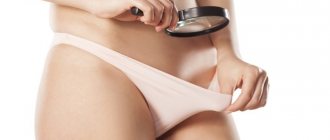
A woman can often detect a lump in the labia on her own, but in order to find out the nature of such a formation, it is still necessary to contact an appropriate specialist, that is, a gynecologist.
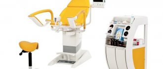
During the history taking, the doctor must conduct a gynecological examination of the external genitalia using a special chair and a magnifying mirror.
During this procedure, the doctor evaluates the condition of the pubis, anus, labia minora and labia majora. In addition, the gynecologist pays special attention to the presence of any neoplasms and the structural integrity of the skin in the intimate area.
Expert opinion
Knyazev Igor Vladimirovich
doctor
The examination is based on palpation (feeling) of the following anatomical structures: the clitoris, the external opening of the urethra, the labia minora, the entrance to the vagina, the anus and the large glands of the vestibule.
If the patient has leucorrhoea (liquid discharge), a sample is additionally taken, after which it is examined under a microscope (cytological examination). If necessary, the uterus and its appendages are examined.
Instrumental diagnostics
Instrumental diagnostic methods are also used to diagnose pathological formations in the vagina. The main ones are:
- lymphography (studying the condition of the nodes of the lymphatic system with the aim of possibly detecting cancer cells in their structure - metastases, which can, along with the bloodstream, enter the lymphatic tissue from other internal organs). The procedure is done using intravenous injection of a contrast agent into the lymphatic vessel. Its movement is then monitored using x-rays;
- ultrasound examination of the pelvis. Today, ultrasound is considered the safest diagnostic technique. During its implementation, the condition of the human genitourinary system is assessed. This procedure has virtually no contraindications and therefore can be used under any circumstances. However, ultrasound scanning is not recommended during menstruation, since at this time the indicators are somewhat distorted, which will significantly complicate further diagnosis;
- computed tomography or magnetic resonance imaging of the pelvic organs. Such diagnostic techniques represent layer-by-layer scanning of anatomical structures and are non-invasive (that is, they do not require direct penetration into the body of the subject through the skin or mucous membranes) and highly accurate procedures. In addition, if the lump on the labia is of a malignant nature, the patient will be prescribed an additional consultation with an oncologist.