Any changes in the condition of the skin can significantly worsen a person’s sense of internal comfort. And neoplasms, such as papules, vesicles and pustules, that arise in the upper layer of the epidermis and are located in the facial area - first of all. A vesicle, being a type of facial skin lesion, can be either a mild cosmetic defect of the skin or a manifestation of infection penetration into the upper layers of the epidermis, causing its stratification.
Today, there are several fairly effective methods of therapeutic action on vesicles. However, in order to choose the most effective treatment regimen, you should first determine the reasons that caused this phenomenon. This will avoid possible relapses after the course of treatment and ensure the longest possible preservation of the result.
What are vesicles
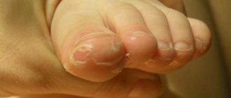
Externally, such a manifestation as vesicles are relatively small neoplasms that protrude above the surface of the skin. Inside they contain liquid contents, which may be transparent or slightly cloudy. Formed in the form of round neoplasms, vesicles have the shape and size of a pinhead or pea.
Vesicles have a certain classification based on both the specific manifestation and the location of the vesicles. In this case, the reasons for their occurrence are usually both the penetration of microbes and infections into the upper layer of the epidermis, and the delamination of its upper layer. Each person’s sensations when they appear may vary somewhat, but for vesicles of different shapes or classes there is a marked similarity in the manifestations and symptoms of the disease.
Accompanied by itching and a slight burning sensation, vesicles can be classified as more of a cosmetic defect. However, their appearance is usually associated with certain internal reasons, so getting rid of them should begin with understanding the reasons that caused this phenomenon.
A specialist will tell you about extracellular vesicles in the video below:
Why does the crust on the wound take a long time to fall off?
We are talking about shallow wounds (abrasions) and cuts.
Healing a wound “under a scab” (that is, with the formation of a crust) is a completely special process. The scab itself is a natural dressing that protects the wound from dirt, bacteria, and simply from additional trauma. A crust does not always form on the wound, but only under the influence of air. That is, if you apply a wet ointment bandage to the wound, a crust will not appear.
Normally, the crust on the wound does not fall off until healing has occurred. As soon as a thin epidermis (surface layer of skin) appears at the wound site, the damaged area begins to itch. First on the sides, and then closer and closer to the central part, the crust begins to separate from the wound. This is an absolutely painless process if healing has occurred completely.
In some cases, the crust on the wound does not fall off for quite some time. This occurs when deep wounds heal “under the scab.” It's no secret that extensive damage takes longer to repair. Another case is the formation of suppuration under a crust. In this case, the scab no longer plays a protective role.
On the contrary, it prevents the separation of pus from the wound - the breakdown product of bacteria. Pus forms under the crust when the wound is treated poorly, when pathogens penetrate the tissue and begin to multiply there. The resulting crust protects them from exposure to oxygen, sun and antiseptics - the main disinfectants.
Classification
Main types
Thanks to the existing classification, it is possible to subdivide such epidermal lesions as vesicles. According to medical statistics, vesicles can occur in people at almost any age, but they are most often diagnosed in adolescence and young adulthood.
Such neoplasms on the upper layer of the epidermis are classified as follows:
- Located inside the upper layer of the epidermis, or intraepidermal . If you try to squeeze them out, there will be no result, since their localization does not allow making the necessary gap in the thickness of the skin.
- Subepidermal , which are located in the upper part of the skin. Inside each vesicle there is a cloudy hemorrhagic fluid. And although it is possible to squeeze them out, the final disposal of this type of vesicle is quite difficult.
- Subthreshold vesicles . Their location is characterized by being in the stratum corneum and directly below it.
- Smallpox vesicles , externally resembling rashes caused by smallpox.
The listed varieties have a number of differences both in manifestations and in the place of probable appearance. The method of therapeutic influence on them also has differences, which make it possible to have a greater effect on neoplasms.
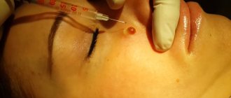
Vesicle on the skin (photo)
Additional varieties
There is also a type of vesicle called throat herpes. It is manifested by the formation of similar spherical neoplasms, which are localized in the throat area. Their appearance and content of cloudy hemorrhagic fluid are similar to the forms described above. Typically, throat herpes causes severe burning and itching of the throat mucosa, which significantly interferes with the patient’s normal life activities.
How to identify a symptom in yourself
To detect a disease such as vesicles, it is recommended that in case of any changes in the condition of the skin and mucous membranes, you immediately undergo a full examination of the body. This will make it possible to timely detect the initial stage of the pathological process, exclude the possibility of the acute form becoming chronic, and begin treatment.
To detect vesicles in yourself, it is enough to regularly examine your mucous membranes for the appearance of tiny bubbles filled with air or cloudy liquid contents. You should also examine your skin, especially in those areas that are more exposed to external environmental influences and often come into contact with clothing: it is in these places that the skin has increased sensitivity, gradually becomes thinner and minor damage can occur on it, becoming a “gate” for infection to enter.
Read below about what diseases can cause the appearance of vesicles in adults and newborns.
Diseases and disorders
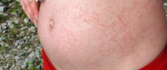
The appearance of vesicles on the surface of the skin or mucous membranes can be a manifestation of some internal lesions in the body. Most often, such synaptic vesicles, vesicles, are formed when immunity decreases, which indicates a general weakening of the body.
Also, vesicles on the skin and mucous membranes can appear due to the following reasons and their combinations:
Also, the appearance of blisters on the body can occur after an insect bite or the use of a cosmetic product that causes an allergy.
Read below about how to deal with ringed and other vesicles on the lips, face, mucous membranes, etc.
Vesicles in chickenpox
How to deal with this symptom
Since the therapeutic effect can bring maximum benefit only if it is correctly selected depending on the type of disease, first of all it is necessary to determine the cause that caused this external manifestation. Therefore, preliminary comprehensive diagnostics will eliminate the risk of unwanted side effects during treatment and achieve a speedy disappearance of the symptoms of the disease.
The most effective treatment measures for the occurrence of vesicles include the following:
- the use of products that soothe the skin and relieve such unpleasant manifestations of the disease as itching and burning;
- when blisters appear on the skin of the face, special drug treatment is usually not required, since they go away on their own after some time;
- the formation of vesicles on the mucous membranes already requires drug treatment. Typically, antibacterial drugs are used here to stop the development of infection inside the body;
- If a vesicle is detected in a child, you should immediately contact a medical institution for examination and prescribing individual systemic treatment.
A rise in body temperature with the formation of such bubbles on the body or mucous membrane also requires medical intervention, since it indicates the activation of a pathological process in the body. The most dangerous cases are considered to be cases of detection of this disease due to scratches from an animal, with shingles, dermatitis and scabies, since there is an increased proliferation of pathogenic microorganisms, which requires immediate medical treatment.
The video below will tell you about transport vesicles:
There are quite a lot of skin diseases, but all this diversity consists of different combinations of the same morphological elements of the rash. All of them are divided into two large groups:
- primary rashes - appear for the first time on clean, healthy skin;
- secondary rashes - appear on the surface of primary elements as a result of their evolution or appear at the site of a disappeared primary rash.
Why does a crust form on a wound and does it need to be removed?
A crust on a wound is a scab that consists of skin cells, dried blood or purulent fluid. Formed as a result of damage to the dermis due to burns, cuts, bites, abrasions, scratches and due to certain skin diseases (chickenpox). The main function of the crust is to protect the hole from the penetration of pathogenic microorganisms from the outside - bacteria, infections.
The crust is formed from tissue fluid that has a yellow tint; it has nothing in common with pus; it has a thicker and more viscous consistency. Doctors believe that the film covering the injured area is a natural defense against infection. The yellow crust helps the skin to heal.
In medicine, there is a classification of the color of formation on a wound to assess the condition. The shades indicate the stages of healing of the damaged area:
- An injured surface with a black scab means the presence of dry necrotic masses. Such damage requires moisturizing dead tissue and stimulating the natural cleaning process.
- In some cases, a yellow crust on the wound means a large amount of pus and indicates the presence of necrotic masses.
- Pink color indicates the epithelialization process, red indicates the granulation phase.
The formation of a film on the damaged surface begins immediately after the injury. To form a crust, you need an influx of fresh air, which dries out the lymphatic fluid. Growths form on superficial skin lesions, with 2nd degree burns, cuts, abrasions and scratches.
If the wound is noticed in time, cleaned of foreign bodies and treated with antiseptic ointment or gel, a scab will form in a short time. There are cases when the wound surface does not crust over for a long time due to the ingress of harmful bacteria that interfere with healing.
If pus has accumulated under the damaged surface and an inflammatory process has begun, you need to remove the scab from the wound. Scabs are removed in the following ways:
- Soak cotton wool or a cotton pad in hydrogen peroxide, apply to the crust, wait 20 minutes. You can use other drugs from the pharmacy. Furacilin in the form of an aqueous solution has antibacterial properties; it contains nitrofural, sodium chloride, and purified water.
- The soaked scab is examined and lifted starting from the edge of the wound with tweezers pre-treated with an antiseptic.
- The free edges of the crust are trimmed with disinfected manicure scissors.
If the central part of the scab does not move away from the surface, it cannot be torn off. Over time it will disappear on its own. After the procedure, the wound should begin to heal better; if this does not happen, you should go to the hospital. The doctor will pump out the purulent contents with a syringe or remove it with a scalpel. The operation is painful but effective.
You can recognize a wound that needs to remove the crust by the following signs:
- appearance of the injured area: the injury appears swollen and red;
- itching;
- painful to touch.
If the wound surface is not cleaned in time, it can lead to increased body temperature, decreased motor activity and general weakness.
You can soften the scab on the wound for subsequent removal in the following ways:
- If a crust has formed on your leg, you can steam it in water with the addition of soda and laundry soap. Then treat the damage with ointment.
- Softening crusts from wounds on the face after acne removal with Chlorohexidine. A sterile bandage is generously moistened in the solution and applied to soften the scab, then removed.
- You can soften the scab with healing ointments, the doctor will tell you the name, when using them you need to follow the instructions.
Wound surfaces take a long time to heal. Damage on the face takes three to five days, in the arms and chest lasts for a week, on the legs up to ten days.
The protective crust, which takes too long to fall off, prevents air from entering the injured area, the wound takes a long time to heal, and after healing, scars remain on the skin that have to be removed using cryodestruction, unless there is a contraindication.
You cannot remove scabs from injured surfaces; you must carefully remove them after softening. If the scab is pulled away from the wound, bleeding may begin again. The injured area will begin to heal more slowly and a scar will remain.
Those that do not heal should go to the hospital. The healing process, which occurs without pathologies, lasts no more than three weeks. Most often, the injured area does not crust over due to germs. Doctors believe that the following methods should be used:
- Treat the damaged area with Chlorohexidine.
- Remove dead cells and purulent contents from the wound. The procedures are carried out with cotton wool and hydrogen peroxide. The affected area is watered with a pharmaceutical solution, dried, rolled into a ball of cotton wool, penetrated deep into the injured surface, and the liquid is removed.
- Vishnevsky's liniment or hydrocortisone ointment is used if there is pus in the crusted wound.
- If the damaged surface is clean, use medicines with herbal ingredients.
- Infected skin is lubricated with preparations containing an antibiotic (Levomekol). To speed up the healing process of the crust, anti-inflammatory medications (Romazulon) are applied.
If the wound heals poorly and slowly, the doctor prescribes antibiotic medications and vitamin complexes.
If the patient wishes, the doctor may allow the use of folk recipes as remedies:
- Fresh cucumber juice. The product has an antimicrobial effect. It is applied to the wound crust in the form of compresses, which are left for a couple of hours.
- Celandine. The leaves of the plant are applied to the surface and secured with a bandage.
- A mixture of burdock and celandine. Add 30 g of crushed burdock roots and 20 g of celandine roots to 100 ml of vegetable oil. The mixture is boiled for 15 minutes, cooled, and filtered. The product is applied to the affected areas of the dermis two to three times a day for a week.
In order for the crust on the wound surface to cope with its protective function and prevent infections from entering the body, it must be properly cared for. The resulting damage is treated with an antiseptic drug. If the scab does not come off from the injured area, it means that the wound has not yet healed completely; the process of removing the scab provokes bleeding.
Papule, vesicle, pustule. What it is?
The primary morphological elements of the rash include:
- vesicle (bubble);
- papule (nodule);
- pustule (pustule);
- nodes;
- spots;
- tubercles;
- blisters.
The first three elements will be described in more detail below.
A vesicle is a spherical vesicle that rises slightly above the surface of the skin and ranges in size from a pinhead to a pea. Inside the bubble is a cloudy or clear liquid. This rash element is located in the superficial layers of the epidermis.
A papule is a small nodule that measures 1-20 millimeters in size. Papules often leave behind peeling and pigmentation.
Pustule is a later form of vesicle. It resembles a small ball in appearance and is yellowish in color. At the top of the ball there is a white spot, which over time bursts, forming a wet surface.
The skin around the pustule is swollen and hyperemic. After opening the abscess, its contents fall on the surface of the epidermis and form a crust. After the tissue under the lesion is restored, the crust disappears, leaving no noticeable marks.
Single pustules do not pose a particular danger to the patient’s health and usually disappear on their own after some time. The presence of a large number of pustules can be a symptom of a number of dangerous diseases (smallpox, syphilis, and so on), so in this case you need to make an appointment with a dermatologist to find out the causes of the rash.
How to remove a crust from a wound?
If the crust on the wound does not fall off, or has fallen off incompletely, and you decide to remove it yourself, do not peel it off dry. The crust should be soaked in an antiseptic solution. Hydrogen peroxide softens scabs best. Sterile gauze should be generously moistened with peroxide and applied to the wound for 15-20 minutes. After this, carefully separate the softened tissue. Peroxide can be replaced with chlorhexidine.
When suppuration occurs, the wound must be opened by a doctor. He will release the pus by softening the crust or by making a small incision with a scalpel or sterile scissors. This is a minimally painful procedure, after which the child’s condition improves immediately.
Vesicles. Photos, reasons for appearance
Vesicles appear on the surface of the skin as a result of pathological stratification of its upper layers.
The reasons for the appearance of vesicles are as follows:
- allergic dermatitis;
- inflammatory processes in the epidermis;
- eczema;
- a bite of an insect;
- some autoimmune diseases;
- penetration of pathogenic microorganisms under the skin;
- decreased immune defense;
- allergies to certain medications;
- hypothermia;
- increased sweating;
- exposure to the epidermis of metal salts, alkalis or acids, which the patient may deal with due to his profession.
Vesicles in the throat
A vesicle in the mouth, the photo of which is posted below, is a small (up to 1 centimeter) round bubble on the mucous membrane.
The vesicle usually contains serous fluid or lymph, but in some cases the vesicle contains pathogenic microorganisms and blood. The surface of the bubbles is thin, so they usually burst quickly, forming erosions and crusts.
Vesicles in the throat are often formed during viral infections, stomatitis, herpes, and chickenpox. With these diseases, the vesicles may contain a large number of viruses, which, after opening the vesicles, can become a source of infection for surrounding people.
Pathological causes of deviation
The causes of ulcers on the hands or other parts of the body often indicate the presence of a pathological process in the body. And the most dangerous thing in this situation is that the disease can be latent, that is, it can not reveal itself in any way for a long period of time.
A brief overview of the pathological causes of ulcers on the human body is given below.
Diabetes
A non-healing skin sore often occurs in patients with diabetes. In this case, the type of disease does not play a role. The wound surface is often wet and very deep. Ulcers in diabetes can be either single or multiple, and have different sizes.
Allergy
An ulcer on the hand also appears when the epidermis comes into direct contact with powerful allergens. But the possibility of such skin defects due to food allergies cannot be ruled out either. The main thing is to respond to such a deviation in a timely manner, otherwise, when an infection enters the wound, it will become much more difficult to fight the disease.
Infectious skin diseases
Sores on the hands that itch can be a harbinger of herpes. The disease develops in stages. The first phase is characterized by the appearance of redness and swelling of the skin at the site of the lesion, itching and burning. It is this kind of sore that you need to pay attention to, since it is followed by the second, vesicular, stage of herpetic pathology, which is more difficult to treat. After opening the vesicles, new sores form on the human skin, which become covered with a scab and gradually heal.
Streptoderma can also cause skin defects. This is an infectious skin pathology caused by streptococcal infection. A person notices redness and swelling, as well as the presence of itchy scabs on the body. This disease can only be treated with antibiotics. During therapy, the patient must be isolated from others, since streptoderma is very contagious.
Another dermatological disease that can lead to the appearance of sores throughout the body is pyoderma. This is a pathology in which ulcers form on the surface of the epidermis. Its danger lies in the fact that the exudate can affect the underlying layers of the dermis, and if left untreated, lead to blood poisoning. When the abscess opens and its contents come out, ulcers form, which gradually scar.
Diseases of the hematopoietic system
The causes of the formation of sores on the face and body associated with pathologies of the hematopoietic system are very dangerous. First of all, we are talking about:
- Blood poisoning. It can occur due to infectious, in particular bacterial, lesions of the skin or internal organs. With sepsis, the outflow of lymph is disrupted, and all toxic substances that penetrate the systemic bloodstream are carried throughout the body, affecting healthy tissues. As a result of such a severe disorder, sores may appear on the leg, arm, face and body, which do not heal and itch.
- Anemia. Iron deficiency or another type of anemia leads to a deterioration in the oxygen supply and nutrition of cells and tissues of the body. Therefore, a person whose leg sore does not heal and itches, and at the same time has symptoms in the form of weakness, dizziness and general malaise, must, first of all, consult a doctor and donate blood for hemoglobin and iron levels in the body.
- Blood cancer. Non-healing ulcers on the skin can form with severe blood cancer. With this disease, all metabolic processes in the body are disrupted, and not only the skin, but also many internal organs suffer.
Important! Blood pathologies should be treated only under the supervision of a hematologist, therapist or oncologist. Self-medication in this case is unacceptable!
Avitaminosis
One of the most common pathological reasons for the appearance of ulcers on the hands is hypo- or vitamin deficiency. With this deviation, a violation of the vitamin balance occurs, and the indicators of both a single substance and several at once can decrease. This condition is corrected with the help of multivitamin complexes.
On a note. Stress, lack of sleep, nervous tension - all these factors lead to severe itching throughout the body. As a result of scratching, ulcers and wounds can form, into which pathogens can easily penetrate. There are many methods for treating such skin damage, but a specific method of dealing with them is selected only after the exact cause of the ailment is determined.
Vesicles in newborns
Within a few hours after birth, the baby may develop rashes on the skin. This is vesiculopustulosis - an infectious disease that affects the sweat glands of a newborn.
This is why rashes appear only in places where there are a lot of sweat glands (scalp, torso, armpits, inguinal folds). The bubbles are no larger than the head of a pin. Inside them there is a clear liquid, which soon becomes cloudy.
Within a few days, the vesicles turn into pustules, which open and dry out, forming a crust that falls off after a while.
The child’s skin in the areas of the rash is swollen and hyperemic, his general condition worsens and his appetite disappears. In severe cases, with multiple rashes, the temperature rises to 38-39 degrees. The child is nervous, anxious and often cries.
Vesicle is a bubble on the skin up to 5 mm in size containing serous or serous-bloody fluid. Occurs due to delamination of the upper layer of the epidermis.
Is it necessary to remove the scab from a wound?
In case of minor damage, the crust should not be removed. With large wounds, the issue remains not fully studied: a number of surgeons claim that healing proceeds faster under the crust, while others believe that the scab in most cases prevents the wound from quickly epithelializing. Today, most doctors share the second opinion.
You should definitely remove the crust from the wound if there is suppuration under the scab. It is not difficult to recognize suppuration: the skin around it becomes red, hot to the touch, painful, and swollen. Sometimes yellowish pus appears under the crust. In severe cases, the child develops a fever. The wound will not heal until the pus is removed!
Causes
The most common causes of vesicle formation on the skin:
- Acute and chronic dermatitis of any origin.
- Insect bites and contact with poisonous plants.
- Viral or bacterial infection.
- Allergic reactions.
- Decreased immunity.
- Autoimmune diseases.
- Mechanical, physical or chemical effects on the skin.
- Vesicles in children often occur as a result of improper care - poor washing of skin folds and insufficient exposure of the child to fresh air.
Scabies
Scabies is one of the common skin diseases. It is observed among all groups of the population, but more often among antisocial, unscrupulous individuals.
The causative agent of the disease is the itch mite. Scabies is very contagious, infection occurs through contact with a patient, through household items. People often become infected in transport and public places. A characteristic symptom is painful itching of the skin.
The rash is visible all over the body, including the head and genitals. Due to itching, patients scratch the skin, damaging and infecting it. This leads to the formation of yellow scabs - scabs. Crusts are usually localized in places of greatest itching - on the stomach, sacrum, between the fingers.
Scabies is treated with a 60% sodium thiosulfate solution or special preparations (Spregal, Invermectin). Scabs can peel off on their own; in advanced cases, treatment of skin lesions with antibacterial ointments is required.
Symptoms
The location and symptoms of vesicles greatly depend on the manifestation of the disease they are. Often, the pathological process begins with the appearance of red itchy spots, which quickly turn into papules, and subsequently into blisters. Once they are opened, crusts form on the skin.
Depending on the structure, there are several forms of vesicles:
- Intraepidermal - the wall is thin and flaccid, easily opened. Such vesicles often contain clear liquid.
- Subepidermal - the wall is thick and strong, the vesicles are tense. They are difficult to open; there is a red liquid inside.
- Smallpox are the size of a pea, have a characteristic umbilical depression in the center, and are found in viral infections.
- Subthreshold - located under the stratum corneum of the skin.
The main causes of skin peeling and crust formation, what to do, which doctor to see
Psoriasis
Peeling in psoriasis manifests itself as pink spots slightly raised above the skin level with a diameter of 5 mm to 5 cm. When scratched, they become covered with silvery scales.
Contact a dermatologist immediately. There is no effective treatment at home.
Atopic and contact dermatitis
Peeling in atopic and contact dermatitis is manifested by the following symptoms. The skin becomes inflamed, itchy, flaky, and sometimes weeping occurs. Atopic dermatitis often affects the elbows and popliteal folds, and contact dermatitis often affects the hands.
For treatment, use hydrocortisone ointment. If there is no improvement after a week, consult a dermatologist. Antibiotic ointment may be needed.
Dermatomycoses
Dermatomycosis - red spots with peeling in the center appear on the skin, which slowly increase in diameter. With inguinal ringworm, scaly pink (sometimes weeping) spots appear in the groin folds and on the inner thigh.
Use antifungal ointment. If there is no improvement, consult a dermatologist.
Seborrheic eczema
Seborrheic eczema is scaly crusts on the scalp, often with an irritating discharge. Peeling may also appear on other areas of the skin.
Wash your hair with an antifungal shampoo containing selenium.
Pityriasis rosea (pityriasis rosea)
Pityriasis rosea are oval red spots 5-20 mm in size raised above the skin, arranged in a herringbone pattern. At first, one large spot usually appears. The spots disappear after a month and a half. Possible cause is viral infection.
There is no specific treatment. Regresses spontaneously.
Treatment
Treatment for vesicles depends on the cause. After eliminating it and isolating the patient from the provoking factor, symptomatic therapy is prescribed:
- Antihistamines.
- Ointments and creams with corticosteroids.
- Antibacterial drugs.
- Sedatives.
- Antiviral drugs.
- When the process is generalized, systemic steroids are prescribed.
- In some cases, the use of physical therapy can help speed up recovery.
Causes
The causes of this insidious disease are striking in their diversity. Most often, eczema in young children occurs due to a genetic predisposition. If someone in your family has had this disease, the likelihood of it occurring in a child increases rapidly. A number of situations can serve as a trigger:
- Allergic reactions to food products, household chemicals, hygiene products.
- Stressful situations that lead to scratching of the skin on the hands.
- Diseases of internal organs (liver, kidneys, stomach).
- Impaired metabolism.
- Creams and soaps whose composition irritates the delicate skin of a child’s hands.
- Weakened immunity.
It can also appear in adolescence due to hormonal changes in the body.