Melanoma is a malignant neoplasmfrom pigment cells, which are designed to protect the body from ultraviolet rays. At some point, degeneration of pigment cells occurs, and they turn from defenders into aggressors. The exact cause of malignant degeneration of melanocytes is unknown.
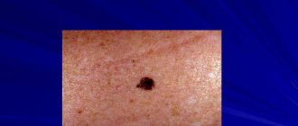
Photo 1. Melanoma is the fastest growing type of cancer. Under no circumstances should you postpone a visit to the doctor. Source: Flickr (wildmeadow)
Indications for melanoma removal
If you suspect a malignant tumor, you should consult a doctor. After a complete examination, the doctor will be able to prescribe the correct tests and treatment. Removal of melanoma is carried out at an early stage of development. This will prevent it from further increasing.
The method for removing melanoma depends on a number of factors. Size, degree of germination, individual parameters of the body - all this influences the choice of treatment. The matter will not be limited to one operation. Later, you will need to consolidate the result with the help of postoperative treatment - you will have to take medications and undergo chemotherapy courses.
In advanced stages of tumor development, cancer cells actively multiply and penetrate into tissues. During this time, treatment is carried out using intensive immunotherapy. To avoid such situations, familiarize yourself with the first symptoms of the disease:
- The mole changes shape, and its boundaries become fuzzy and blurred.
- The size of the tumors increases.
- The pigment changes color - it becomes bright, red dots appear.
- These areas are itchy and itchy.
- Places with moles bleed.
If you notice a similar symptom, consult a doctor as soon as possible.
Melanoma symptoms
Symptoms of melanoma include new-onset skin lesions or those that arise from a pre-existing pigmented nevus (mole):
- It is often observed that in the immediate vicinity of these lesions thickenings form, changes in the existing surface, color and changes at the edges of the mole.
- You should also be concerned about sudden itching or the appearance of bleeding or bloody discharge.
- An inflammatory reaction associated with a mole is also a warning sign of melanoma.
- One symptom to look out for is blue and black moles. The occurrence of this type of pigmentation should be resolved as soon as possible by consulting a doctor.
For early diagnosis of melanoma, monitoring changes in moles, carried out by the patient individually, is of great importance.
If there have always been pigment changes on the skin, the following should alert you:
- sudden itching;
- bleeding;
- increase in lesion;
- changes in the structure of the mole.
Particularly noteworthy is the fact of uneven pigmentation, which contains various shades of brown and pink. It is also worth paying attention to the new changes.
Removal methods
There are two ways to eliminate a malignant tumor: using a laser or a scalpel. Removing melanoma with a laser is painless and is performed without anesthesia. The procedure does not leave large scars or complications as a result. This method only affects malignant epidermis. Healthy areas are not affected. The laser is prescribed exclusively for small tumor sizes - 5-8 mm in diameter. And the device does not have the ability to reach the deep layers of the epidermis.
And using a scalpel allows you to do this. This is the only way to penetrate deep into the skin. Although the scalpel is effective, it is not without its drawbacks. The instrument leaves a large scar. This occurs as a result of the process creating a large, slow-healing wound. It will need to be processed periodically.
There is a risk of bleeding during surgery. Since this is a serious intervention in the body, infection can be introduced through the wound. Special care is prescribed. Plus, incomplete removal of melanoma does not guarantee recovery - the risk of recurrence of the formation remains high.
The second option is popular. Doctors prefer to work with a scalpel. In their opinion, this is an extremely effective way to eliminate melanoma.
Contraindications to laser use
Both methods are used in medical practice. But laser melanoma removal is not for everyone. This is the penetration and influence of rays. You should be careful. The use of laser is prohibited:
- During pregnancy and during breastfeeding.
- The presence of chronic heart and vascular diseases, pre-stroke and pre-infarction conditions.
- Very sensitive skin.
- New occurrence of melanoma after elimination.
Although the procedure is painless, side effects are possible. Doctors do not prescribe it to all patients.
Specifics and methods of surgical intervention
The essence of surgical removal of melanoma is that the cancer cell is removed along with surrounding healthy cells. This prevents the spread of malignant cells to nearby areas of the skin. The depth of the cut depends on the size of the tumor. In medicine, 4 methods of eliminating tumors have been created:
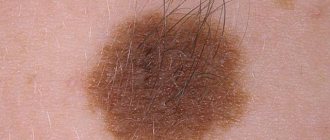
Melanoma on the hand
- Easy removal. Painkillers are injected into the area around the tumor, then the melanoma is cut out with a scalpel. And the wound is sewn up with threads and then removed. A scar remains on the area of skin.
- Wide excision. The method is carried out in advanced stages of melanoma. A marker is used to outline the area around the tumor with a margin of 2 cm in each direction. This helps reduce the possibility of a new formation appearing. The resulting wound is sutured.
- Amputation. This method is used in cases where melanoma appears on the extremities. At an early stage, parts of the fingers or toes are removed. If the tumor form is neglected, you can lose a limb.
- Removal of lymph nodes. Due to melanoma, metastases quickly appear in the lymph nodes nearby. Getting rid of them involves removing the lymph nodes. This reduces the risk of their distribution throughout the lymphatic structure.
For a routine operation, anesthesia is used. Sometimes the procedure is performed under local anesthesia. Amputation of limbs and lymph nodes is performed under general anesthesia. This will help to overcome the painful shock.
Dermatoscopy
Dermatoscopy is an examination in which a dermatologist uses a dermatoscope. This is an instrument that combines a magnifying glass 10 or 20 times, magnifying the fragment under study, equipped with a standardized light source. In such conditions, the doctor may initially assess skin changes.
Dermatoscopy
Videodermatoscopy is a study that, in addition to the techniques used in dermatoscopy, uses visualization on a computer monitor. This allows the clinician to store the displayed image in the device's memory, which can be useful for comparative purposes at a later stage in treatment. In practice, several dermoscopic methods are used to diagnose melanoma.
The doctor, based on the appearance of the lesion, will evaluate whether there is a reason to remove it along with a margin of healthy skin, and send the material after the procedure for histopathological examination.
The specialist studies the characteristic features of the mole, in particular:
- Color – uneven color, darker and lighter areas or pink and red areas are abnormal;
- Torn edges – moles with smooth edges are less dangerous to health;
- Asymmetrical shape;
- Size greater than 5 mm - smaller lesions are usually not a cause for concern, but if they are dynamically growing or show other abnormalities, they need to be removed;
- Bleeding, ulceration, presence of discharge.
Diagnosis that confirms or excludes a skin neoplasm, which is melanoma, is possible on the basis of a histopathological examination, which is carried out after removal of the entire lesion.
Forecast and consequences
Major surgery carries a high risk of complications. If the form of the tumor is initial, then the consequence will be only scars or cicatrices. But you have to be careful. If an infection gets into the wound, a purulent complication may occur. This rarely happens in medical practice.
The prognosis of melanoma development plays an important role in successful treatment. It is worth calculating all the risks and possible consequences. Based on the information, the doctor prescribes treatment that is effective in a particular case. Talk to the doctor.
Forecast
It is good if the tumor can be found in the initial stage. The likelihood of a favorable treatment outcome is high. The main threat of melanoma is that during the first year of therapy there remains a high risk of relapse. Comprehensive treatment will eliminate melanocytes and reduce the likelihood of its development in the body.
For advanced stages of cancer, palliative therapy is carried out. This helps maintain the vitality of the body. The rate of spread of malignant cells directly affects life expectancy. The smaller the metastasis, the better.
The age of the patient is also important. A young body has more strength and a chance to recover quickly. After tumor removal, life expectancy varies among people. A person is capable of living to a ripe old age. You must follow your doctor's recommendations and follow his instructions.
Removal of lymph nodes contributes to the occurrence of lymphostasis. The movement of lymph will be disrupted. The result is swelling of the affected limb, which leads to an increase in its volume. There is also a high risk of developing seroma. This is a situation when lymph begins to accumulate in the area of the removed nodes. It is dangerous because it later begins to rot. And this excess liquid must be removed.
Possible complications
Any method, both surgical and laser, carries a risk of complications. If you have any concerns about your condition getting worse, you should tell your doctor.
Laser removal of melanoma
This situation needs to be discussed and a further solution must be reached. There are 4 groups of complications:
- Getting infected. This situation occurs when treatment is carried out under unsterile conditions or wounds are poorly treated. As a result, the patient experiences burning and itching around the wound.
- The appearance of a new tumor. It may occur near the site of a previous excision. This is the result of an incorrect procedure to eliminate melanoma. As a result, the neoplasm is also removed along with healthy epidermal cells.
- The appearance of scars. They appear when large tumors are eliminated. The areas need to be sutured, which is why visible scars remain.
- Long wound healing and bleeding. It depends on the individual abilities of the body. Or it is a sign of dysfunction of the lymphatic system.
Melanoma can only be removed in a medical facility. Other methods pose a danger to the health and life of the patient. The tumor is eliminated by removing the root; special equipment is required. An incorrect operation will cause the cancer cell to grow at a faster rate.
How to recognize melanoma?
To diagnose melanoma, it is necessary to carry out very extensive diagnostics. Of course, the basis is the medical interview conducted during the first visit. The information obtained from the patient concerns mainly information about the condition of the skin, with particular emphasis on changes in moles on the skin or the appearance of new ones.
Information regarding factors that have been identified as increasing the risk of melanoma is also very important to the dermatologist. This group usually includes sunburn or the use of tanning beds. It is also very important for the doctor whether there have been cases of skin melanoma in the patient’s family.
A detailed interview is the basis for further action. The most important element of diagnosis is the skin examination, which the dermatologist performs during the visit.
Postoperative treatment
Rapid removal of melanoma increases the likelihood of a long life expectancy to 85-90%. It all depends on the area where cancer cells spread. After simple tumor removal, it is necessary to undergo annual examination by a dermato-oncologist for the first 5 years. The doctor examines the body completely.
If you undergo a procedure to remove lymph nodes, you need to be checked once every 3 months for the first 2 years. The examination process includes an ultrasound of the abdominal cavity, a CT scan of the chest and an MRI of the brain. Later, the frequency of visits to the medical facility will decrease. Over the next 3 years, diagnostics are carried out every six months, and then annually.
Unfortunately, the statistics are not encouraging. If you start treatment on time, follow the instructions and follow the doctor’s instructions, the chance of getting rid of melanoma is high. Postoperative therapy is prescribed depending on the stage of tumor development. Treatment is divided into 3 groups:
- Radiation therapy helps reduce the likelihood of metastases after surgery.
- Immunomodulatory therapy – designed to fight cancer cells.
- Chemotherapy helps destroy cancer cells.
After the procedures, patients are advised to avoid direct sunlight. Wear long, full-body clothing and wide-brimmed hats. And don't forget to use sunscreen.
Early detection of melanoma will increase the chances of a favorable treatment outcome. To avoid severe forms of the tumor, regularly examine your moles. If you have the slightest suspicion, consult a doctor. It’s better to be safe than to suddenly discover that you have a dangerous disease.
Types of melanoma
Skin lesions diagnosed as melanoma do not always look the same. Thus, there are several types of melanomas, which define different clinical subtypes of the disease.
- Superficial melanoma
. It is indicated that the most common type of melanoma, which accounts for up to 60-70% of all cases of the disease, is superficial melanoma. This group of melanomas is most often based on pigmented lesions of a dysplastic nature. - Nodular melanoma
. Indicated as the most severe form of the disease, affecting 10-30% of patients. This group, in turn, includes two clinical subtypes. The first is lentiginous melanoma, which is a malignant form. Occurs in 5-15% of cases. It most often affects older people, especially in the neck or face. Lentiginous melanoma is caused by prolonged and prolonged exposure of the skin to sunlight. The second subtype is acral melanoma, which is observed on the inner surface of the skin of the arms and legs. - Colorless melanoma
. The disease also includes pigmented melanoma. Only 2% of patients suffer from it. This is a characteristic type of skin lesion in which pigment is absent. In this case, the symptoms are also quite specific, and the not very characteristic but enlarging pink-red papule should be of concern.
Superficial melanoma
Nodular melanoma
You should also pay attention to the so-called Spitz moles, which are a juvenile form of melanoma. Most often, scientists indicate that this is an active nevus, usually single, and occurs in young people.
The surface of a Spitz mole is very well demarcated from the surrounding area and has a smooth, red or bluish color. Most often it occurs on the face. When observing the lesion, there is no inflammatory reaction. Spitz nevi rarely become cancerous, although it is recommended to remove them before puberty.
Why chemotherapy doesn't help with melanoma
Melanoma is not sensitive to either chemotherapy or radiation therapy. The treatment of melanoma does not have an algorithm; it simply does not exist. For example, for lung cancer there are algorithms, there is an understanding of what drug regimens (protocols) to prescribe. In melanoma, the tumor can change the configuration of its proteins in a short time.
The proteins of melanoma cells are protected from the penetration of drugs, so the use of chemotherapy and interferons brings results in no more than 11% of cases and only for a short time - up to 6 months. Further, melanoma progresses significantly when immune function decreases. Treatment of melanoma with chemotherapy suppresses the immune system, stopping the synthesis of antibodies, monoclonal bodies, and lymphocytes. Lymphocytes' main function is to protect the body from microbes and foreign tumor cells. But if the patient suppresses the activity of lymphocytes using chemicals and prevents them from multiplying, then there is no one to protect the body.
When is melanoma dangerous?
Metastasis always occurs when the cancer process is far advanced and cancer cells damage subsequent internal organs of a person. First, stage III is indicated when regional lymph node metastases are diagnosed. To treat melanoma, it is important how many lymph nodes are affected by cancer cells.
It is indicated that a much more serious disease is the occurrence of macrometastases, that is, changes that indicate tumor foci in a significantly enlarged, clinically palpable lymph node. Particular danger and difficulties in treatment are indicated for those patients who are diagnosed with infiltrates outside the lymph node capsule.
Melanoma, as a malignant tumor, can also metastasize to distant organs. Then the progress of the disease and the effectiveness of treatment depend on the organs in which the metastases are located and the general condition of the patient.
Stage 0
At stage zero, melanoma has not yet spread beyond the epidermis - the uppermost part of the skin.
During diagnosis, the doctor cannot know for sure whether it is melanoma or another skin disease. There are methods that make it possible to determine with a high degree of probability that it is a malignant rather than a benign tumor. But you can still be 100% sure of this only by receiving material for histological examination.
A biopsy of melanoma is usually not performed before it is removed. The formation is excised and only then sent for histology. At the zero stage, the operation is usually carried out to a minimum extent.
The doctor removes the skin lesion. But after it is examined and the doctor receives a histological conclusion that it is melanoma, the scope of the operation will be expanded. The doctor will perform a second surgery. He will remove pathologically changed tissue, increasing the distance from the edge of the tumor.
Melanoma is a malignant skin tumor
Melanoma is one of the types of malignant skin tumors. The test that confirms or excludes the diagnosis is a biopsy, which confirms the diagnosis based on microscopic examination. The results of the performed biopsy provide information about factors that have a significant impact on the prognosis of the patient's treatment. This information allows you to determine the further course of the treatment process.
Melanoma biopsy
Examination of removed tumors for malignancy is carried out on the basis of macro- and microscopic examination. Among the factors that play a significant role in assessing the patient's health prognosis, primary outbreaks are of great importance. If there are no metastases in the progression of the disease, the change in thickness (Breslow scale) and the presence of ulcers observed in the primary lesion are assessed.
Due to the fact that melanoma is classified as a malignant neoplasm, some patients experience metastases to other organs. Therefore, during diagnosis and treatment, the surrounding lymph nodes are also checked.
What to do after tumor removal
When skin tumors are removed, it is important to properly care for the wound. The doctor explains all the details and invites you for a routine examination and dressing. The stitches are removed after one and a half to two weeks.
After laser removal, a crust remains at the removal site, which should fall off on its own within one to two weeks. Until complete healing, it is advisable to carry out all hygiene procedures with extreme caution so as not to damage the wound with a washcloth or towel.
If the disease recurs after the operation, this indicates the spread of metastases to other organs and systems. In this case, immunotherapy, radiation therapy and local or general chemotherapy are prescribed simultaneously. And although such treatment has significant negative consequences, they can be considered a lesser evil compared to a relapse of the disease.