Author: Dmitry Beinusov.
Source: https://beinusov.ru/info/kak-chasto-byvaet-rak-posle-udaleniya-rodinki/.
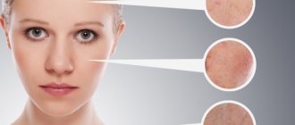
When communicating with loved ones or patients, this topic is raised, you can often hear the following statements: “cancer developed after removing a mole,” “he removed a mole and died,” and so on. Every time these relationships are presented to me, my first question is: “Are there documents?”
Histological examination? Extracts from hospitals, CT, MRI, PET? But there are no documents. But for some reason we believe the storyteller, although 99% of such stories always reach us in the form of rumors, retellings, through third parties and are not at all supported by facts.
In this article, we'll take a closer look at where these stories come from, whether they have any basis in reality, and what the remaining 1% consists of.
Here are a few scenarios that may suggest that there is a connection between mole removal and death from cancer.
Causes of mole recurrence
Indications for removal are pain when touched, changes in size, color, and shape. If the growth often catches or is injured, the growth should be removed to avoid infection. Moles are removed from the body using the following methods:
- Surgical - allows you to get rid of moles along with the root. If, after excision, a tumor grows again in the area, the root has been partially removed.
- Cryotherapy involves freezing the mole with liquid nitrogen. A painless method, the risk of a new mole remaining remains.
- Electrocoagulation - consists of the use of high-frequency current that burns out the nevus. After the procedure, a slight scar is left that disappears over time.
- Laser is effective and leaves no traces after the procedure. The action is aimed directly at education and does not affect healthy skin.
- The radio wave method helps remove moles without leaving any traces, without leaving minor scars after the procedure. Healthy skin is not exposed to radiation and does not become infected.
After eliminating the problematic mole, you may notice that it has grown again. Relapse after mole removal is quite real and occurs for the following reasons:
- During surgery, errors were made in determining the depth of pigment spots, and the excision was partial.
- A dangerous method of eliminating growths has been chosen. Freezing with liquid nitrogen brings a lot of trouble. The procedure results in a burn that does not heal well and becomes infected.
- Improper care of the wound after surgery or failure to comply with a doctor’s prescription leads to infection.
- During the process of removal, the mole degenerates into melanoma.
- Removing a nevus at home without observing all sterility conditions is fraught with the penetration of pathogenic bacteria. Partial removal is a potential threat of recurrence of the nevus.
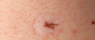
The red spot does not go away
Sometimes, even after successful removal of a mole by laser or other methods, a red or pinkish spot remains on the skin, which is not a recurrence of the nevus. This mark may appear:
- If the skin healing process is still ongoing. It may take about six months or even more until the color of the epidermis is completely evened out.
- Soon after the crust falls off. In this case, a pinkish spot is observed on the skin - an area of young, delicate epidermis.
- If you tear off the crust ahead of time (accidentally or intentionally). Such a dark spot at the site of a removed mole can remain for life; to get rid of it, you must definitely use means to activate regenerative processes (silicone ointments and patches, Contractubex, Solcoseryl, etc.).
- With improper skin care. In particular, reddish spots may appear when the young epidermis is exposed to sunlight. In this case, they indicate a skin burn and can subsequently lead to pigmentation of such an area.
- Due to the penetration of infectious agents. In this case, the problem area may swell, itch, and even be noticeably painful. The formation of an abscess is also possible.
If strange spots appear on the skin after removing a mole, it is better to play it safe and consult a specialist. This will help reduce the risk of developing melanoma, a skin cancer.
What to do if a mole grows back after removal
After removing the growth, a lump or dark dot appears; do not do anything on your own. You should examine the area on your skin daily. If a scar or scar appears, visit a specialist. He will examine and prescribe medications that accelerate the healing process and resorption of the skin defect.
Before the procedure, think carefully and weigh all the pros and cons. Improper removal or introduction of infection will lead to serious consequences and the development of cancer. Consider contraindications: the presence of chronic diseases, cardiovascular disorders and problem skin.
Contraindications for specific methods:
- Surgical is contraindicated in case of exacerbation of herpes, inflammatory or infectious diseases.
- Cryotherapy is not recommended for malignant tumors.
- Laser removal cannot be carried out:
- if you are allergic to the sun;
- if the tumor is malignant;
- during exacerbation of herpes;
- during pregnancy;
- during menstruation in women;
- increased body temperature;
- for inflammatory diseases.
When exposed to radio waves, there are the following contraindications:
- during the degeneration of education;
- during pregnancy or while breastfeeding a baby;
- diabetes;
- presence of a pacemaker.
What to do if a new spot appears at the site of a removed mole?
If a brown spot or bump appears again at the site where the mole was removed, nothing needs to be done. The main thing is to examine the problem area on the skin every day. If a scar or scar forms at the site where the nevus was removed, you can consult a doctor who will prescribe medications that promote rapid healing of the wound and resorption of the skin defect.
In the event that a recurrent formation causes concern and manifests itself with unpleasant signs, you should immediately consult a doctor. When a malignant nevus recurs, the following signs appear:
- if a mole appears and hurts when touched;
- severe itching of the skin appears;
- the surface peels off and becomes covered with small cracks;
- blood oozes from under the growth;
- the mole grows in size;
- the growth changes color and structure.
Recurrence of a nevus at the site of a previously removed one requires repeated removal.
If a person has all of the above signs, this can lead to unpleasant consequences. The doctor will conduct the necessary studies to determine the nature of the malignancy. When cancer is detected at an early stage, it is important to start therapy on time. The operation carried out at the initial stage will be successful. The surgical treatment method involves excision of melanoma and nearby tissues. In the case when the metastases have managed to spread throughout the human body and infected vital organs, alas, the treatment will be ineffective.
Dear Lyudmila!
Of course, pain at the site of nevus removal cannot persist for 3 months, and also normally cannot recur several months after the intervention and certainly should not be of an increasing nature. Most likely, we are talking about complications after the operation, the occurrence of which depends on the professionalism of the doctor who performed the removal, on the chosen method, on the type and size of the mole, and, of course, on how well you followed the recommendations for caring for the wound.
Nerve damage
One of the most serious consequences of mole removal is nerve damage, although we must admit that this is a fairly rare occurrence. Most often this happens if the surgeon made an incision too deep with a scalpel or did not direct the laser correctly. The damaged nerve begins to die, and this is accompanied by pain of various types. Moreover, this process is quite lengthy - from several weeks to several months. The death of nerves occurs especially painfully on the face and in the joint area.
Infection
Infectious contamination of the wound at the site of mole removal is observed quite often if sterility rules were violated at the medical center where the operation took place, or recommendations for wound care were poorly followed. To avoid infection, experts advise treating the removal site twice a day with antiseptic solutions - brilliant green or potassium permanganate; in some cases, antibiotic-based ointment may be needed. Normally, painful sensations persist for up to 48 hours, during which time the wound becomes covered with a crust. As a rule, a feeling of discomfort, itching and slight occasional pain may be present during the first two weeks, but then the scab disappears safely, which means that healing has occurred successfully.
An infection in the wound is indicated by redness of the skin, swelling, painful twitching or stabbing sensations, discharge of pus, and an increase in temperature. If you experience any of the symptoms, you should urgently seek medical help, as a serious bacterial infection can lead to sepsis.
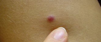
Relapse
A fairly common complication after mole removal is its recurrence. A tubercle appears at the site of the operation or in close proximity to it, gradually increasing in size. This formation may cause itching or discomfort. A mole that appears in the same place can grow to the same size or even larger. Relapses are dangerous because there is a risk of malignancy of the formation, i.e. degeneration of healthy tissues into malignant ones. Gently feel the mole removal site and the tissue around it. If you find any bumps or bumps, contact your doctor immediately to have a biopsy of the new growth and determine a course of action.
Scar formation
If the mole was removed surgically (the most traumatic way), there is a risk of disruption of the process of fusion of connective tissue fibers if the incision was made incorrectly. Scarring of the tissue may be accompanied by minor pain or itching. However, within a few months the discomfort should have disappeared and the scar should have fully formed. If it continues to hurt, then it means that nerve endings have gotten into the scar tissue and repeated surgery may be necessary.
Best regards, Ksenia.
Relapse Prevention
After diagnosis and confirmation of the benign quality of the formation, you should consult a doctor with experience in this field. The country's leading dermatologist-oncologist Igor Evgenievich Sinelnikov faces similar problems every day. He offers various ways to remove the growth without complications. To avoid consequences and re-growth, you must follow these rules:
- In the first week, do not allow water to enter the area after removing the growth.
- Read the instructions for proper wound treatment. Wash your hands thoroughly before handling and monitor the area for two weeks to avoid infection. Will help avoid inflammatory processes and relapse.
- After the procedure, a crust will appear on the wound; it is not recommended to remove it yourself. Over time, it will disappear, leaving healthy pink skin.
- It is not recommended to sunbathe in the sun or in a solarium. Ultraviolet radiation negatively affects the skin, stimulates the growth of old and new formations, and the appearance of birthmarks. If you cannot hide from the sun, cover the area with an adhesive plaster or cover it with clothing.
Simply following the rules and carefully caring for the skin area is the key to a quick recovery. preventing relapse. The use of medicinal ointments and creams will hide cosmetic defects.
What is surgical excision of a nevus? Is it worth removing this formation? It is best to discuss such questions with your doctor, since the answers to them depend on the characteristics of the formation itself, which only a specialist can study.
What does it show?
Carrying out histological analysis provides information not only about the presence of cancer cells, but also about the features of the detected neoplasm:
- Allows you to determine the type of cancer.
- Detects the thickness of the tumor.
- Allows you to determine the presence of ulceration.
- Provides information about infiltration (growth into surrounding tissues).
- Allows you to determine the presence of vascular invasion (penetration of the tumor into the vessels) and its degree.
- Provides information about the exact size of the tumor (its edges).
- Determines the mitotic rate (the growth rate of abnormal cells) or Clark levels (how deep the tumor has grown into the skin).
Even in the laboratory, they can make assumptions about possible damage to the lymph nodes, the presence of necrotic changes, cell regression and the level of neurotropism (features of the growth of the formation relative to the nerves).
Performing an in-depth histological analysis to identify all of the above points is possible only when examining a removed nevus. Studying a biopsy is not so informative.
Ways to eliminate moles
A nevus is a growth on human skin. Its other name is mole. Nevi can be either congenital or acquired.
As a rule, doctors do not recommend removing moles. An exception can only be made in certain cases:
- if excision of the mole is recommended by a medical report;
- if the nevus causes physical discomfort to a person and there is a threat of damage.
Today, the most effective method of removing a nevus is with a laser, but such an operation is performed only if there is indications from a dermatologist.
Getting rid of nevi on your own at home can result in cancer. If the formation brings discomfort, as, for example, if it is located in the lip area, then you need to find a specialist who will conduct an examination and prescribe removal of the nevus.
Mole removal
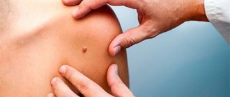
There are moles on every person's body, some have more, some have less. They do not always decorate the body with their presence. Their ability to degenerate into malignant tumors leads to severe cancer. Therefore, when they discover new birthmarks or convex growths, people run to the doctor, demanding that the suspicious mark be removed.
Removing moles is a serious procedure that requires a high level of professionalism from the doctor. A mole needs to be removed if it bothers a person, for example: it has increased in size, hurts when touched, changes color and structure. If the tumor is located in a place that is often subject to friction or clinging, then it is better to remove it too, so as not to damage it or cause infection.
Removal of nevus by surgical and radio wave methods
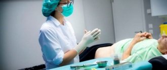
Surgical removal of nevus is the oldest method. This method helps with deep and large formations on the skin. However, this procedure has a drawback: it leaves scars and scars on the skin.
When resection takes place, the growth is cut out, including healthy skin. The manipulation itself takes place under local anesthesia and lasts 30 minutes. Once the formation is removed, the wound is sutured so that the scar is less noticeable.
After the operation, the person goes through a recovery period. The main thing is to care for the place where the manipulation was performed. It should be treated with anti-inflammatory drugs for a week after the stitches are removed.
Surgical removal of the growth is an outdated method; it has been replaced by more modern options. For example, removing a mole with Surgitron. Surgitron is a radio wave surgical device for getting rid of tumors. Its use is considered the safest to date. The procedure takes place without the electrode coming into contact with the skin. The main advantage is that there are practically no postoperative complications, and the rehabilitation period is easy.
Technique and duration of the procedure
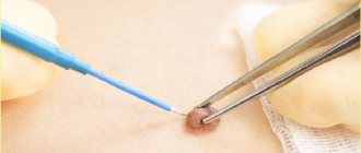
Transportation of the nevus for histological analysis is carried out before the formation is removed or after surgery. Medical institutions remove nevi using various methods:
- Cryodestruction method. The mole is burned off using liquid nitrogen. The temperature at which the formation is removed exceeds 180 degrees.
- Excision with a scalpel. The patient is given local anesthesia. The surgeon removes the growth with a scalpel and applies stitches. The method is not practical for removing a mole located on a visible area of the body.
- Radio wave removal. The nevus is excised using a radio wave knife. The advantage of the method is the absence of scars after surgery.
- Electrocoagulation method. Using a hot metal tip, remove the mole. After three weeks, there will be no traces of the surgery left.
- Laser excision. The growth is removed using a laser. The method is in demand because it does not leave scars.
The study of a removed nevus occurs in several stages:
- Collection of material. The tissue of the formation is taken with special instruments.
- Fixation. The resulting fabric is placed in a container filled with water. Next, ethanol is poured instead of water. An alternative to ethanol is formaldehyde. These substances help preserve the structure of the formation and prevent cellular decay.
- The material must be hard. To do this, pre-dry and place in paraffin.
- The hardened tissue is divided into plates using a special device.
- Paraffin residues are removed.
- The cut plates are placed in a container with dye, which has been prepared in advance. This procedure helps determine cellular structure.
- The painted plates are placed on glass and placed in a container that closes tightly. This allows you to store them for a long time.
- Microscopic examination. A histologist examines the resulting plate using an optical instrument.
- The received data is recorded in order to transmit it to the doctor.
After receiving laboratory data, the attending physician performs a decryption. How much histology of a mole is done depends on where the study is performed. There are cases when the material needs to be transported to a laboratory located in another locality.
The time required for histological analysis ranges from five to seven days.
If a severe form of cancer is suspected, the attending physician will prescribe emergency removal of the nevus. In this case, histological examination is done in an accelerated manner. It involves freezing the excised material, which after three hours is suitable for laboratory study.
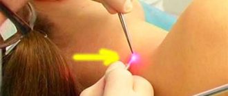
Nevus removal with laser
One of the most effective methods for eliminating moles is also laser removal. The laser removal surgery is performed under local anesthesia. This method is also one of the safest.
The procedure takes no more than 20 minutes, and healing occurs within 2 weeks. After this time, any traces that indicate the operation being performed disappear, there are no scars. This is very important if the operation is performed on the face, for example in the same lip area.
As a rule, the doctor recommends this method in the presence of benign moles or shallow formations. Removal of a malignant or giant nevus occurs only surgically.
One of the features of the laser procedure is that the likelihood of the nevus reappearing is excluded.
After surgery, it is recommended to avoid the sun for a week. At the site where the mole was, the skin should be treated with an antiseptic for the first 3 days.
It is also possible to remove a mole with a neodymium laser. This method is used only in aesthetic medicine and cosmetology. It is effective if a mole needs to be removed from the face, such as the eyelids, nose, lips or cheeks.
Despite the fact that neodymium laser is safe, it has a number of contraindications. It should not be used in certain cases:
- during pregnancy and breastfeeding;
- for purulent diseases;
- if the moles are malignant;
- if there are cracks, scratches or cuts at the impact site.
The essence of the procedure
The histology of a nevus consists of examining a small part of it. The examination will reveal the presence or absence of cancer cells in the mole.
Histology is especially necessary if there is a border nevus. It belongs to a more dangerous formation. To undergo the test, a referral from an oncologist or the person’s own desire is required. A biopsy is taken using the following methods:
- A piece of biomaterial intended for research is taken during surgery. The excised material is transported to the laboratory in a special container.
- Using the needle method. An area of nevus tissue is excised using a dermopanch. Before the procedure begins, the person is given an anesthetic.
- The excisional method is performed during surgery. The entire body of the mole is removed. The intradermal nevus is sent for histology along with the healthy cells that were located near it.
After collecting the material or completely removing the mole, it is immersed in a container with formaldehyde, where it can be stored for up to 25 days. The duration of storage does not affect the quality characteristics of the material. The condition for this is the amount of formalin in the container: it should be several tens of times greater than the volume of the nevus.
Histology of the mole after removal is possible after freezing the nevus. The method is used in case of urgent need to study a mole for the presence of malignant cells. If an urgent examination reveals melanoma, the doctor will change the tactics of surgical intervention.
In addition to taking part of the nevus, there is a cytological method for studying the tumor. The surgeon scrapes the surface of the spot. Using glass, which is pressed against the mole, material is taken. He is sent for laboratory examination.
Consequences of nevus removal
Sometimes, after removal, a nevus recurs, that is, it reappears. This happens if the nevus cells were not completely removed during surgery. At the site of excision, when the crust falls off, a small dark spot may appear. It is worth knowing that this does not lead to cancer, but medical supervision is necessary. If the patient wishes, the nevus can be removed again. But in this case, mandatory histological examination will be required.
The person decides for himself whether to remove a mole or not. And if a decision is made about surgical intervention, it is necessary to take care of choosing a specialist who will carry out this manipulation.
The main thing is that the doctor has experience in carrying out such manipulations. You should always remember that removal that was carried out incorrectly can cause serious consequences, in particular, the risk of developing cancer increases. And if a mole does not bother a person and does not cause him aesthetic or physical discomfort, it should not be removed.
If a mole still bothers you, be sure to consult a doctor. Don't try to remove it yourself. The specialist, after examining the formation, will suggest surgical, laser or radio wave removal. Be sure to listen to the recommendations.
Doctor's answer
Unfortunately, some types of nevi (moles) after surgical removal can recur, that is, recur. This is not only unsightly: in certain cases, there may be a risk of malignancy of the mole, that is, its malignant degeneration.
Recurrence of the nevus appears in rare cases and looks like a small brownish spot. It is at the same level as the skin and practically does not increase over time. The brown color of the nevus is due to the presence of melanin pigment, produced by melanocyte cells.
The main reason for the recurrence of a nevus is its incomplete removal, that is, when its cells remain in the skin. Over time, they intensively produce melanin. In rare cases, a relapse may occur at the site of complete removal of the skin lesion. To find out whether such a nevus is dangerous, the patient must undergo a histological examination. If the nevus is confirmed to be benign, then the reappearance of the mole is not dangerous.
But if the removal was not combined with histological examination, then the risk of malignancy increases significantly. It is always necessary to remember that the risk of malignancy increases in proportion to the amount of time lost.
Often, a re-occurring nevus can grow very quickly, which is typical for melanomas. However, the good news is that the risk of melanoma is very low.
It is possible to accurately determine the nature of a newly appearing mole on the leg after it has been removed using a histological examination of a fragment of its tissue. In the case of a benign course, you must independently monitor the relapse. The doctor determines the necessary diagnostic methods: scraping, puncture, dermatoscopy.
The risk of relapse increases if a mole was removed, and then it quickly increased to its previous size and even became larger. If this happens, then you need to go not only to a dermatology clinic, but also to an oncologist: this, alas, can be a dangerous skin disease.
You can get rid of melanoma only when the patient consults a doctor as early as possible. There is no need to put off and try “folk” methods of removing moles. This is very harmful and can contribute to their rapid growth. There is also no need to try to remove a recurrent nevus, cauterize it or perform other dangerous actions.
Answered by Maria Fedorova
(Moscow), Oncologist
I decided to fantasize a little here and become a theorist (well, based on the latest facts, of course). I will try to show you one of the options for the appearance of local recurrence of skin melanoma after removal of a mole. With “jokes and jokes”, because everything ended very positively.
It turned out to be a very clear example and I am very grateful to one wonderful girl who sent me a piece of paper, otherwise the first part (before) is a standard phenomenon, but for some reason most people have “problems” with the second (after).
Briefly
: a diagnosis of “
dysplastic nevus
” was made, which, after re-checking with the Israeli histological sorcerer Dr. Vaknin, turns into “
melanoma
0.25 mm thick”.
I skip all correspondence (very short) and leave only the essence
22.01.2017
Again about melanoma. ***** (Nsk is a town not inferior to Baghdad)
Uncle Vadik, hello! I studied your entire blog - there is a lot of optimism, the support is colossal, at least my mother wiped away her tears. The main question is this. 10 days ago, a “bad” mole was removed on my back. But alas, they cannot yet make an accurate diagnosis (either melanocytic dysplasia, or superficial spreading melanoma...) They sent the glasses to another laboratory, an answer will come on Tuesday. In general, there is no trust in doctors. And after reading your posts about incorrect diagnosis... it becomes scary. We are planning to go to Israel. If you are interested, I will describe the whole story in detail. I really hope for your answer. The trip is planned for the coming week.
After a short conversation, it was decided to slow down a little and first wait for the results of the local review and “dance from there”
.
05.02.2017
Vadim, good morning! In general, my mother’s story has a super positive ending))))) Repeated histology in Nsk gave a positive answer: dysplastic nevus
.
We contacted Israel and decided to send the material to Dr. Vaknin for review
. The day before yesterday the answer came: everything is fine, but several atypical cells were still found, and therefore Professor Gutman recommends a wide excision, and does not insist on surgery in Israel at all.
I didn’t quite understand about a few cells, but the main thing is this:
That's it, don't get sick!
*************************
Z.Y. “While the issue was being typed up.” Continuation of the banquet. No comments
Good morning! I didn’t finish my “thought” yesterday. Weren't you denied surgery?!
Kindest! We were refused, citing a conference that took place last month specifically on melanoma. All our luminaries seemed to perform there. I’ll try to quote: scientists have proven that wide excision after 21 days from the moment of the first operation does not affect a person’s life expectancy. If the cells were destined to spread throughout the body, this happened.
And yes, Israeli papers are not the basis for registration with our cancer clinic. We were told to go to Blokhin, where I tried to call all day yesterday, give them the material for repeat histology, and if they confirm melanoma, then they will register it and maybe do a wide excision.
*****************
Now that's all. I bow to the wisdom of oncologists, even though this all seems like some kind of surreal nonsense to me.
If anyone knows something about this study, please send me the link
12.02.2017
Wide excision was performed at the Blokhin Regional Cancer Research Center, i.e. The diagnosis made in the laboratory wilds of Israel was completely confirmed. Rezal Dr. Baryshnikov
:
After a long conversation and another dialogue with Israel, the opinion was unanimous: the histology of a wide excision is unlikely to show anything, because a maximum of 15 sections are made from the entire mass of removed material and examined. Therefore, the probability of finding a saboteur cell is extremely low. But it is necessary to remove its potential - this is what Israel calls preventive medicine.
The doctor removed everything exactly the way Gutman removes it in your video - with a scalpel. Mom said that despite the size of the incision in half her back, she felt much more comfortable compared to the operation with an electric knife in Voronezh.
Doc left a very good impression.
By the way, about the timing of wide excision. In Blokhin, the time frame is 2 months after the first operation. Why 2 months? They don’t really answer, but they definitely shrug their hands about 21 days and say that they’ve never heard of it.
Z.Y. Not a single one of our oncology centers, including Blokhin, has given even approximately such a detailed decoding of histology as Israel did. In Blokhin, the result sounded like this: Zlokach. melanoma less than 1 mm thick. All.
**********************
THAT'S EXACTLY ALL NOW.