A birthmark is a benign growth formed on the skin. It is subject to the mechanical influence of external factors that provoke the transformation of healthy cells into cancer cells. This process occurs at lightning speed, so if there is the slightest change in the structure, color, or shape of the formations, you need to consult a doctor. He will prescribe a set of examinations to take treatment measures. One of them is the histology of the mole.
The importance of histology in the early diagnosis of cancer
Histological examination of a growth on the skin is carried out on the basis of a referral issued by a physician, or on the basis of the personal will of the patient. The procedure is only permissible within the walls of the clinic. For analysis, preliminary collection of biomaterial from the growth area is required.
There are 3 examination methods:
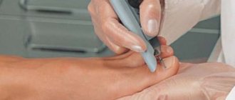
- using a needle (in this case, part of the tissue structure is excised using a specialized instrument, anesthesia measures are first carried out);
- incious method (a part of the material being studied is taken and several fragments of it are sent to the laboratory);
- excision method (used during surgery, however, to the full depth of the skin with excision of healthy elements that are located near the growth, the subject of study is the complete body of the nevus, which was surgically removed).
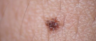
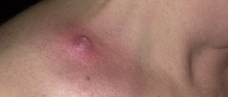
Small in size and regular in shape, moles do not pose any threat. At least this lasts until changes occur in them. If the slightest abnormalities are noticed, you should not delay a visit to a doctor, as they may indicate the initial stage of a dangerous pathology - melanoma. It is a skin cancer. If detected early, it responds well to treatment (traditionally removal is used).
Summary or briefly about the main thing:
When removing a mole without histology by a non-oncologist, I recommend repeated excision with a scalpel with mandatory histological examination. If the operation was performed by an oncologist, in my opinion, it is enough to simply tell the doctor about your concerns.
Other articles:
- How to remove a mole without pain?
- Mole injury - what to do
- Are moles with jagged edges dangerous?
- Breaking the pattern: 5 myths about mole removal!
Useful article? Repost on your social network!
Indications for histology of a mole
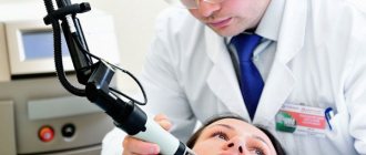
During the visual examination, the doctor has the right to refer the patient for additional tests.
These include:
- study at the molecular level,
- skin examination,
- radioisotope type scanning,
- CT,
- histology.
It is the latter option that is considered the most acceptable and preferable. This is due to the fact that the technique helps to identify malignant cells and prescribe timely treatment measures.
Here are the indications, when they occur, the doctor will refer you to a histological procedure:
- intensive growth of the tumor;
- changes in structure;
- the appearance of tubercles;
- the appearance of a shiny crust on the mole;
- excessive bleeding;
- dark plaque or darkening of the mole;
- spreading of the stain over the surrounding skin areas;
- the occurrence of pain due to mechanical impact.
If glomus angioma is suspected, tactics are rarely performed; it is appropriate only if a cancerous tumor is suspected.
What will the analysis results tell you?
The result will tell the oncodermatologist whether the birthmark was benign or vice versa. Do not try to decipher it yourself - this will be done by the doctor who gave the referral for the study. Poor histology will indicate whether the mole turns out to be melanoma, which can save a person’s life. Oncology develops in 4 stages; early stages are easier and more effective to treat.
If the histology is positive, measures are taken to remove the malignant spot as quickly as possible. Even if the nevus is safe, it is better to eliminate it. This operation is performed in cancer centers or clinics. Removal can be done using surgery, laser removal, or electrocoagulation. It happens that a mole comes back again - you need to have it checked again by a doctor.
Methods of histology of moles
The lion's share of medical institutions conduct histological examinations free of charge. As a result, it becomes possible to make a final verdict and confirm or refute the fact of cancer or glomus angioma. The procedures are carried out within the walls of the laboratory according to classical schematics.
It looks like this.
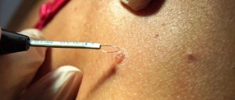
- The doctor removes the problematic tumor.
- After removal, he takes the biomaterial for himself.
- The growth is put into a special solution and taken to the laboratory.
- The material is applied to the glass and must be painted.
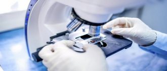
- After a certain period of time, the doctor examines the cells under a microscope.
The research process can be carried out in several ways.
- Freezing the medication. This method is appropriate in a situation where an urgent study of a mole is required. For example, during surgical treatment. If the results of the analysis do not correspond to the doctors’ assumptions, it remains possible to change the course of the operation.
- Paraffinization of biomaterial. This technique makes it possible to preserve the removed growth for a long time. This is required for it to be studied by several specialists at once.
Using modern equipment, it becomes possible to remove and examine tumors of any complexity, and the result is guaranteed.
The classic scheme of an event consists of a number of stages. Let's look at them in more detail.
- Sampling (direct excision of biomaterial).
- Fixation (placing the sample in a container with water, which is subsequently replaced by ethanol, formalin). This measure helps prevent cellular decay and preserve the intravital structure of the mole.
- Wiring. In order to give the taken “raw material” the optimal shape and structure for examination, it must be dried and filled with paraffin.
- Cutting. When the material hardens, it is cut using special techniques until thin plates are obtained. Excess paraffin must be removed.
- Coloring. At this stage, the plates are lowered into a special solution with dyes that were prepared in advance. This helps determine the cellular structure.
- Microscopic examination. The most important stage of the entire procedure. Modern optical instruments are taken, and the laboratory assistant uses them to obtain a series of data that subsequently needs to be transferred to the attending physician.
Carrying out the technique requires about 7 days.
If it is necessary to deliver the material to another locality, the period is extended for the entire transportation time.
If a physician suspects aggressive types of pathology, urgent surgical treatment is necessary in order to save human life. For this purpose, an accelerated method is relevant.
Stages of histological examination: timing and reasons for delays
There are several stages of histological examination. I won’t go into detail about their details, I’ll just list them and briefly explain the essence:
1. Biopsy, i.e. removal of a mole. The main thing I want to note here is that there is never “too little” material for research. They often write to me: “The doctor said that the formation was too small, there would be nothing to send for examination, so I did not have a histology.” In my practice, there have been cases when a normal conclusion was received for a mole up to 1 mm in size. Therefore, no matter how huge a “soldering iron” a mole is removed, it is important to send a fragment of the largest possible size for histology. This is certainly worse than examining the entire nevus, but much better than nothing.
2. Fixation of the material. To prevent the mole from decomposing while it is being transported to the laboratory, it is necessary to destroy microorganisms and deactivate the enzymes of the mole cells. This is done using special solutions. Immediately after removal, the mole is placed in a formaldehyde solution. You can replace it with alcohol, but this option is less common