It's rare to see a person without small dark marks on their body. Is it worth paying attention to these points? Only a doctor will distinguish between dangerous and normal moles - malignant melanoma or harmless nevus - and give recommendations on what to do with them. Is it worth worrying about the appearance of new formations, when immediate contact with specialists is required, what are the signs of cancer development - the answers to these questions remain to be found out. No one is immune from disaster, and early diagnosis will protect you from severe consequences.
Are moles on the body dangerous?
There are a wide variety of types of nevi - they differ in color, size, and shape. Some of them are flat, others are convex. Some do not cause any inconvenience to the owner, others interfere and get caught all the time. Before answering the question of what to do if a mole comes off, let’s figure out why damage to it is dangerous?
No matter what a mole looks like and no matter when it appears, its main danger is that it can develop from a benign neoplasm into a malignant tumor - melanoma. The ability to transform into oncogenic-dangerous formations is distinguished by nevi, which are easily susceptible to injury - damage to a mole increases the risk of blood infection with pigmented cells of which it is composed. Therefore, when a tumor on the body is damaged, a feeling of anxiety and fear always arises - a person who has torn off or injured a nevus is lost and does not know what to do?
Nevus formation
Research into the formation of nevi has not yet reached a common opinion as to why exactly they degenerate. Nevertheless, several factors that provoke the formation of nevi:
- Heredity. Data about melanocytes, which subsequently transform into moles, are found in DNA. Often moles are located in the same places as those of parents and grandparents. If a mole of this particular type becomes inflamed, you should be very careful and contact a specialist.
- Hormones. The endocrine gland (pituitary gland), which is responsible for growth, development and metabolism, produces and releases a certain hormone to the body during puberty. This hormone activates the release of melanin. This leads to the production of pigment, which tends to accumulate in melanocytes. In fact, everything is ready for the formation of new nevi. A similar process occurs in the body of a pregnant woman. Pregnancy is a huge burden on a woman's health. Therefore, during its course, it is necessary to pay attention to all the little things, which include the formation of new moles. The only difference between the surge of hormones during pregnancy and puberty and the formation of nevi is that moles can form during maturation. And during pregnancy, they not only form, but also disappear.
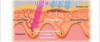
- Ultraviolet radiation. Simply put, sunlight. Staying in the sun for a long time leads to active production of melanin. Therefore, an inflamed mole is a completely normal reaction of the body to excessive sunbathing.
- Cuts, scratches - in general, any mechanical impact on the area of skin where the mole is located. Through damaged skin, infection can enter the mole. The mole begins to hurt, itch, and sometimes peel off. These are just the main factors that trigger the process of inflammation of nevi. Some scientists are inclined to believe that both X-rays and radiation also contribute to the formation of nevi.
A mole is torn - what to do?
Most often, it happens that a person partially damages a mole: he tore it off a little, injured it, caught it or tore it - that is, the nevus did not fall off completely, but remained hanging. In this case, the wound may bleed or there may be no blood at all. What to do in these cases?
An injured mole is bleeding
The very first measures to be taken if damage to the nevus causes bleeding is to disinfect the wound and stop the bleeding. To do this, you will need a clean bandage and an antiseptic - hydrogen peroxide (3%), an aqueous-alcohol solution of chlorhexidine (0.5%).
- The injured nevus needs to be washed generously with peroxide - literally “drown” the damaged area in a bandage soaked in peroxide. You need to keep this compress for at least 15 minutes - this time should be enough to stop the bleeding. In addition to the fact that peroxide acts as an antiseptic - it disinfects the wound, it helps stop bleeding;
- Next, it is advisable to wash the wound with chlorhexidine. This is a stronger antiseptic - the disinfecting effect lasts longer than with peroxide. After the bleeding has stopped, blot the wound with a bandage soaked in chlorhexidine;
- If chlorhexidine is not at hand, you can lubricate the edges of the wound with iodine;
- A sterile bandage should be applied on top of the mole and secured with a sterile plaster;
- It would be a good idea to see a doctor, who will give an objective assessment of the wound and make a decision to remove or preserve the damaged nevus.
A damaged nevus does not bleed
If the nevus was not severely damaged (for example, a woman slightly scratched the mole with a manicure, clothing, or a man caught it while shaving), and there was no or little blood, then it is not necessary to see a doctor. It is enough to wash the wound with an antiseptic and apply a sterile bandage for 15 minutes.
What to do if a mole is torn and hanging?
If a mole falls off and remains hanging, you should not put off visiting the doctor. Most often, they will advise you to get rid of it so that it does not interfere and does not cause a feeling of discomfort - this will be better than waiting until it disappears on its own or trying to remove it at home.
You cannot bandage a torn mole with thread or hair, as is recommended in folk methods; it is better to trust a doctor with this problem. The process of removing a damaged mole must be carried out in conditions where all disinfection measures will be observed. In addition, the doctor will evaluate the type of mole and the location of the damage. If necessary, he will order additional laboratory tests to determine whether the torn mole was dangerous for melanoma.
Today, various methods are practiced with which you can get rid of a nevus, ranging from excision of the mole with a scalpel and ending with more gentle methods - laser, liquid nitrogen, radio knife.
How quickly does melanoma develop from a mole?
The transformation of a nevus into a cancerous formation can occur in different ways. The process depends on the stage of the disease and the type of tumor. Instant metastases are dangerous. Begins:
- growth of cancer (oncological) cells in the deep layers of the epidermis;
- their entry into the blood and lymph;
- penetration into the lungs, liver, kidneys;
- growth in these organs;
- complete damage to the body;
- death.
The growth phases of pigment cells are observed, along which melanoma develops from a mole. There are varieties:
- horizontal - damage to the upper layers of the skin occurs, lasting up to 10 years, but metastases do not appear;
- vertical – accompanied by the spread of cancer cells throughout the organs, can last two years, has an unfavorable prognosis;
- nodular - especially dangerous - characterized by deep spread within two months.
The mole has come off completely - what to do?
If the nevus comes off and falls off completely, this should not be ignored. What to do with the wound? It should be disinfected and the bleeding stopped, as described above. Next is the work of a surgeon or oncologist, who should be contacted immediately.
- You must take with you the torn mole, which must first be placed in saline solution. To do this, a sterile bandage is well moistened in saline solution and a nevus is placed in this bandage;
- If you do not have saline solution, the nevus can be placed in alkaline mineral water for a short time, or you can prepare a saline solution at the rate of 1 teaspoon per liter of water - but this is in extreme cases, when there is no saline solution at hand;
- First aid and examination will be provided at the hospital. A torn mole or its remains on the skin, which the doctor must remove during examination, is sent for histological analysis to check the nevus for the presence of oncogenic cells.
A mole can be damaged in different ways - completely torn off or torn. The main thing is not to get confused and know how to provide first aid in order to prevent serious consequences and not cause complications.
A visit to the doctor cannot be delayed for a long time due to injury to a nevus - it is important to understand that if a mole bleeds, this is not normal. It’s one thing when it gets caught in underwear or a person accidentally damages it with a fingernail, which also shouldn’t be done, but another thing is further prolonged bleeding, which can indicate a dangerous disease.
What moles can be removed
Often nevi cause trouble for women when they are in a visible place - the face, neck. Even if they do not bother you, using removal will be the right decision - the appearance will improve significantly. After the procedure, the doctor must send the tissue for histological analysis to decide whether the mole is malignant or not. If the neoplasm is not dangerous, does not bother you, and does not change in size, surgery is not required. What moles cannot be removed? Experts believe:
- there are no contraindications;
- It is important to choose the right excision technique.
You should be careful about skin growths; it is unacceptable to remove them yourself. Only the doctor will determine whether a nevus is dangerous or not and decide what to do with it. You can delete it if:
- injured from clothing - on the neck, in the groin area, under the armpits;
- cause pain when touched;
- are located under the hair on the head and can be damaged when combing or cutting;
- change color, shape, outline;
- significantly increase in size;
- characterized by the presence of burning, itching;
- accompanied by inflammation and bleeding.
Many readers of the older generation have probably heard the song “I will dream of Karelia for a long time...” The performer of this song, popular in the 60s, was also remembered by fans for the piquant mole on her swan neck. Rumor blamed this fatal spot for the singer’s tragic departure from the stage. They say, trying to achieve perfection, the beauty asked surgeons to remove the mole. And this small flaw concealed salvation from cancer. Having removed the mole, the pop singer withered away and died...
Don't believe these scary legends! Such stories are reborn in the consciousness of those cases that actually happened in medical practice when the patient came to the appointment too late. It can be very bitter to realize that you could have saved a person if he had paid attention a little earlier to a tiny spot that had degenerated into a serious cancer - melanoma, and had not listened to harmful advisers: “Don’t touch the mole out of harm’s way.”
Melanoma can form from almost any pigmented growth on the skin. In recent years, oncologists have been sounding the alarm: the thinning ozone layer increases the ultraviolet radiation of the Earth, which entails an increase in the incidence of malignant neoplasms. Therefore, any moles must be hidden from direct sunlight.
Surprises can be expected both from a black mole, called nevus in Latin, and from a relatively light one. Although the darker the mole, the more melanoma cells it contains and the higher the risk of malignancy. Watch your moles. There are signs by which you can recognize the beginning of unpleasant changes. These signs cannot be missed. A red inflammatory rim appeared on the skin around the nevus.
The color of the mole has changed - it has become lighter or darker. The mole began to grow quickly. An adult will quickly notice this: the size of the birthmark that has been customary for many years has suddenly changed. But in a child, the nevus grows along with the body. Therefore, excessive enlargement of a mole, inadequate to the growth of the baby, should be a cause for alarm. If you have doubts: the mole has enlarged or it just seemed, you can transfer the outline of the nevus onto parchment, and after a week or two, apply tracing paper to the child’s body and compare.
On the surface of the nevus, crusts appeared that looked like dried blood or stearic scales that looked like dandruff. Blood or other fluid is released from the mole. If you notice one of these symptoms, don't panic. Don't immediately suspect the worst. Just see a specialist as soon as possible, preferably a dermatologist, oncologist or surgeon.
The second most common reason for the development of a tumor at the site of a mole is its trauma. Therefore, it is best to remove moles that create the possibility of injury in childhood, before the onset of puberty. It is definitely necessary to remove nevi if they are on a boy’s cheeks and chin, so that after a few years they will not be touched by a razor blade. It is also necessary to part with birthmarks located on the head under the hair - they are easily damaged when washing, combing or styling your hair. It is necessary to remove moles on the back - a person spends about a third of his life lying on it. Moles are also removed in places of contact with the seams of clothing - on the neck, in the armpits, in the groin, in the waist area. You can remove a mole in almost any cosmetology clinic. A few years ago, doctors removed nevi using liquid nitrogen or a special grinding floating knife. Now doctors are increasingly using laser successfully. The simple operation is performed under local anesthesia. The scar heals like a regular wound in about ten days. With the correct removal technique, a virtually invisible scar remains, slightly different from the surrounding skin only in color. This flaw can easily be hidden with camouflage cosmetics.
A watery mole is a sign of degeneration into a malignant formation that requires immediate treatment. You should not self-medicate; you need to seek help from specialists.
Why shouldn't moles be injured?
Oncologists identify two main reasons that all owners of moles should know:
- any injury to the nevus can lead to melanoma (malignant formation);
- the growth of new moles around the injured one, which increases its size;
- the occurrence of an inflammatory process.
If the second and third reasons are not dangerous, but simply require attention, then the first can lead to the death of a person. Cancer is a very insidious disease; it may not make itself felt for a long time, and when the patient feels serious deterioration, medicine is not always able to help.
If a mole bleeds, and later even slightly changes its shape, color, or spots appear around it, medical consultation and examination are also required.
Types of injuries
Many people mistakenly believe that injury can only be dangerous if the nevus is cut off. However, medical removal is much safer since all parts of it are surgically removed. The following are considered household injuries that have bad consequences:
- regular rubbing of the mole (for example, with underwear);
- scratching (in cases where a person scratched himself unsuccessfully);
- constant pressure (belt, tight clothing, shoes);
- cutting off nevus (during shaving, if they are on the head, face, or armpits).
A person does not always notice that he has accidentally caused damage, so when he sees changes on the body, he does not attach any importance to them. It is important to monitor the pigmentation of moles and consult a doctor immediately.
Blood from a mole - why and how to check it?
Why does a birthmark or nevus bleed? People who notice a similar phenomenon on their body often panic, fearing that they have already contracted a deadly disease. Many people don’t even understand how this happened? A nevus can bleed only in two cases:
- his injury, as already mentioned;
- At the cellular level, the mole degenerates into a malignant one, so it fills with blood. Sometimes it may ooze.
Both reasons require testing and consultation with oncologists - the appearance of blood does not always mean the presence of the disease. Only a doctor can determine whether there are skin problems. It will also indicate the level of risk of developing melanoma in a particular case.
Check moles as follows:
- external examination, including assessment of the shape and color of the problematic nevus, as well as palpating it for pain;
- carrying out dermatoscopy with a special device, thanks to which you can see all the processes and changes in the mole.
Doctors may recommend a biopsy to find out how dangerous the mole is. However, such methods should be avoided, since collecting material in the laboratory will injure the nevus. What this can lead to is already known - to oncology.
Never agree to a biopsy, but histology can give a complete picture of the mole. It is carried out after removing a mole using any of the methods. If a mole begins to bleed, the patient will most likely be asked to cut it out for histological examination.
Are weeping moles dangerous?
If a mole on the surface of the skin begins to become weeping, this is a sign of abnormal behavior of a benign tumor; it may begin to transform into melanoma. Dangerous symptoms:
- fluid, ichor or blood oozes from under the formation;
- the formation of wet blisters on the surface of the formation and the area around it;
- changes in the size, shape and color of the nevus;
- inflammatory process with redness and swelling of the formation. The neglected condition leads to the release of pus with an unpleasant odor;
- long-healing wound;
- itching, burning, pain when touched;
- loss of hair from the birthmark, increase in the area of pigmentation.
If treatment is not started, characteristic symptoms may develop:
- increased fluid secretion;
- increased pain;
- rapid growth of nevus, change in skin color around;
- enlarged lymph nodes;
- the formation of ulcers on the surface, which can produce an unpleasant odor and pus;
- painful formations appear;
- bleeding;
- compacted structure of formations.
Melanoma grows by metastases, their signs:
- chronic obsessive cough;
- a sharp decrease in body weight;
- headache;
- muscle spasms.
The nevus begins to become wet due to the development of malignant melanoma, which manifests itself in different shades and asymmetrical shapes of nevi measuring more than 6 mm. Timely treatment will help avoid serious complications that can lead to death. The tumor can spread quickly, growing through several layers of the skin, traveling through the blood and lymphatic vessels. The liver, brain, and lungs are often affected.
Fighting the bleeding
A mole is bleeding - what to do? Indeed, if nevus injury is so dangerous, it is possible that improper treatment can lead to negative consequences. Leading oncologists advise not to panic and take a number of the following measures:
- treat the wound with hydrogen peroxide. In this case, you should definitely wash your hands with soap, and even better, wear sterile gloves, if you have them in your first aid kit. The cotton wool must be well moistened with the solution so that its fibers do not remain in the wound;
- then proceed to stop the bleeding - tightly fold the gauze or bandage (sterile), apply it to the injury site and press it lightly. Take a comfortable position and wait until the bleeding stops;
- after the bleeding has stopped, treat the wound with any antiseptic - brilliant green, iodine.
Moles sometimes bleed for a long time, and this is not unusual, since each of them consists of a large number of blood vessels that are damaged. In fact, bleeding is absolutely not scary compared to the consequences.
Red moles require special attention - they do not bleed, they can simply be vascular nevi. However, it is better to check them, especially if recently they were the usual brown color.
Can the crust fall off on its own?
When a mole becomes covered with a dry crust, you need to find out the reasons for this process and consult a doctor. This may be a sign of melanoma development.
The crust along with the mole may fall off in some cases:
- damage has occurred from regular contact with synthetics;
- temperature effect;
- lack of the required amount of nutrients;
- prolonged exposure to ultraviolet rays.
A hanging mole can easily be touched, twisted, thereby cutting off the blood supply and causing it to fall off. New growths occur due to hormonal imbalances. When the hormones return to normal, the mole with the top layer disappears.
The most dangerous case is when the birthmark peels off, causes itching, and a dry crust. Then, over time, the top layer falls off, which may indicate the beginning of melanoma formation.
After an injury, a scab often appears that falls off on its own. This process cannot be accelerated. If you accidentally hit someone and start bleeding, you need to urgently perform the following manipulations:
- Take a piece of cotton wool or gauze, soak it in hydrogen peroxide and place it on the wound. The injured area will not become inflamed and bleeding will stop;
- After the bleeding has stopped, you need to remove the cotton wool and cover the wound with a bandage, and then seal it with a plaster;
- The detached layer with the growth should be placed in a sterile vial or wrapped in a bandage and taken to the laboratory for examination. By conducting a histological analysis, specialists will be able to verify its benign nature.
What to do next?
After diagnosis, doctors usually prescribe removal of suspicious moles, and every injured nevus becomes just that. Modern medicine offers gentle and safe methods, so you don’t have to worry about the consequences.
The simplest method is surgical excision. It is performed with a scalpel, and the patient is first given local anesthesia. If you follow all the doctor’s recommendations and properly care for the wound, do not rip off the crust and treat the removal site in a timely manner, any inflammatory processes will not affect the patient.
Laser removal is more often used, through which moles are cut off layer by layer to the very base. This procedure takes several minutes. The healing process also occurs quickly and unnoticed.
Based on the results of the histological examination, the doctor will suggest a further strategy of behavior and prescribe treatment, if necessary.
What to do if fluid comes out of a mole
You can identify a water mole by regularly examining your body. If fluid is released from a mole, it is recommended to contact a clinic to diagnose the formation for malignancy. Watery discharge is one of the alarming signs of degeneration. It is not recommended to self-medicate: this will cause pain relief, not healing. Lack of proper treatment will contribute to the development of cancer.
During the examination, the doctor will find out the reasons for the changes. If cancer is suspected, the patient will have to undergo examination. First, material is taken for histological examination to determine the nature of the formation. If oncology is confirmed, a series of tests are prescribed that reveal the spread of metastases affecting the lymphatic system and organs. Based on the test results, the doctor prescribes treatment. It is recommended to remove a water mole.
Modern medicine has several methods of removal:
- Surgical intervention. A safe method for removing nevi in the presence of cancerous signs. Excision of the tumor and its roots along with the surrounding skin to prevent spread. Surgery removes large, flat growths and deep-growing ones.
- Cryodestruction – removal by freezing with liquid nitrogen. Exposure to nitrogen causes tissue necrosis, a bubble with watery content appears on the surface, which bursts on its own. A crust forms, after which it peels off leaving a pink spot. The disadvantage of this method is the inability to calculate the depth of nitrogen exposure. In the case of malignant formation, further development of melanoma is possible.
- Laser therapy helps remove small nevus. In case of metastasis, the method is ineffective - it is impossible to remove the tumor completely.
- The radio wave method is effective in removing small benign formations. A small tumor can only be removed at the first stage.
Prevention
All people have moles, including young children who are restless. Considering the seriousness of the situation, you should adhere to a number of preventive measures that will help moles remain unchanged throughout your life:
- avoid frequent exposure to the sun;
- do not get carried away by visiting the solarium;
- monitor hormonal metabolism in the body;
- do not wear tight, chafing clothes;
- carefully shave, wash and perform other daily manipulations so as not to damage moles;
- If necessary, cover nevi with adhesive tape, especially for children.
You should not remove “torn off” nevi yourself; such procedures should be done by a doctor. It is important to remember that all types of damage can provoke the degeneration of a nevus.
Moles are benign neoplasms that appear on the skin due to the accumulation of a large number of melanocytes, the pigments of the skin. If a mole is small in size and flat in shape, then most often it does not cause any problems to its owner. But sometimes moles can become crusty, bleed and peel. You will learn how dangerous this is in our next article.
Could the appendage be a cancerous tumor?
A malignant tumor that appears due to the activity of melanocytes can be recognized by its appearance:
- superficial location in the form of a process on the skin or mole;
- knotty - the most dangerous and aggressive. It looks like a nodule and can appear on a pigmented area of the skin;
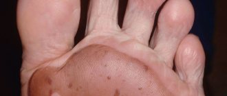
- subungual melanoma - affects the lower extremities, in the area of the nail plate of the big toes, soles.
Any growth on the surface of a mole may turn out to be a cancerous tumor that requires immediate treatment.
Moles are congenital or acquired spots on the body of different sizes, structures and colors. They don't bother you until they start behaving dynamically. Sometimes they change shape or increase in size so that clothes begin to touch them. If a crust appears on a mole, this may signal a modification of hormones in the body that adversely affect health.
What is a mole and what do we know about them?
The presence of moles (nevi) on the body, of course, is not a reason to worry. However, in order to avoid unpleasant consequences, you must treat them carefully. And in order to know why moles are dangerous, it is useful to familiarize yourself with this phenomenon in more detail. Moles are congenital and acquired pigmented formations on the body; the most common ones look like a brown spot or a pea. The medical name for a mole is nevus, which refers to a pigmented formation on the skin. This includes moles and age spots that appear on the skin after birth.
What dangerous moles look like
What do nevi that are subject to pathological changes look like? Only a doctor can correctly distinguish between non-dangerous tumors. Dangerous formations look like this:
- blue – seals under the skin with clear boundaries, with dimensions no more than 10 mm;
- nodal – round, flat in shape, color – brown, black;
- skin – often pale, convex;
- halo nevus - pigment surrounded by a light or white rim;
- Spitz - looks like a dome-shaped tumor of pink shades, with the possible presence of a hole through which blood and fluid leaks;
- connective – connect individual formations into a whole.
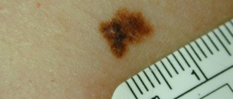
Mole with jagged edges
One of the signs of a non-hazardous formation turning into a dangerous one is a change in contours. It often has blurred edges and scalloped borders. There are non-dangerous types of nevi - dysplastic. Only a specialist can make a correct diagnosis. A mole with uneven edges can be dangerous if there are additional signs of melanoma:
- accelerated changes in size;
- the presence of clearly defined asymmetry;
- the appearance of highly indented boundaries.
Rough mole
Such a neoplasm is harmless if its diameter is no more than 5 mm and remains constant in size. Often its appearance signals a lack of vitamins and nutritional disorders. Doctors advise coming for a consultation if it is discovered that:
- the smooth nevus turned into a rough one;
- bothered by burning, itching, tingling;
- irregularities and compactions appeared in the middle;
- areas with different shades formed;
- diameter has increased significantly.
A dangerous rough mole requires immediate examination if:
- the appearance of bleeding;
- development of the inflammatory process;
- rapid change in size;
- formation of asymmetry;
- formation of purulent discharge;
- the occurrence of painful sensations when touched;
- the emergence of an irregular shape, blurred boundaries, along the edges of the neoplasm.
Large moles
Large formations on the skin are pigment spots. When they remain unchanged and do not cause inconvenience, this is not a dangerous phenomenon. It is important to constantly monitor their appearance, color, and size. To eliminate worries, you need to consult a dermatologist. During the visit, the specialist will conduct a diagnosis and give a forecast of the risk of developing a malignant neoplasm. Large moles become dangerous if they:
- injured;
- thickened;
- started to itch;
- were unsuccessfully removed independently;
- changed in size, shape;
- are bleeding.
Causes of moles
A nevus consists of pigment cells - melanocytes, which in normal condition are located between two layers of skin: the upper layer - the epidermis and the inner layer - the dermis, and are already there at birth. Congenital nevi can be divided into three categories based on size:
- small, with a diameter of 0.5 - 1.5 centimeters
- average, from 1.5 - 10 centimeters
- large moles, more than 10 centimeters, in rare cases they occupy entire anatomical areas, then they are called giant.
What types of moles are there?
One person may discover very different tumors. Types of moles are classified according to several criteria. This helps in correct diagnosis in case of changes. They differ in:
- origin – congenital, newly acquired;
- structure – pigmented, vascular;
- place of formation - in depth, on the surface, in the boundary layer;
- raised above the skin - flat - even, protruding as a hemisphere, pedunculated, larger birthmarks;
- potential threats - dangerous, degenerating into melanoma, non-dangerous.
Benign formations and tumors
As an independent phenomenon, moles are a benign formation, which in some cases can transform into a small ticking bomb, ready to turn into a tumor.
The reasons for this behavior include:
a large number of sunburns at a young age;
- frequent exposure to the sun - for example, from eleven o'clock in the morning to four o'clock in the evening. Those who like to get a good tan are at risk. Ultraviolet radiation produced by the sun causes many skin diseases;
- with a love for the solarium. This is also a very serious risk, since a few minutes in it can be equal to a whole day in the sun;
- nevus injury. Even a simply accidentally touched mole can turn into a tumor if damaged;
- disturbances in the functioning of organs responsible for the body’s hormonal levels.
People of Aryan appearance, with fair skin and red hair are most at risk. In pregnant women, the skin is susceptible to the development of new nevi. In general, anyone who has a large number of freckles and moles should be careful going out into the sun. Those whose relatives were sick with a similar disease, as well as those who have suffered many severe sunburns or who have moles that are darker in the center than the edges, should be constantly examined by specialists.
Crust on a mole
A mole is a completely safe spot on the skin that does not cause discomfort. Over time, it may become covered with a thick crust or change color. In this case, any changes are recorded in the body, the nature of which must be established. Quite often, a crust on a mole forms in the event of hormonal changes that negatively affect the condition of the tissue.
The appearance of a tumor on the skin is not always harmless. A person should know in what cases it is necessary to be extremely attentive to changes of this type on the skin.
Among the symptoms that should alert a person when a nevus is detected are:
This list is not complete. If something worries you, it is better to consult a doctor, especially in the cases listed above or if the mole has changed in any way.
What to do
Under no circumstances should the crust on a mole be torn off . It should fall off on its own, naturally.
If it so happens that in an attempt to get rid of the crust, it comes off (perhaps along with the mole), it is necessary to take urgent measures:
- sterile cotton swab or disk with a pharmaceutical solution of hydrogen peroxide and apply to the site of injury . Peroxide prevents inflammation and bleeding.
- After making sure that blood is not oozing, remove the cotton wool, and in its place, stick a small piece of sterile bandage , folded several times, with a bandage.
- Wrap the detached mole in a sterile napkin, which can be made from a bandage, or even better, place it in a clean bottle with saline solution (one teaspoon of salt in a liter of water), and rush to the clinic. There, see a doctor and submit the mole for histological analysis . Such an analysis will make sure that the neoplasm is benign. This will relieve you from unnecessary worries regarding possible oncological consequences.
If a mole has partially come off, only a dermatologist can give recommendations on its further fate - remove it or treat it without using methods of destruction.
It is best to get an appointment with an oncologist-dermatologist , but such specialists usually work in specialized clinics at oncology hospitals. But there are ordinary dermatologists in almost every ordinary clinic.
What not to do
If a crust appears on a mole, you should not:
- scratch, rub and try to remove it;
- apply to the surface and lubricate with ointments to get rid of the crust . The use of such “treatment” without the consent of a doctor may be not only useless, but also harmful;
- stay in the open sun for a long time . However, exposure to direct sunlight is also dangerous for those who have no problems with moles. And even for those who don’t have them at all.
Features of the degeneration of moles into a malignant formation
Melanoma or cancer poses a serious threat to human life and health. In medical practice, recovery is recorded only in 50 cases out of 100. Today, this type of cancer accounts for approximately 10% of the total statistics. Unfortunately, according to worldwide data, 40,000 people die from this disease every year. To prevent the development of a dangerous pathology, it is recommended to be attentive to the condition of the skin and, if abnormalities are detected, seek advice from a doctor.
A person should know the following symptoms that indicate the start of the rebirth process:
- The structure of the nevus begins to slowly change. One formation may contain several shades and colors at once. When palpated, compaction or excessive softness is noticeable. In this case, the condition is negative and requires immediate consultation with a doctor.
- The outlines lose clarity, and the formation begins to protrude above the surface of the skin. Additionally, it should be noted that a mole in its normal state should have symmetrical edges.
- The mole is covered with a crust, on which cracks appear over time. The formation may also ooze fluid and blood.
- Pigmentation changes the character and causes discomfort to a person.
- The nevus disappeared, and after that a new formation appeared in its place.
- Hair began to fall out around the mole.
A crust can also form on a mole if it has been injured. The situation arises in case of regular scratching. In this case, the formation of a crust is considered completely normal.
After a certain period of time, it will heal or become covered with pink skin. If the patient had a burn before, the crust becomes black. A person can get such a burn in a bathhouse or sauna. It is important to prevent the development of infection, because due to it the nature of the disease can be aggravated.
What are the dangers of injuring moles?
The main problem and concern for any person is the trauma of birthmarks. As medical practice shows, this phenomenon can lead to death. In the event that a cancerous disease is started (without timely treatment of bleeding moles of a malignant nature), metastasis to the bone tissue may occur. Rubbing the nevus, scratching, squeezing and cutting can provoke the development of severe inflammation and bleeding.
Necessary examination
Adequate treatment can be selected only after a correct diagnosis. It involves taking a smear in the area of formation. Additionally, secretions are taken for examination and a biopsy of the removed area is performed. Oncomerkers are of no small importance. Thanks to them, a conclusion is made about the malignancy of the formation.
To monitor the condition of moles in medical practice, the ACCORD system is used. This method was first proposed in America, and only over time became widespread in our country. The system can be used at home without the help of a specialist in this matter. Thanks to this, the patient will be able to recognize a negative situation at the initial stage of development. During the examination, attention should be paid to the following characteristics:
- presence of asymmetry;
- the nature of the ends of the mole;
- presence of blood stains;
- nature of color distribution;
- changes in size;
- growth intensity.
Under what conditions should you go to the hospital immediately?
It is important to consult a surgeon or dermatologist if a mole bleeds. The cause of the blood will be determined by the doctor after examining the patient. You should definitely visit an oncologist in order to identify the form of the formation. If the mole was malignant, then pathogenic microorganisms will begin to multiply throughout the body. If blood flows without injury, a strong inflammatory process develops in the body. In such circumstances, it is important to consult a doctor. You should visit your doctor immediately if:
- bleeding is systematic and profuse;
- moles have changed color;
- education is growing and its form is changing;
- there is a feeling of burning and itching;
- the mole is filled with blood;
- nevus causes severe discomfort.
Under such conditions, self-medication at home is not only not recommended, but also dangerous.
Features of mole removal
It is imperative to get rid of a malignant nevus. Otherwise, metastases may penetrate the lymphatic system. It is recommended to remove a mole only if it is absolutely necessary. Otherwise, the appropriateness of the procedure is called into question. To excise a mole, the following procedures can be used:
- laser treatment of tumors;
- freezing using liquid nitrogen.
- excision with a radio knife;
- cauterization using electricity;
- operation with surgical supplies.
The procedure for removing moles must be carried out according to all the rules. After this, you should not wet the skin in this area for seven days. The sun's rays can also have a negative effect. That is why it is recommended to seal the area when going outside. Additionally, it is advisable to apply sunscreen. During the recovery period, you should avoid applying cosmetics. When a crust forms, you cannot tear it off yourself. It should just fall away. In any case, it is necessary to obtain advice from a specialist in this field, since the risk of malignancy remains.
When to go to the doctor
Consultation with a specialist is necessary in case of any damage to a mole .
A visit to the doctor is mandatory if, in addition to the formation of a crust on the mole, the following occurs:
- the neoplasm began to increase in size and changed shape , acquiring irregularities on the surface and signs of asymmetry;
- that grew on the mole began to fall out
- nevus causes pain when touched ;
- blood or ichor oozes from the mole ;
- ulcerations appeared.
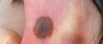
Photo 2. Any moles should be protected from mechanical injury and exposure to sunlight. Source: Flickr (bree)
A growth on a mole is a dangerous sign of degeneration of the formation, requiring a visit to the clinic and examination. Ignoring the problem will lead to serious consequences, which will take a lot of effort to eliminate.
Mole removal methods
Electrocoagulation.
Removal of moles by this method is carried out using a special surgical coagulator. Local anesthesia is used. The mole is cut off by high frequency current, which simultaneously solders the tissue. This avoids bleeding and infection of the wound. After electrocoagulation, the cut material can be examined. A small crust forms at the site of the mole, falling off after 7-10 days, sometimes leaving a barely noticeable scar. The method is effective for removing small moles on the face and body.
Cryodestruction.
For moles that involve only the upper layers of the epidermis, removal with liquid nitrogen - cryodestruction - is suitable. Removal of flat moles is carried out with a cotton swab, which is pre-wetted in liquid nitrogen. The application lasts no more than 3 minutes. During the procedure, a slight numbness is felt at the site of treatment, but there is no pain. In case of deeper germination of moles, a cryodestructor is used. A special ultra-thin needle with a temperature sensor is inserted deep into the skin. After insertion, the needle is cooled to a predetermined temperature and removed almost immediately. In the first few days after the procedure, a rejection reaction of the treated pigmented tissue begins. A crust forms, which falls off and heals within a month and a half. The procedure is contraindicated in case of individual intolerance to cold.
Radio wave removal of moles.
Removing moles with a radio knife is a non-contact, non-traumatic method based on the possibility of coagulation and tissue cutting with high-frequency radio waves. It is characterized by rapid healing - the crust disappears after a week. Contraindications to radio wave removal of moles are the presence of a pacemaker in the patient, as well as herpes and a feverish condition.
Laser removal of moles.
The tool for removing a mole in this case is a powerful light beam, which destroys its cells and evaporates them from the skin. Neighboring tissues are not affected, so burns are excluded. The procedure is bloodless and is performed under local anesthesia. Laser flow parameters must be selected individually. The tissue evaporation treatment is carried out in layers and is completed to achieve healthy skin without touching it. A crust forms at the treatment site after a few days, which disappears after one to two weeks.
Surgical removal of moles.
The most reliable method, suitable for removing large formations against which other methods are powerless, as well as “hanging” moles. Removal of dangerous moles is best done surgically. The procedure has virtually no contraindications and is performed under local anesthesia. The mole is excised with a scalpel and removed, including a small area of healthy skin. The surgery may leave a large scar that will take several weeks to heal. Although the procedure is quick, the wound requires care for a long time. To remove small moles on the face, it is wiser to choose other methods, if possible.
"Folk" methods.
Currently, many different natural-based ointments are advertised, designed to eliminate skin defects - warts and moles. They can be bought without a prescription or a doctor's prescription, but the responsibility for the effect produced will lie solely with you. It is better not to risk using such a product to remove moles, but to consult a doctor. It is strictly forbidden to cut or burn moles yourself.
What is important to know?
If a mole on the face bleeds, it is forbidden to use a traditional treatment method (applying a compress or lotion), since such remedies can provoke the development of cancer cells and cause serious complications.
It is forbidden to touch a bleeding mole with your hands, as this can cause infection. It is forbidden to rip off the scabs - this can cause excessive bleeding.
If the mole has fallen off, you should place it in a sterile container and take it to the doctor for a histological examination. It is important to consult a doctor promptly if blood from a mole appears systematically.
The healing process of the nevus should be monitored by a doctor. If the formation is malignant, then cancer cells will begin to actively multiply throughout the body, which will provoke a worsening of the disease.
If severe pain occurs, you can take a painkiller.
Recommendations from experts
Among other things, if you decide to have an operation to remove a mole, then be prepared for the fact that it will first become covered with a crust. This is a normal reaction to such a procedure. At first, the mole will darken almost to black, but literally in two weeks, when it has completely healed, there will be no trace of it.
Never remove a mole at home! It is very important. The results of such attempts are very disastrous: with the best outcome, you can earn yourself a burn. Rely on specialists who are sufficiently qualified to solve such problems.
Possible complications after removal
If you neglect the process of removing a mole, then melanoma, a scar, and a scar may appear at the site of the formation. Excessive bleeding is also possible. To avoid complications, it is important to follow the specialist’s recommendations and periodically treat the wound with an antibacterial agent prescribed by the specialist. It is important not to pick off the scabs and not visit the solarium until complete recovery.
Traditional methods of treatment can greatly harm and aggravate the problem. In order not to provoke the development of complications, the neoplasm should be diagnosed in a timely manner. People with a family history of cancer should regularly visit an oncologist, since many diseases (or rather, a tendency to them) are transmitted genetically.
It is very rare to meet a person who does not have moles on his body. Small spots ranging from light to intense brown in color may appear on different parts of the body. They usually do not cause discomfort. Therefore, if suddenly changes color, becomes inflamed, begins to ooze or crust over, this immediately causes concern, and not without reason : the consequences can be very serious.
Photo 1. A crust on a mole is not a reason to panic, but a reason for careful observation. Source: Flickr (Donq question).