Share on social media networks:
Lumps and lumps are a common problem that many people experience. They can develop on any part of the body - face, arms, legs, phalanx, hand, back, etc. Sometimes they seem not to be visible, since they are hidden in the folds of the skin and in the hairline. In most cases they are harmless, but some require immediate treatment. Lumps, small lumps that cause pain and discomfort, are most often the result of infection. The skin over them turns red, the ball can be white, blue and is accompanied by fever, headache, and weakness. With timely treatment, all symptoms quickly disappear. Malignant skin tumors are much less common. They need to be recognized in time and consult a doctor immediately. If you have a ball under the skin on your hand and white balls roll when pressed, what should you do? Let's look at the main culprits for such problems.
Causes of seals
There are many different diseases that can cause a hard ball to appear under the skin on your arm or elsewhere.
Hygroma
If you have a dense, inactive ball under the skin on your hand on the side of the wrist, then it is likely that it is a hygroma. It does not hurt or cause harm, and when located in rarer places, for example, on the palm, it interferes with normal operation.
Important! With a strong blow, the hygroma can disappear on its own, since it is an accumulation of fluid between the fibers of the tendon and bursts under strong mechanical stress.
Dermatofibroma
It is a dense red-brown ball consisting of connective tissue with a fibrous structure, therefore it is absolutely safe.
Neurofibroma
This is a stationary fleshy ball, which is a pathologically grown soft tissue. May be located in deep dermal layers.
Important! This ball is dangerous because it can develop into cancer over time.
Lipoma or wen
If you have a ball under the skin on your finger, it could be a lipoma. This is a benign and harmless tumor of fat cells. It is a soft formation with clear boundaries. The skin over the wen is of normal color and easily folds.
Important! When reaching large sizes it can cause pain. Easily removed surgically.
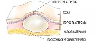
Atheroma
It is often confused with a wen, but, in fact, it is a cyst, that is, a stretched, clogged sebaceous gland. Such a lump consists of sebum, which gradually accumulates, stretching the capsule of the gland. In appearance, it is a dense, round formation with clear boundaries. The skin over the atheroma cannot be folded.
Important! Such a lump can become inflamed and fester. If necessary, it can be removed surgically.
Nodules on the joints
Various joint diseases, such as arthritis and arthrosis, are often accompanied by the appearance of small, hard, immobile nodules under the skin. They are called rheumatoid nodules. They are treated under the supervision of a doctor.
Warts, papillomas, condylomas, soft fibromas
All these formations can be of various shapes, yellowish, brown or flesh-colored, and have a smooth or flaky surface. They can be caused by various factors, such as a viral infection, mechanical injury or hormonal disorder.
Sometimes a wart or papilloma appears for no apparent reason and can be located anywhere. In most cases it is completely safe. So don't be alarmed if you have a ball under the skin on your finger, it could be a wart.
Important! The variety of their shapes, sizes and colors does not allow one to independently distinguish benign and safe condyloma from malignant diseases. Therefore, if a suspicious growth appears, you should consult a specialist.
Skin inflammation and ulcers
A whole group of formations on the skin may be the result of an infection:
- The most common cause of suppuration is staphylococcus bacteria. In these cases, the skin turns red and lumps of various sizes appear. May be accompanied by an increase in temperature.
- Sometimes inflammation quickly spreads across the skin, covering large areas. This is characteristic of erysipelas.
- Phlegmon is a purulent inflammation of the subcutaneous fatty tissue.
- Carbuncles and boils are formed when the hair follicles and sebaceous glands are damaged.
Important! Surgeons treat such diseases. If balls, swelling and redness appear on the skin, you should immediately contact a specialist.
Hernia
A hernia forms in the navel, sometimes in the groin or on the inner thigh. It feels like a soft protrusion under the skin, and can be very painful when palpated. Sometimes you can push it back in with your fingers.
Important! A hernia is formed by the internal organs of the abdomen, which fall out through weak spots in the abdominal wall when intrauterine pressure increases.
The appearance of balls on the legs
Typically, this problem occurs in women due to the following factors:
- When wearing uncomfortable or tight shoes, which provoke poor circulation and joint damage. As a result, a deposit of salts occurs that looks like a ball.
- Due to varicose veins of the legs, thick blood accumulates in places where the walls of the veins are weak, resulting in the formation of a soft and mobile ball of a bluish-violet hue.
- Erythema nodosum is an inflammation of small blood vessels and fatty tissue.
Which doctor should I contact with a lump or lump on the skin?
If you are concerned about formations on your body, find a good doctor using the NaPravku service:
- a dermatologist - if the lump looks like a wart or papilloma;
- surgeon - if surgical treatment of an abscess or benign tumor is required;
- oncologist to rule out a tumor.
If you think that another specialist is needed, use our help section “Who Treats It”. There, based on your symptoms, you can more accurately decide on the choice of a doctor. You can also start with an initial diagnosis from a therapist.
Malignant tumors
Compared to other skin diseases, malignant tumors are rare. Initially, a small nodule appears, which gradually grows.
Signs of a malignant tumor:
- Fuzzy and uneven tumor boundaries.
- Enlargement of nearby lymph nodes.
- The rapid growth of such a ball.
- Bleeding and adhesion to the skin surface.
- Inactivity when palpating the formation.
Important! If you suspect a malignant tumor, you should immediately contact an oncologist.
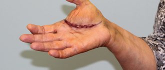
Many people consider new growths on the skin to be simply an unpleasant cosmetic defect until this lump or lump begins to hurt. However, such a hard bulge in a person often indicates the development of a dangerous pathology. If a lump appears under the skin on your hand, you should consult a doctor. It is very important to undergo a comprehensive diagnosis, since the etiology of such a neoplasm may be associated with oncology.
Palpation examination of a pathological area of the patient's upper limb
What is a lump under the skin
The etiology of the appearance of lumps under the skin is very diverse. This may be a hygroma, boil, atheroma, abscess, etc. Often a lump under the skin in the form of a ball on the arm is associated with professional activities.
Due to the anatomical and physiological characteristics of the upper extremities, the joints are often deformed, salts accumulate in them, which cause such formations. The process of salt accumulation is accompanied by pain. It is also worth noting that there are other types of neoplasms, the causes of which are significantly different.
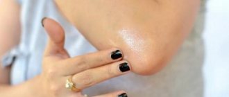
Hygroma
A dense ball is clearly visible, which can be easily felt upon palpation. Most often it is localized on the hands and wrist joints, does not differ in mobility, does not hurt, and does not in any way affect the general state of health. The main cause of concern is cosmetic discomfort.
Appearance of hygroma of the wrist joint
Lipoma
It is a movable ball under the skin on your arm. This tumor is often called a lipoma. It is easily palpable, painless, and has clear outlines. The skin above it folds easily and has a natural color. Most often, lipomas are localized on the outside of the arm, where there is more hair. As the size increases, it may cause some discomfort. The lipoma does not penetrate into the surrounding tissue, so it is easily removed surgically.
Diagnosis of pathology
Undoubtedly, the formation of a swelling or lump is a sufficient reason to consult a doctor. You should not try to determine the cause of the disorder yourself by examining the lump. Even an experienced specialist cannot always make a diagnosis based on the results of the examination. There is a certain diagnostic algorithm that helps to accurately assess the situation.
The list of research methods consists of a number of procedures:
- survey (identifies the main complaints, their severity and how long ago the lump appeared);
- visual examination (determining the location of the lump, damage to the right and left sides or only one of them, its size, the condition of the skin above it, the presence of redness);
- palpation (allows you to find out what the lump feels like to the touch, its density, degree of pain, presence of mobility or adhesion to surrounding tissues);
- puncture (detects the presence of fluid inside and its nature);
- ultrasonography;
- laboratory techniques (blood testing, serological tests);
- biopsy and histological examination (used to clarify the diagnosis in case of suspected tumor).
Etiology of compactions under the skin
The appearance of painful or painless lumps on the arm under the skin can be due to a number of reasons:
- long-term insolation;
- metabolic disorders;
- injuries;
- weak immunity;
- unfavorable environment;
- infections;
- poor body hygiene;
- exacerbation of any disease;
- emotional stress.
Advice. The etiology of subcutaneous neoplasms is quite extensive, so it is sometimes very difficult to establish the true cause that provoked the development of pathology. To make a correct diagnosis, you need to undergo a comprehensive diagnosis.
Cyst
Causes of cysts
Epidermal cyst (atheroma) is a fairly common phenomenon. It is a tumor of benign etiology that forms on the surface of the skin. Its size and location may vary. If removed in a timely manner, the pathology does not pose a health hazard. Cysts form for various reasons.
Infections
Cysts often form against the background of the development of furunculosis. The disease is provoked by two types of staphylococcus: aureus and epidermal. The second type of microorganisms causes suppuration when they enter the deeper layers of the skin from the surface. About 75% of humanity are carriers of staphylococci, so the transmission of microorganisms occurs constantly, however, for the development of the disease, it is necessary for the infection to enter favorable conditions: into an organism with weakened immunity, various diseases.
Blockage of the sebaceous glands
A blockage of the sebaceous gland is a mobile, painless formation. It is characterized by clear contours. In some cases, it grows almost imperceptibly, sometimes its size does not change at all for several years. Sometimes blockage of the sebaceous gland is accompanied by suppuration with pain, swelling, redness and fever. In many cases, pus breaks out in combination with fatty contents. Increased sweating, which manifests itself mainly during hormonal changes characteristic of young age, also contributes to the development of atheromas.
Complications after piercing and tattoo
Piercing and tattoos as an element of subculture are unlikely to go out of fashion. However, their popularity still fell; mass demand was observed in the late 90s - early 2000s. After piercing, the puncture site must be constantly treated with antiseptic agents, otherwise the wound may become infected. If seals form at the puncture site over time, you should consult a cosmetologist or dermatologist. Epidermal cysts are very often diagnosed after piercings and tattoos.
Cyst treatment
Treatment of cysts can be carried out using both conservative and radical methods. The method of treatment depends on the size of the tumor. If the epidermal cyst is small in size and does not cause concern to the patient (including aesthetics), then it does not need to be treated. In this case, doctors recommend monitoring the dynamics of tumor development. If a cyst bothers a person, then it needs to be removed. The following methods can be used in treatment.
Crushing the subcutaneous bladder
Often a cyst is mistaken for a common pimple and they try to squeeze it out. However, this method will not lead to anything good, except that it will begin the inflammatory process.
Pumping out liquid
Ineffective method of treatment. During this procedure, there is a high risk of wound infection and the development of inflammatory processes. You should absolutely not squeeze out the cyst. During destruction, the contents of the cyst can get under the dermis, leading to an abscess. If this has already happened, you should immediately go to the doctor.
Surgery
All manipulative actions are performed under local anesthesia. The epidermal cyst, which is located above the outer skin, is removed within 20 minutes. After injecting a solution of novocaine or another anesthetic, the doctor makes an incision in the center of the cyst and removes its contents, capturing the capsule, then scrapes out the cavity and cleanses the skin. In case of relapse, repeated surgical intervention is prescribed.
Lipoma
Causes of lipoma
Lipoma is a polyetiological disease that develops against the background of various endo- and exogenous factors.
Hormonal disbalance
Lipomas are most often diagnosed in adolescence and in women during menopause. Steroid hormones (androgens and estrogens) affect the functioning of the sebaceous glands, regulate the proliferation of skin cells, and the secretion of sebum. If androgens predominate in the body of women (taking hormonal drugs, age, menopause, tumors), the tissue of the sebaceous glands grows with an increase in secretion production.
Genetics
It has been proven that the formation of lipomas has a hereditary predisposition. Multiple lipomas (lipomatosis) are often traced in several generations, regardless of gender. In a study of twins with this pathology, data were obtained on the presence of this pathology in both children. Vertical inheritance is traced in 99% of cases.
Note. The development of lipoma is often observed against the background of alcoholism, malignant tumor processes of the upper respiratory tract, and diabetes mellitus.
Schematic representation of a lipoma
How to remove wen at home
At home, you can use both pharmacy and folk remedies. Before using any product, it is necessary to perform a sensitivity test. A small amount of the drug used is applied to the skin in the wrist area. If allergic reactions (rashes, itching, hyperemia) are absent, the product can be used.
Doctors recommend using the following medications:
- We see. The product is applied to the skin twice a day, after which the problem area is covered with a plaster. The duration of therapy is determined by the doctor individually.
- Vishnevsky ointment - the substance is generously applied to the lipoma, covered with a napkin and sealed with a band-aid. As the product dries, the “compress” is changed. It usually takes 4 to 5 days to remove the wen.
Advice from doctors
At the first signs of a subcutaneous neoplasm, it is imperative to carry out diagnostic measures and undergo the necessary course of treatment. You do not need to make a diagnosis yourself and prescribe a treatment regimen. This should be done by professionals.
There are many reasons that provoke the formation of lumps, lumps, growths, and other types of neoplasms. Some of them are harmless and can be ignored. However, there are those that are malignant in origin, and they need adequate therapy. You should not engage in self-diagnosis and self-medication; it is better to trust your health to a qualified specialist.
Video
No one is immune from the appearance of foreign tumors on the body - rashes, wen, acne, moles, papillomas, etc. Some of them are absolutely safe and do not pose any harm to health, while others can provoke the development of quite serious diseases, even cancer .
Subcutaneous bumps can appear anywhere: on the legs, arms, face, including the cheeks and other parts of the body. As a rule, their occurrence is noticed after the neoplasm reaches a large size.
Myogeloses
A special form of myalgia (muscle pain) is myogelosis, which is painful subcutaneous lumps in muscle tissue. They occur when a muscle is subjected to excessive stress or due to direct injury. Clinically, movable subcutaneous lumps the size of a hazelnut are palpable, sharply limiting the function of the limb due to pain.
The mechanism of myogelosis formation is a local contraction, an increase in the tone of muscle fibers, caused by a violation of neuromuscular signal transmission. In the treatment of myogelosis, deep massage is used. This is practically the only case when the manipulations of a massage therapist will be painful, but will bring a therapeutic effect. In addition, local anesthetic blockades and physical therapy are used.
Types of subcutaneous bumps
This is a seal that comes in several varieties. Some of them appear almost instantly - within a few hours, others are characterized by slow growth, so their increase in size can be noticed only after a certain amount of time. In any case, if you notice a thickening under the skin, you need to monitor its behavior and, if necessary, consult a doctor. This symptom should not be ignored, since a subcutaneous lump may be the first sign of an incipient disease.
The most common types of subcutaneous neoplasms are:
- A fatty tumor or lipoma is a benign neoplasm that consists of fat cells. It has clear boundaries and is soft to the touch. It can appear on any part of the body (back, shoulders, elbows), without having any effect on the internal organs. The tumor has its own capsule.
- Hygroma. This is an absolutely safe lump, one of the types of cysts. The lump on the hand under the skin (on the wrist) looks like a ball or lump. Hygroma is the result of the accumulation of fluid between tendons that are constantly under tension, and therefore most often appears on the hands.
- Lumps on the leg under the skin. If such a lump appears, you should immediately consult a doctor and undergo an examination. A neoplasm on the lower leg may indicate the development of quite dangerous pathologies.
- Joint nodes or growths. These are immobile, hard neoplasms that accompany the development of gout or arthritis.
- Cyst or atheroma. It is formed as a result of blockage of a violation of the outflow of secretions secreted by the sebaceous glands. The fluid gradually accumulates and forms a lump, shaped like a cyst. The neoplasm has a capsule filled with a thick mass. Most often, atheroma appears in places where sebaceous glands accumulate and near the hairline. May spread throughout the body. Threatens the development of the inflammatory process and suppuration.
- Painful lumps. Appear within a few hours due to an insect bite, bruise or infection under the skin (through a cut, scratch). In this case, there may be an increase in body temperature. Such bumps are treated quite quickly. If the lumps appear as a result of an impact, long-term therapy may be required.
Hernia
Hernial protrusion can be detected under increased loads. It manifests itself:
- on the stomach;
- in the umbilical zone;
- in the groin area;
- on the inner thighs.
In a calm state and lying down, it becomes invisible.
Such formations under the skin are easily palpable and lead to severe pain. In some cases, it is possible to straighten them using your fingers.
A hernia manifests itself by squeezing out internal organs through a weakened peritoneum. The process is especially acute in the case of increased pressure as a result of coughing or physical exertion.
Neoplasms on the back
Lumps on the back under the skin can have different origins. Therefore, a specific treatment regimen is determined for each type. Each type has its own symptoms and characteristics.
Lipoma
A neoplasm consisting of adipose tissue and benign in nature. The lump is soft to the touch, mobile, and forms under the skin on any part of the back.
There are the following causes of lipoma:
- blockage of the sebaceous bed;
- disruption of metabolic processes in the body;
- unfavorable environmental conditions;
- gradual mechanical impact;
- unbalanced diet and unhealthy lifestyle.
This pathology most often affects people in working professions (porter, loader). Lipoma often appears in women under the age of 30.
The size of the cone can vary from a small pea to the size of a child's head.
Features of formations on the hands
A harmless neoplasm most often appears on the hand, in particular on the wrist, called a hygroma. It usually develops near tendons and joints, in places that are often subject to injury. In some cases, hygroma develops due to hereditary characteristics. Most often, the disease affects young women aged 20–30 years. Experts attribute this to the constant stress on the hands of a young mother when she is carrying a baby.
If the cyst is hidden (under the ligaments), it can only be detected in the clinic, where the patient comes with complaints of pain in the wrist joints that occurs when flexing the hand.
Basically, subcutaneous tumors in this area do not cause pain; pain can only appear with pressure or as a result of mechanical impact.
Hygroma often occurs in the following areas:
- On the palm, near the thumb. In such cases, pain is noted when squeezing and loading.
- Near the base of the fingers. As a rule, such neoplasms are small in size and quite painful.
- On the finger phalanges (one or two). Since there are many nerve fibers in this area, the seals here are quite painful.
- On the front of the foot near the shin or on the back side.
Causes of lumps on the hand
Soft, dense tumors are most often found near small and large joints. They can form as a result of mechanical impact (impact, bruise, etc.), prolonged monotonous load on these areas, or an inflammatory process occurring in them.
In older people, such formations can develop against the background of an accumulation of connective tissue fragments near tendons or joints.
Lumps usually appear on the outer surface of the hand, which is constantly in a tense working condition. This may be due to heavy physical labor, as well as constant work on the computer.
If you shine a flashlight on a subcutaneous lump in complete darkness, you can discern some iridescent substance resembling a gel.
Symptoms of hygroma
The tumor develops quite quickly. First, a small compaction appears, which soon turns into one or several bumps located close to each other. The process may be accompanied by mild pain, which is often characterized as a dull ache. If the lump presses on tendons, nerve fibers or blood vessels, the pain may intensify, which significantly impairs the quality of life. The dimensions of the neoplasm reach 3 cm.
Other signs include:
- the surface of the hygroma is dense and rough or completely smooth;
- the tumor is motionless, as it clings to neighboring tissues;
- as a rule, at the initial stage of tumor development, there is only liquid in the hygroma capsule;
- In the future, blood clots may appear, like small balls.
Although this is an absolutely safe neoplasm that does not develop metastases, it is still better to cure it. Firstly, it looks rather unaesthetic, and secondly, it still causes some discomfort that interferes with normal life activities.
Therefore, it is better not to postpone a visit to the clinic, especially if the lump begins to increase in size.
Contact a specialist
If a lump under the skin appears on your stomach, legs and arms, buttocks or back, you should definitely visit a doctor and undergo an appropriate examination. If necessary, the surgeon may refer the patient to a dermatologist or oncologist.
In such a situation, you should never self-medicate, as this can lead to serious complications, the development of inflammatory processes, as well as severe irreversible consequences.
Cyst
A cyst can form as a result of skin infection or blockage of the sebaceous gland ducts. Inside the formation there is a sac filled with pus or liquid.
A special type is the epidermoid cyst, which is also called sebaceous. It looks like a round subcutaneous sac formed from a hair follicle. There is a black center inside the formation.
The growth can be found on the genitals, back and chest. It must be removed and then treated with antibiotics. Often the growth does not cause pain and goes away on its own. But in case of inflammation, removal in the surgeon's office is required.
Cysts on the palms may indicate various diseases
Treatment of neoplasms
Often people turn to the doctor when a tumor that has appeared under the skin begins to hurt. After all, it is quite difficult to notice the moment the lumps appear: at first the tumors are small in size and do not bother the owner in any way.
Although there are many recommendations for getting rid of subcutaneous tumors, the most effective and reliable method is removal. The fact is that non-surgical methods of treating such tumors bring only temporary relief, after which the pathology reappears.
There are the following methods for removing subcutaneous bumps:
- Radio wave method. The formation is removed on an outpatient basis, the scars remaining after the operation resolve within 2.5-3 months.
- Using a laser. The subcutaneous seal is instantly removed, while the integrity of the surrounding tissue is not compromised. The advantage of this method is the absence of a rehabilitation period for scars.
- Liquid nitrogen is used to cauterize lipomas. This leaves a scar.
If the pathological compaction has reached a large size, it will have to be removed only in a hospital setting, using a regular scalpel. Before surgery, the doctor prescribes anti-inflammatory therapy for atheroma to prevent pus from entering the bloodstream. Naturally, after such an operation a long rehabilitation period will be required. Open intervention is also indicated for the formation of a malignant tumor.
At the first signs of a subcutaneous neoplasm, it is imperative to carry out diagnostic measures and undergo the necessary course of treatment. Do not try to determine the type of tumor and prescribe therapy yourself. The diagnosis should only be made by a specialist based on the studies performed.
Atheroma
Cystic benign neoplasm - appears against the background of blockage of the sebaceous duct. The diameter of the ball is on average from 0.5 cm to 7 cm.
In the inner part there is a mixture of epithelium, sebum, cholesterol and hair. When pressed, fat is released from the center of the atheroma.
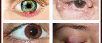
The growth of cysts is provoked by cosmetics based on fatty oils. Doctors consider the eye area to be a dangerous location.
The inflammatory process for about a century causes difficulties, up to surgical intervention. Do not neglect qualified help and trigger the development of atheroma.
Sarcoma, neurofibromatosis - a malignant formation, reaches the size of a walnut (3-4 cm in diameter).
At the initial stage it does not bother a person; over time, the color of the seal changes from light to brown.
The process of suppuration begins, then decomposition. The compaction causes pain.